Abstract
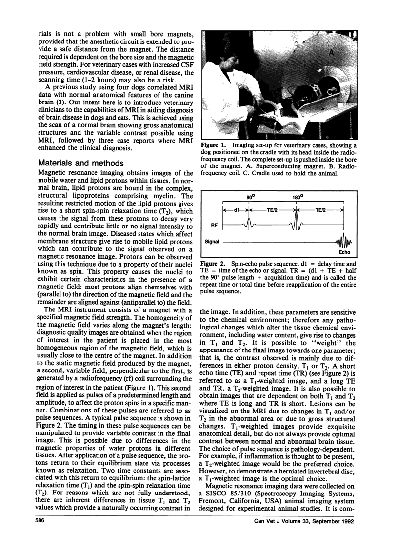
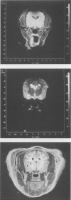
Magnetic resonance imaging (MRI) data were correlated with clinical and cerebrospinal fluid (CSF) findings in one cat and two dogs with brain lesions. In all three cases, localization of the lesions, as determined clinically, was confirmed using MRI. Magnetic resonance imaging also helped us to define the full extent of the lesion(s) in each case. In one case, the lesion would have been diagnosed as purely inflammatory based on the abnormalities in the CSF. The MRI study, however, showed a homogeneous mass with circumferential changes characteristic of peritumoral edema or inflammation. In two cases, the MRI findings were confirmed at necropsy. An MRI study was also done on a normal dog, demonstrating the variable contrast and anatomical detail possible using this technique. We also discuss difficulties in identifying tumor type using MRI.
Full text
PDF


Images in this article
Selected References
These references are in PubMed. This may not be the complete list of references from this article.
- Bydder G. M., Kingsley D. P., Brown J., Niendorf H. P., Young I. R. MR imaging of meningiomas including studies with and without gadolinium-DTPA. J Comput Assist Tomogr. 1985 Jul-Aug;9(4):690–697. doi: 10.1097/00004728-198507010-00006. [DOI] [PubMed] [Google Scholar]
- Felix R., Schörner W., Laniado M., Niendorf H. P., Claussen C., Fiegler W., Speck U. Brain tumors: MR imaging with gadolinium-DTPA. Radiology. 1985 Sep;156(3):681–688. doi: 10.1148/radiology.156.3.4040643. [DOI] [PubMed] [Google Scholar]
- Graif M., Bydder G. M., Steiner R. E., Niendorf P., Thomas D. G., Young I. R. Contrast-enhanced MR imaging of malignant brain tumors. AJNR Am J Neuroradiol. 1985 Nov-Dec;6(6):855–862. [PMC free article] [PubMed] [Google Scholar]
- Hesselink J. R., Healy M. E., Press G. A., Brahme F. J. Benefits of Gd-DTPA for MR imaging of intracranial abnormalities. J Comput Assist Tomogr. 1988 Mar-Apr;12(2):266–274. doi: 10.1097/00004728-198803000-00015. [DOI] [PubMed] [Google Scholar]
- Kilgore D. P., Breger R. K., Daniels D. L., Pojunas K. W., Williams A. L., Haughton V. M. Cranial tissues: normal MR appearance after intravenous injection of Gd-DTPA. Radiology. 1986 Sep;160(3):757–761. doi: 10.1148/radiology.160.3.3488563. [DOI] [PubMed] [Google Scholar]
- MacKay I. M., Bydder G. M., Young I. R. MR imaging of central nervous system tumors that do not display increase in T1 or T2. J Comput Assist Tomogr. 1985 Nov-Dec;9(6):1055–1061. doi: 10.1097/00004728-198511000-00010. [DOI] [PubMed] [Google Scholar]
- Spagnoli M. V., Goldberg H. I., Grossman R. I., Bilaniuk L. T., Gomori J. M., Hackney D. B., Zimmerman R. A. Intracranial meningiomas: high-field MR imaging. Radiology. 1986 Nov;161(2):369–375. doi: 10.1148/radiology.161.2.3763903. [DOI] [PubMed] [Google Scholar]


