Abstract
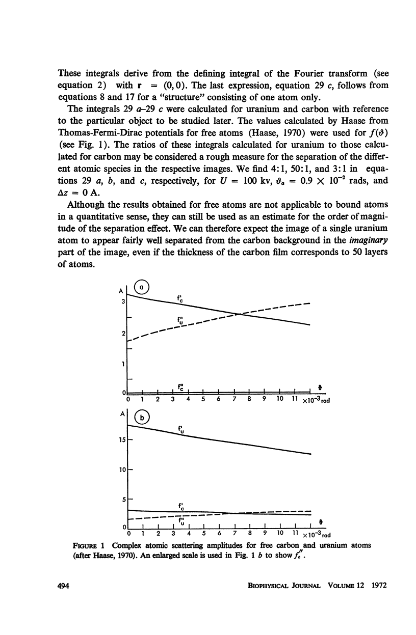
The Z dependence of the phase angle of the complex atomic scattering amplitude can be used to separate the image due to the heavy atoms from that due to the light atoms of the object structure. The linear theory of image formation applied to a focus series of bright-field images leads to Schiske's formula for the calculation of the structure factor. A program system is described which uses this algorithm for computing both images from a set of digitized electron micrographs of a focus series of uranyl-stained DNA on a thin carbon film.
Full text
PDF








Images in this article
Selected References
These references are in PubMed. This may not be the complete list of references from this article.
- BEER M., MOUDRIANAKISEN Determination of base sequence in nucleic acids with the electron microscope: visibility of a marker. Proc Natl Acad Sci U S A. 1962 Mar 15;48:409–416. doi: 10.1073/pnas.48.3.409. [DOI] [PMC free article] [PubMed] [Google Scholar]
- Crewe A. V., Wall J. A scanning microscope with 5 A resolution. J Mol Biol. 1970 Mar;48(3):375–393. doi: 10.1016/0022-2836(70)90052-5. [DOI] [PubMed] [Google Scholar]
- Fiskin A. M., Beer M. Autocorrelation functions of noisy electron micrographs of stained polynucleotide chains. Science. 1968 Mar 8;159(3819):1111–1113. doi: 10.1126/science.159.3819.1111. [DOI] [PubMed] [Google Scholar]
- MOUDRIANAKIS E. N., BEER M. BASE SEQUENCE DETERMINATION IN NUCLEIC ACIDS WITH THE ELECTRON MICROSCOPE. 3. CHEMISTRY AND MICROSCOPY OF GUANINE-LABELED DNA. Proc Natl Acad Sci U S A. 1965 Mar;53:564–571. doi: 10.1073/pnas.53.3.564. [DOI] [PMC free article] [PubMed] [Google Scholar]
- Ottensmeyer F. P. Macromolecular finestructure by dark field electron microscopy. Biophys J. 1969 Sep;9(9):1144–1149. doi: 10.1016/S0006-3495(69)86441-6. [DOI] [PMC free article] [PubMed] [Google Scholar]
- Williams R. C., Fisher H. W. Electron microscopy of tobacco mosaic virus under conditions of minimal beam exposure. J Mol Biol. 1970 Aug 28;52(1):121–123. doi: 10.1016/0022-2836(70)90181-6. [DOI] [PubMed] [Google Scholar]
- ZOBEL C. R., BEER M. Electron stains. I. Chemical studies on the interaction of DNA with uranyl salts. J Biophys Biochem Cytol. 1961 Jul;10:335–346. doi: 10.1083/jcb.10.3.335. [DOI] [PMC free article] [PubMed] [Google Scholar]


