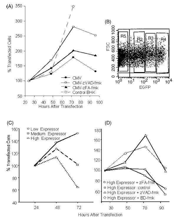
Fig. 1.

Induction of apoptosis by a conventional DNA vaccine: (A) Reduced cell proliferation after transfection with a conventional DNA plasmid is—at least in part—due to caspase-dependent apoptosis and cell survival can be prolonged with a caspase-inhibitor (zVAD-fmk). (B) Sorting of BHK-21 cells transfected with a conventional, EGFP expressing plasmid into three groups 24 h after transfection. Shown is the setting of the sorting windows in an FSC-FL1 scattergram. Medium-expressor cells are defined as cells expressing EGFP levels comparable to cells transfected with the replicase-based EGFP plasmid [23]. (C) Sorted cells are cultured and their survival is monitored. (D) Cell survival in the high-expressor group can be significantly prolonged by the addition of two different caspase inhibitors (zVAD-fmk, BD-fmk). Shown are the average values of quadruplicate wells (96-well plate) based on the number of cells seeded (set to 100%).
