Abstract
The abilities of cysteine-containing compounds to support growth of Bordetella pertussis and influence pertussis toxin transcription, assembly, and secretion were examined. Cysteine is an essential amino acid for B. pertussis and must be present for protein synthesis and bacterial growth. However, cysteine can be metabolized to sulfate, and high concentrations of sulfate can selectively inhibit transcription of the virulence factors, including pertussis toxin, via the BvgAS two-component regulatory system in a process called modulation. In addition, pertussis toxin possesses several disulfide bonds, and the cysteine-containing compound glutathione can influence oxidation-reduction reactions and perhaps disulfide bond formation. Bacterial growth was not observed in the absence of a source of cysteine. Oxidized glutathione, as a sole source of cysteine, also did not support bacterial growth. Cysteine, cystine, and reduced glutathione did support bacterial growth, and none of these compounds caused modulation at the concentrations tested. Similar amounts of periplasmic pertussis toxin were detected regardless of the source of cysteine; however, in the absence of reduced glutathione, pertussis toxin was not efficiently secreted. Addition of the reducing agent dithiothreitol was unable to compensate for the lack of reduced glutathione and did not promote secretion of pertussis toxin. These results suggest that reduced glutathione does not affect the accumulation of assembled active pertussis toxin in the periplasm but plays a role in efficient pertussis toxin secretion by the bacterium.
Pertussis toxin is a major virulence factor of Bordetella pertussis, the causative agent of whooping cough. Pertussis toxin is a member of the AB5 family of toxins, which also includes cholera toxin, Escherichia coli heat-labile toxin, and Shiga toxin. Pertussis toxin has the most complex structure of any bacterial toxin (18, 20, 24). It is assembled from six subunits encoded by five genes, ptxS1 to -S5. ptxS1 encodes the structural gene for the A-subunit, which is an ADP-ribosyltransferase. S1 modifies mammalian G-proteins, which play a critical role in cellular signaling, and disruption of signaling by these proteins impairs development of the immune response to B. pertussis. Unlike the other AB5 toxins where the five B-subunits are identical, pertussis toxin has four different subunits (encoded by the ptxS2 to -S5 genes) in the B-pentamer.
Assembly and secretion of pertussis toxin is a complex process requiring many accessory factors. Each of the five subunits possesses a secretion signal peptide, which directs secretion to the periplasm (18, 20). In the periplasm, the signal peptide is removed and the subunits fold and assemble (5, 9, 10, 21, 26). Assembled pertussis toxin is released from the periplasm by the Ptl secretion complex, a member of the type IV secretion systems (7, 8, 10, 21, 29). Each of the nine ptl genes (ptlA to -I) are essential for extracellular secretion (7). Periplasmic enzymes have also been shown to be essential for pertussis toxin assembly (26). Mature pertussis toxin contains 11 intramolecular disulfide bonds. The Dsb family of enzymes catalyzes disulfide bond formation in the periplasm. DsbA forms disulfide bonds (1) and is essential for assembly of pertussis toxin (26). DsbC catalyzes the exchange of disulfide bonds (22, 31). DsbC has been shown to be essential for pertussis toxin secretion, but not for assembly of pertussis toxin in the periplasm (26).
The pertussis toxin structural genes and the ptl secretion genes are encoded in a single operon that is regulated at the transcriptional level by the BvgAS two-component regulatory system (13, 17, 29). Under permissive conditions, transcription of the B. pertussis virulence factors, including pertussis toxin, is activated. Under nonpermissive conditions, such as in the presence of high levels of sulfate ions, transcription of the B. pertussis virulence factors, including pertussis toxin, is repressed (19). Recently, it has been demonstrated that cysteine in the culture medium can serve as a precursor for sulfate production, which then modulates expression of the Bvg-regulated genes (2). Cysteine has been reported to be an essential amino acid for growth of B. pertussis, an observation that is supported by the presence of defective copies of the genes for cysteine biosynthesis in the preliminary genome sequence of B. pertussis that are intact in the preliminary genome of Bordetella bronchiseptica. These sequence data were produced by the Bordetella Sequencing Group at the Sanger Centre and can be obtained from ftp://ftp.sanger.ac.uk/pub/pathogens. Two sources of cysteine are available in the defined medium, Stainer-Sholte broth (SS), cystine and reduced glutathione (23). Cystine is a cysteine dimer formed by disulfide bonds. Reduced glutathione contains the amino acids glutamic acid, cysteine, and glycine, and oxidized glutathione is the dimerized form. Both reduced and oxidized glutathione are present in the endoplasmic reticulum of eukaryotic cells and play an important role in oxidation-reduction reactions and disulfide bond formation (11). In addition, reduced glutathione is present in high concentrations in the respiratory tract and serves as an important defense against oxidative damage (3, 15, 27).
In this study we were interested in systematically examining the ability of different cysteine-containing compounds to support the growth of B. pertussis, as well as their influence on pertussis toxin transcription, assembly, and secretion. Reduced glutathione appears to play a unique role in promoting pertussis toxin secretion.
MATERIALS AND METHODS
Bacterial strains and growth conditions.
B. pertussis strains were maintained on Bordet-Gengou agar (Difco, Detroit, Mich.) containing 15% sheep's blood (Colorado Serum, Denver, Colo.). When necessary, the following antibiotics at the indicated concentrations were added to the media: nalidixic acid, 30 μg/ml; gentamicin, 30 μg/ml; and kanamycin, 50 μg/ml. B. pertussis strain BP338 (28) was used as the wild-type strain. BPM3171 is a derivative of BP338 which contains an insertion of transposon Tn5 lac in the ptlC gene (29, 30) and serves as a transcriptional reporter. BPM3171 fails to efficiently secrete pertussis toxin, but active toxin accumulates in the periplasm (29). BPM3171::pKC113 was generated by introducing the suicide plasmid pKC113 into the adenylate cyclase toxin gene as previously described (5). Plasmid pKC113 encodes the entire ptxptl operon and restores pertussis toxin secretion (5).
Toxin synthesis and secretion were assessed for bacteria grown in SS broth or modified SS differing in cysteine-containing compounds. SS contains 40 mg of cystine/liter and 100 mg of reduced glutathione/liter. In modified SS, the concentration of the cysteine-containing compounds was the same on a per-gram basis as the original SS, or the molar concentration of disulfide forms of the nutrient was half that of the reduced form. The modifications were as follows: H2O supplements contained water instead of cystine and glutathione; CSH (reduced cysteine) supplements contained cysteine; CS-SC (oxidized cysteine) supplements contained cystine; GSH supplements contained reduced glutathione; and GS-SG supplements contained oxidized glutathione. In other experiments, the non-cysteine-containing reducing agent dithiothreitol (DTT) was added at 3 mM, or 2-mercaptoethanesulfonic acid (MESNA) was added at 10 mM. Chemicals were purchased from Sigma Chemical Company (St. Louis, Mo.).
To assay de novo production of pertussis toxin, strains were passed twice on Bordet-Gengou agar containing 40 mM MgSO4 to modulate the bacteria and turn off transcription of the pertussis toxin operon (12, 19). Bacteria harvested from 24-h plates were suspended in SS containing 40 mM MgSO4 to an optical density at 600 nm (OD600) of 0.1 and grown at 37°C with shaking at 150 rpm for 48 h to generate seed cultures. Bacteria were suspended into SS or modified SS to an OD600 of 0.1, and 50 ml of each suspension was grown statically at 37°C for 48 h in Roux bottles to stationary phase. Our initial growth studies suggested that B. pertussis grows with similar kinetics in the modified SS media that support appreciable growth, despite modest differences in the final ODs obtained. It has been previously observed that functional toxin accumulates in the periplasm and remains constant throughout log phase, while secretion is maximal from mid- to late-log phase with no further secretion in stationary phase (6, 7). By reporting values for stationary phase, 48-hour cultures, the possibility of growth phase differences between the cultures has been eliminated. Pertussis toxin transcription was monitored using β-galactosidase (β-Gal) as a reporter as previously described (19). Data were collected from at least three independent growth assays for all experiments.
Pertussis toxin production.
The amount of secreted and periplasmic pertussis toxin was determined as previously described (5-7). Briefly, the bacteria were harvested by centrifugation. The culture supernatants were filter sterilized for determination of secreted toxin. The cell pellets were suspended to the original volume in phosphate-buffered saline, and the OD600 was determined. Periplasmic toxin was released from the cell suspensions by treatment with lysozyme and EDTA and filter sterilized for determination of cellular pertussis toxin.
CHO cell assay.
The Chinese hamster ovary (CHO) cell assay was used to determine pertussis toxin activity as previously described (5, 14, 29). Pertussis toxin-treated CHO cells lose contact inhibition and clump together. The limit of detection for purified pertussis toxin (List Biological Laboratories, Campbell, Calif.) was approximately 1 to 2 ng/ml, and the last positive well for an unknown sample was assigned that value. Each sample was assayed in duplicate. Student's t test was used to analyze the data. Tissue culture media, antibiotic supplements, and fetal bovine serum were acquired from Invitrogen Life Technologies (Carlsbad, Calif.).
SDS-PAGE and immunoblotting.
Sodium dodecyl sulfate-polyacrylamide gel electrophoresis (SDS-PAGE) and immunoblotting were performed as previously described (26). B. pertussis cells were grown and harvested as described for the secretion assay. SDS-PAGE was performed by suspending the cells in phosphate-buffered saline to an OD600 of 8, adding an equal amount of loading buffer containing 4% β-mercaptoethanol, and boiling for 7 min. To assess secreted S1, 325 μl of culture supernatant was precipitated with an equal volume of 40% trichloroacetic acid by incubating on ice for 1 h. The precipitate was collected by centrifugation, washed twice with acetone at −20°C, suspended in loading buffer, and boiled as described elsewhere. Pertussis toxin was detected by probing with monoclonal antibody 3CX4 or X2X5 to S1 (16). Pertactin and Vra-b were detected with monoclonal antibodies BB05 (4) and 7H1A1 (25), respectively. Peroxidase-conjugated goat anti-mouse immunoglobulin G secondary antibody was purchased from Cappel (West Chester, Pa.). Antibody binding was visualized by chemiluminescence by using the Renaissance Western blotting kit (Perkin-Elmer, Boston, Mass.) according to the manufacturer's recommendations. Apparent molecular weights were determined by comparison with a BenchMark prestained molecular weight ladder (Invitrogen Life Technologies) and purified pertussis toxin. SDS-PAGE reagents were obtained from Bio-Rad Laboratories (Hercules, Calif.).
RESULTS
Bacterial growth and pertussis toxin secretion.
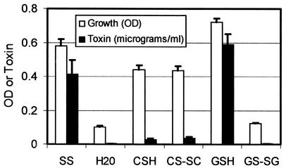
The defined minimal medium, SS broth, contains two potential sources for the essential amino acid cysteine, cystine and reduced glutathione. Bacterial growth was observed when the bacteria were grown in SS broth (Fig. 1, SS), but not in the absence of any source of cysteine (Fig. 1, H2O). The abilities of the bacteria to grow on a single source of cysteine, cystine (oxidized cysteine, CS-SC), reduced cysteine (CSH), oxidized glutathione (GS-SG), or reduced glutathione (GSH) were examined (Fig. 1). When cysteine was supplied as oxidized glutathione (Fig. 1, GS-SG), the OD of the culture did not increase, suggesting that the bacteria could not utilize this chemical form of cysteine. In contrast, reduced glutathione as the sole source of cysteine supported slightly more growth than SS (P < 0.04), while either cysteine (P < 0.02) or cystine (P < 0.03) supported slightly less growth than SS (Fig. 1). On a per-gram basis, all of the forms of modified SS have less cysteine than SS, and these results suggest that the chemical form of cysteine influences growth more than the absolute amount of the amino acid in the medium. This is especially apparent for oxidized glutathione, which fails to support growth.
FIG. 1.
Influence of cysteine source on growth and secretion of pertussis toxin. B. pertussis strain BP338 was grown in SS broth or modified SS containing various sources of the essential amino acid cysteine for 48 h as a static culture in Roux bottles. Growth was assessed by determining the final OD, and the amount of secreted pertussis toxin was determined using the CHO cell assay. SS, broth (containing cystine and reduced glutathione); H2O, modified SS with no source of cysteine; CSH, modified SS containing cysteine; CS-SC, modified SS containing cystine; GSH, modified SS with reduced glutathione; GS-SG, modified SS with oxidized glutathione. Error bars represent standard errors of the means.
The amount of pertussis toxin secretion under the various growth conditions was also examined (Fig. 1). As expected, under conditions that failed to support growth (the absence of cysteine or oxidized glutathione), no pertussis toxin secretion was detected (Fig. 1, H2O and GS-SG). High levels of pertussis toxin secretion were detected from cultures grown in SS or reduced glutathione (Fig. 1, SS and GSH). However, secretion of pertussis toxin was significantly reduced (about 12-fold) compared to SS for the cultures with cysteine (P < 0.003) and cystine (P < 0.02). The basis for the differences in pertussis toxin secretion was examined further.
Influence of cysteine source on expression of pertussis toxin and other Bvg-regulated genes.
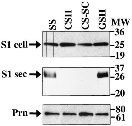
Sulfate has been shown to modulate, or down-regulate, transcription of the Bvg-regulated genes, including pertussis toxin (19) and, recently, bacterial conversion of cysteine to sulfate has been reported to promote modulation (2). Assembled pertussis toxin accumulates in the periplasm prior to secretion. The A-subunit of pertussis toxin, S1, has been shown to be greatly stabilized by its incorporation into holotoxin (9, 10, 21, 26) and reflects the presence of assembled pertussis toxin. To determine if the source of cysteine was affecting pertussis toxin secretion by down-regulating expression of Bvg-regulated genes, accumulation of periplasmic S1 was assessed by Western blotting. Similar levels of periplasmic antigenic toxin were detected in the cells regardless of the cysteine source in the growth medium (Fig. 2, upper panel).
FIG. 2.
Influence of cysteine source on expression of Bvg-regulated genes. B. pertussis strain BP338 was grown in SS broth or modified SS containing various sources of the essential amino acid cysteine as described in the legend to Fig. 1. Accumulation of S1 in cells and culture supernatants was assessed by Western blotting. Top panel, cellular S1 (S1 cell); middle panel, secreted S1 (S1 sec); lower panel, cellular pertactin (Prn), a Bvg-induced product. Migration of molecular weight (MW) markers is indicated on the right.
The amount of secreted S1 was also assessed. Antigenic pertussis toxin was only detected in the culture supernatants of cells grown in the presence of reduced glutathione (Fig. 2, middle panel, SS and GSH), similar to what was seen in Fig. 1 in the functional toxin assay. Expression of pertactin, another Bvg-regulated protein, was also assessed. Equivalent amounts of pertactin were observed under all growth conditions tested (Fig. 2, lower panel).
These results suggest that modulation, or a global down-regulation, of virulence factor expression could not account for the decreased pertussis toxin secretion observed for cells grown in the absence of reduced glutathione, since similar levels of cell-associated pertactin and pertussis toxin were present under all growth conditions.
Transcription of the pertussis toxin operon.
To determine if a selective, down-regulation of pertussis toxin transcription was occurring, we examined transcription of the ptxptl operon by using a β-Gal reporter fusion. Mutant BPM3171 contains a Tn5 lac insertion in the ptlC gene (19). Mutant BPM3171 produces active periplasmic pertussis toxin but fails to secrete toxin (29). A derivate of BPM3171, BPM3171::pKC113 was generated by inserting a suicide plasmid into the adenylate cyclase toxin locus. This plasmid contains the intact ptxptl operon and restores pertussis toxins secretion (5). β-Gal activity for BPM3171::pKC113 was determined at 24 h, when bacterial growth was still occurring, and after 48 h in culture, when bacterial growth was complete. Very similar patterns were observed at both times, and only the data for the 48-h cultures are presented.
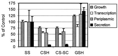
As observed in previous studies (19), modulating conditions (SS with 40 mM MgSO4) resulted in a low level of β-Gal activity, fivefold less than that observed for cells grown in SS (data not shown). Transcription of the ptxptl operon when reduced glutathione was the sole source of cysteine was not significantly different from transcription in SS (Fig. 3). Transcription of the ptxptl operon when cysteine (P < 0.02) or cystine (P < 0.02) was the sole source of cysteine was moderately, but significantly, reduced compared to the SS control (Fig. 3).
FIG. 3.
Influence of cysteine source on transcription of the ptxptl operon and accumulation of periplasmic toxin. B. pertussis was grown in SS broth or modified SS containing various sources of the essential amino acid cysteine as described in the legend to Fig. 1. For comparison, results of growth and pertussis toxin secretion from Fig. 1 are also plotted. Strain BP338 was used to assess growth, periplasmic toxin, and toxin secretion. Strain BPM3171::pKC113, a derivative of BP338 containing the mutant ptxptl operon with β-Gal reporter as well as a wild-type copy of the ptxptl operon, was used to assess pertussis toxin gene transcription. Values are expressed as a percentage of the control value obtained by growth in SS broth, where 100% growth was a final OD of 0.582, transcription was 759 U of β-Gal for the ptxptl operon, periplasmic toxin was 71.2 ng/ml, and secreted toxin was 412 ng/ml. Error bars represent standard errors of the means.
Periplasmic pertussis toxin.
Synthesis and assembly of the toxin in the periplasm can be uncoupled from secretion. Assembled toxin is secreted by the Ptl complex, a type IV secretion system (7, 8, 10, 21, 29). Mutations in the Ptl genes prevent pertussis toxin secretion, but functional pertussis toxin accumulates in the periplasm (7, 29). Similarly, pertussis toxin secretion but not assembly is blocked by mutations in the DsbC gene (26). We examined the amount of functional, periplasmic pertussis toxin as a quantitative measure of toxin folding for bacteria grown in the presence of different sources of cysteine (Fig. 3). The level of assembled pertussis toxin in the periplasm was not statistically different for any of the growth conditions tested.
The combined results from Fig. 2 and Fig. 3 suggest that the failure to efficiently secrete pertussis toxin in the absence of reduced glutathione is not due to a failure to express and properly assemble the pertussis toxin subunits into active holotoxin.
Role of reducing agents in pertussis toxin secretion.
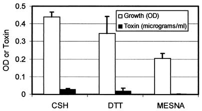
The role of glutathione in oxidation-reduction reactions, and the previously described role for DsbC in pertussis toxin secretion but not assembly (26), suggested the possibility that reduced glutathione could be necessary for pertussis toxin secretion because of a need for a reducing agent. Bacterial growth and pertussis toxin secretion were determined for bacteria grown with cysteine in the presence of the membrane-permeable reducing agent DTT, or the inner-membrane-impermeable reducing agent MESNA. The presence of DTT did not affect growth and did not promote pertussis toxin secretion (Fig. 4). However, MESNA inhibited both growth and pertussis toxin secretion.
FIG. 4.
Influence of reducing agents on pertussis toxin secretion. B. pertussis strain BP338 was grown in modified SS containing cysteine (CSH), modified SS containing cysteine supplemented with DTT, or modified SS containing cysteine supplemented with MESNA. The amount of secreted pertussis toxin was determined by the CHO cell assay. Error bars represent standard errors of the means.
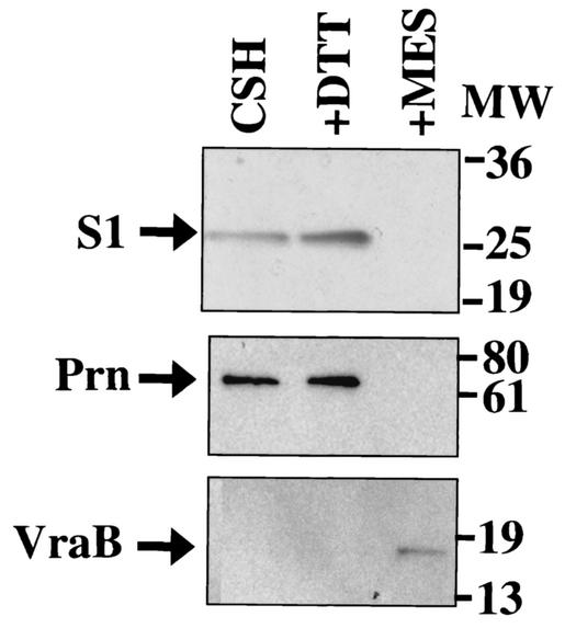
The reduced levels of pertussis toxin secretion in the presence of MESNA suggested that this compound was modulating the bacteria. Western blotting was used to examine the state of modulation of the culture (Fig. 5). The pertussis toxin S1 subunit and another Bvg-regulated protein, pertactin, were detected in cells grown in reduced cysteine, in the presence or absence of DTT (Fig. 5, CSH and CSH+DTT). Neither S1 nor pertactin was detected when the cells were grown in MESNA, suggesting that the bacteria had been modulated under these conditions. This was verified by performing Western blotting using a monoclonal antibody that detects the product of a Bvg-repressed gene, Vra-b. This protein is only expressed when the bacteria are modulated (25). Expression of Vra-b was only seen when the bacteria were incubated with MESNA (Fig. 5) and not under any other growth condition tested unless MESNA, or the known modulator MgSO4, was present (data not shown).
FIG. 5.
Influence of reducing agents on Bvg regulation of virulence gene expression. B. pertussis strain BP338 was grown in modified SS containing cysteine (CSH), modified SS containing cysteine supplemented with DTT (DTT), or modified SS containing cysteine supplemented with MESNA (MES), as described in the legend to Fig. 4. Expression of Bvg-regulated gene products were assessed by Western blotting. Top panel, pertussis toxin subunit S1, a Bvg-induced protein; middle panel, Prn, a Bvg-induced protein; bottom panel, Vra-b, a Bvg-repressed protein. Migration of molecular weight (MW) markers is indicated on the right.
DISCUSSION
Sulfur-containing compounds could influence pertussis toxin expression at several levels, including affecting bacterial protein synthesis and growth, as well as transcription, assembly, and secretion of pertussis toxin. In this study we undertook a systematic analysis of the influence of different sulfur-containing compounds on pertussis toxin expression. Cysteine is a necessary nutrient for B. pertussis. These studies have shown that the growth requirement for cysteine can be fulfilled by cysteine, cystine, or reduced glutathione. In contrast, oxidized glutathione as a sole source of cysteine cannot support bacterial growth, and this further suggests that B. pertussis cannot convert oxidized glutathione to reduced glutathione. The physiologic source of cysteine in human infection is likely to be reduced glutathione. Glutathione is present at high concentrations in the respiratory tract, with greater than 95% present in the reduced form. Reduced glutathione serves to protect the body from damage due to reactive oxygen and nitrogen species (3, 15, 27).
A previous study reported that cysteine in the growth medium can be metabolized to sulfate and that sulfate accumulation can modulate the bacteria or shut down transcription of the Bvg-regulated genes (2), including pertussis toxin. However, we did not observe modulation by any of the cysteine-containing compounds under the growth conditions used in this study (which differed from those used in the original report), as evidenced by transcription of the ptxptl operon and expression of periplasmic pertussis toxin and another Bvg-regulated protein, pertactin. The reducing agent MESNA was able to modulate the bacteria, but the reducing agent DTT did not. It is possible that MESNA (HSCH2CH2SO3) resembles the well-described modulating agent sulfate.
Moderately reduced transcription of the ptxptl operon was observed when cysteine or cystine was the sole source of this essential amino acid. The modest reduction in transcription did not appear to be specific for the pertussis toxin operon, since it paralleled the modest reduction bacterial growth under these conditions. In contrast, pertussis toxin secretion was severely depressed when the bacteria were grown without reduced glutathione. This secretion defect was observed by measurement of the amount of toxin using antibody detection, as well as measurement of active toxin in a biological assay, indicating there is not a folding defect that results in secretion of a biologically inactive toxin. In addition, our results suggest that the reduction of extracellular pertussis toxin is due to a secretion defect per se and not a failure to assemble toxin, since equivalent levels of cell-associated antigenic toxin and biologically active toxin are produced by bacteria grown with or without reduced glutathione. One might predict that the amount of periplasmic toxin would increase when secretion is blocked. However, previous studies (7, 26, 29) have also shown that the amount of periplasmic pertussis toxin does not increase as a result of failure to secrete the toxin, suggesting that the periplasm has a finite capacity for toxin and some feedback mechanism must be operating.
The present study reveals that reduced glutathione is essential for pertussis toxin secretion in a way that can be uncoupled from its role in supplying the essential nutrient, cysteine. It is possible that the antioxidant or reducing properties of glutathione play a role in secretion of pertussis toxin. However, it does not appear that the requirement for reduced glutathione is simply due to the need for a reducing agent, since the reducing agents DTT and MESNA do not restore pertussis toxin secretion in bacteria grown in the presence of cysteine. In a previous study, we observed that the periplasmic enzyme DsbC is also necessary for pertussis toxin secretion, but it is not essential for assembly of pertussis toxin in the periplasm (26). DsbC catalyzes exchange of disulfide bonds. Future studies will be directed at determining where DsbC and reduced glutathione act in the pertussis toxin secretion pathway.
Acknowledgments
This work was supported by NIH grant ROI AI23695.
Editor: J. T. Barbieri
REFERENCES
- 1.Bardwell, J. C., K. McGovern, and J. Beckwith. 1991. Identification of a protein required for disulfide bond formation in vivo. Cell 67:581-589. [DOI] [PubMed] [Google Scholar]
- 2.Bogdan, J. A., J. Nazario-Larrieu, J. Sarwar, P. Alexander, and M. S. Blake. 2001. Bordetella pertussis autoregulates pertussis toxin production through the metabolism of cysteine. Infect. Immun. 69:6823-6830. [DOI] [PMC free article] [PubMed] [Google Scholar]
- 3.Cantin, A. M., S. L. North, R. C. Hubbard, and R. G. Crystal. 1987. Normal alveolar epithelial lining fluid contains high levels of glutathione. J. Appl. Physiol. 63:152-157. [DOI] [PubMed] [Google Scholar]
- 4.Charles, I. G., J. Li, M. Roberts, K. Beesley, M. Romanos, D. J. Pickard, M. Francis, D. Campbell, G. Dougan, M. J. Brennan, C. R. Manclark, M. A. Jensen, I. Heron, A. Chub, P. Novotny, and N. F. Fairweather. 1991. Identification and characterization of a protective immunodominant B cell epitope of pertactin (P.69) from Bordetella pertussis. Eur. J. Immunol. 21:1147-1153. [DOI] [PubMed] [Google Scholar]
- 5.Craig-Mylius, K. A., T. H. Stenson, and A. A. Weiss. 2000. Mutations in the S1 subunit of pertussis toxin that affect secretion. Infect. Immun. 68:1276-1281. [DOI] [PMC free article] [PubMed] [Google Scholar]
- 6.Craig-Mylius, K. A., and A. A. Weiss. 2000. Antibacterial agents and release of periplasmic pertussis toxin from Bordetella pertussis. Antimicrob. Agents Chemother. 44:1383-1386. [DOI] [PMC free article] [PubMed] [Google Scholar]
- 7.Craig-Mylius, K. A., and A. A. Weiss. 1999. Mutants in the ptlA-H genes of Bordetella pertussis are deficient for pertussis toxin secretion. FEMS Microbiol. Lett. 179:479-484. [DOI] [PubMed] [Google Scholar]
- 8.Farizo, K. M., T. G. Cafarella, and D. L. Burns. 1996. Evidence for a ninth gene, ptlI, in the locus encoding the pertussis toxin secretion system of Bordetella pertussis and formation of a PtlI-PtlF complex. J. Biol. Chem. 271:31643-31649. [DOI] [PubMed] [Google Scholar]
- 9.Farizo, K. M., S. Fiddner, A. M. Cheung, and D. L. Burns. 2002. Membrane localization of the S1 subunit of pertussis toxin in Bordetella pertussis and implications for pertussis toxin secretion. Infect. Immun. 70:1193-1201. [DOI] [PMC free article] [PubMed] [Google Scholar]
- 10.Farizo, K. M., T. Huang, and D. L. Burns. 2000. Importance of holotoxin assembly in Ptl-mediated secretion of pertussis toxin from Bordetella pertussis. Infect. Immun. 68:4049-4054. [DOI] [PMC free article] [PubMed] [Google Scholar]
- 11.Frand, A. R., J. W. Cuozzo, and C. A. Kaiser. 2000. Pathways for protein disulphide bond formation. Trends Cell Biol. 10:203-210. [DOI] [PubMed] [Google Scholar]
- 12.Gross, R., and R. Rappuoli. 1989. Pertussis toxin promoter sequences involved in modulation. J. Bacteriol. 171:4026-4030. [DOI] [PMC free article] [PubMed] [Google Scholar]
- 13.Gross, R., and R. Rappuoli. 1988. Positive regulation of pertussis toxin expression. Proc. Natl. Acad. Sci. USA 85:3913-3917. [DOI] [PMC free article] [PubMed] [Google Scholar]
- 14.Hewlett, E. L., K. T. Sauer, G. A. Myers, J. L. Cowell, and R. L. Guerrant. 1983. Induction of a novel morphological response in Chinese hamster ovary cells by pertussis toxin. Infect. Immun. 40:1198-1203. [DOI] [PMC free article] [PubMed] [Google Scholar]
- 15.Kelly, F. J. 1999. Gluthathione: in defence of the lung. Food Chem. Toxicol. 37:963-966. [DOI] [PubMed] [Google Scholar]
- 16.Kenimer, J. G., K. J. Kim, P. G. Probst, C. R. Manclark, D. G. Burstyn, and J. L. Cowell. 1989. Monoclonal antibodies to pertussis toxin: utilization as probes of toxin function. Hybridoma 8:37-51. [DOI] [PubMed] [Google Scholar]
- 17.Kotob, S. I., S. Z. Hausman, and D. L. Burns. 1995. Localization of the promoter for the ptl genes of Bordetella pertussis, which encode proteins essential for secretion of pertussis toxin. Infect. Immun. 63:3227-3230. [DOI] [PMC free article] [PubMed] [Google Scholar]
- 18.Locht, C., P. A. Barstad, J. E. Coligan, L. Mayer, J. J. Munoz, S. G. Smith, and J. M. Keith. 1986. Molecular cloning of pertussis toxin genes. Nucleic Acids Res. 14:3251-3261. [DOI] [PMC free article] [PubMed] [Google Scholar]
- 19.Melton, A. R., and A. A. Weiss. 1989. Environmental regulation of expression of virulence determinants in Bordetella pertussis. J. Bacteriol. 171:6206-6212. [DOI] [PMC free article] [PubMed] [Google Scholar]
- 20.Nicosia, A., M. Perugini, C. Franzini, M. C. Casagli, M. G. Borri, G. Antoni, M. Almoni, P. Neri, G. Ratti, and R. Rappuoli. 1986. Cloning and sequencing of the pertussis toxin genes: operon structure and gene duplication. Proc. Natl. Acad. Sci. USA 83:4631-4635. [DOI] [PMC free article] [PubMed] [Google Scholar]
- 21.Pizza, M., M. Bugnoli, R. Manetti, A. Covacci, and R. Rappuoli. 1990. The subunit S1 is important for pertussis toxin secretion. J. Biol. Chem. 265:17759-17763. [PubMed] [Google Scholar]
- 22.Rietsch, A., D. Belin, N. Martin, and J. Beckwith. 1996. An in vivo pathway for disulfide bond isomerization in Escherichia coli. Proc. Natl. Acad. Sci. USA 93:13048-13053. [DOI] [PMC free article] [PubMed] [Google Scholar]
- 23.Stainer, D. W., and M. J. Scholte. 1970. A simple chemically defined medium for the production of phase I Bordetella pertussis. J. Gen. Microbiol. 63:211-220. [DOI] [PubMed] [Google Scholar]
- 24.Stein, P. E., A. Boodhoo, G. D. Armstrong, S. A. Cockle, M. H. Klein, and R. J. Read. 1994. The crystal structure of pertussis toxin. Structure 2:45-57. [DOI] [PubMed] [Google Scholar]
- 25.Stenson, T. H., and M. S. Peppler. 1995. Identification of two bvg-repressed surface proteins of Bordetella pertussis. Infect. Immun. 63:3780-3789. [DOI] [PMC free article] [PubMed] [Google Scholar]
- 26.Stenson, T. H., and A. A. Weiss. 2002. DsbA and DsbC are required for secretion of pertussis toxin by Bordetella pertussis. Infect. Immun. 70:2297-2303. [DOI] [PMC free article] [PubMed] [Google Scholar]
- 27.Sutherland, M. W., M. Glass, J. Nelson, Y. Lyen, and H. J. Forman. 1985. Oxygen toxicity: loss of lung macrophage function without metabolite depletion. J. Free Radic. Biol. Med. 1:209-214. [DOI] [PubMed] [Google Scholar]
- 28.Weiss, A. A., E. L. Hewlett, G. A. Myers, and S. Falkow. 1983. Tn5-induced mutations affecting virulence factors of Bordetella pertussis. Infect. Immun. 42:33-41. [DOI] [PMC free article] [PubMed] [Google Scholar]
- 29.Weiss, A. A., F. D. Johnson, and D. L. Burns. 1993. Molecular characterization of an operon required for pertussis toxin secretion. Proc. Natl. Acad. Sci. USA 90:2970-2974. [DOI] [PMC free article] [PubMed] [Google Scholar]
- 30.Weiss, A. A., A. R. Melton, K. E. Walker, C. Andraos-Selim, and J. J. Meidl. 1989. Use of the promoter fusion transposon Tn5 lac to identify mutations in Bordetella pertussis vir-regulated genes. Infect. Immun. 57:2674-2682. [DOI] [PMC free article] [PubMed] [Google Scholar]
- 31.Zapun, A., D. Missiakas, S. Raina, and T. E. Creighton. 1995. Structural and functional characterization of DsbC, a protein involved in disulfide bond formation in Escherichia coli. Biochemistry 34:5075-5089. [DOI] [PubMed] [Google Scholar]


