Journal of Virology logoLink to Journal of Virology
letter
. 2006 Jul;80(13):6716–6719. doi: 10.1128/JVI.00386-06

The Egress of Herpesviruses from Cells: the Unanswered Questions

Gabriella Campadelli-Fiume 1,2,*, Bernard Roizman 1,2
PMCID: PMC1488928  PMID: 16775362

The presence of herpes simplex virus (HSV) capsids attached to invaginated cytoplasmic vesicles led Stackpole (15) to propose that capsids undergo envelopment at the inner nuclear membrane, de-envelopment at the outer nuclear membrane, and finally reenvelopment at cytoplasmic membranes. Over the years this model attracted numerous adherents principally on the basis of evidence that the extracellular virions lack proteins present in intracellular virions accumulating in the perinuclear space (11, 14). An alternative hypothesis was recently presented by Wild et al. (17) on the basis of the observation that nuclear pores become grossly enlarged in cells infected with wild-type virus. Two hypotheses have emerged. The first is that HSV virions undergo envelopment at the inner nuclear membrane, de-envelopment at the outer nuclear membrane or extensions thereof, and reenvelopment in cytoplasmic organelles (double envelopment model). The second hypothesis is that virions mature and egress the cell via two pathways. A minority, primarily early in infection, becomes enveloped at the inner nuclear membrane and is transported in vesicles to the extracellular space. The majority enters the cytoplasm late in infection through enlarged nuclear pores and becomes enveloped at cytoplasmic membranes, mainly Golgi and post-Golgi.

Challenges to existing theories are the fabric of science, and no other recent controversy has generated as much discussion as the challenge to the double envelopment hypothesis. The letter by Mettenleiter and Minson and the response by Wild (10) sustain the respective models but do not define the problems associated with each model or the data necessary to reaffirm or reject them.

The double envelopment model requires that virions accumulating in the perinuclear space fuse with the outer nuclear membrane or extensions thereof. HSV encodes a quartet of glycoproteins that execute the entry of virus into cells by fusion of the envelope with the plasma membrane and cell-to-cell fusion (4, 6, 9, 13, 16). Thus, glycoprotein D (gD) interacts with either the herpesvirus entry mediator or nectin 1 receptor, whereas gH, which contains structural elements shared with viral fusion proteins (7), and gB and gL execute the fusion (5). This quartet plays no role in the de-envelopment of virions, since mutants lacking any one of the four glycoproteins, while not infectious, nevertheless egress the cells in a manner similar to that of wild-type virus.

Other viral proteins, notably gK and UL20, are involved in viral egress, in that in their absence virus particles accumulate in the perinuclear space or cytoplasm. These proteins, however, appear to inhibit fusion performed by the quartet (2), and their presence in the viral envelope is debated. SNARE proteins, responsible for fusion of cellular vesicles, by their topology and necessary cytoplasmic cofactors cannot perform the fusion in the perinuclear space required by the double envelopment model. A common strategy of HSV is to sequester and redirect cellular proteins to perform at times novel functions (e.g., the binding of protein phosphatase 1α by γ34.5, the binding of cdc2 by UL42 [1, 8]). Putative cellular protein partners capable of fusing viral envelope with the outer nuclear membrane or vesicles derived from its extensions in the absence of one or more of the glycoprotein quartet have not been identified. In essence, the evidence required to support the double envelopment model is the definition of the mechanism by which enveloped virions accumulating in the perinuclear space could become de-enveloped, resulting in the release of capsids into the cytoplasm.

The egress of capsids though the nuclear pores proposed by Wild et al. (17) challenges the de-envelopment step of the Stackpole pathway. Capsids could egress through enlarged nuclear pores passively along with nuclear components and organelles or actively by engaging the nuclear export machinery of the cell. Unregulated egress of proteins or RNA from nuclei of infected cells has not been reported. On the other hand, while the evidence supports a specific association of capsids with the pores on the cytoplasmic side of the nuclear membrane, there is no evidence for an association with pores at the nuclear side. Thus, capsids encoded by two diverse viral mutants accumulate in large numbers at nuclear pores on the cytoplasmic side, but not juxtaposed at random to nuclear membranes (3, 11). In contrast, capsids frequently line the nuclear side of the inner nuclear membrane but not specifically at nuclear pores. At present, only mutants lacking the US3 protein kinase fail to transport capsids from the nucleus to the perinuclear space (12), but the effect of US3 may be on the integrity of the nuclear envelope and not specifically on the mechanism of capsid transport. The key evidence required to support the single envelopment model is identification of nuclear transport proteins that could interact with capsids and demonstration that the transport is abolished by mutagenesis of the capsid partner or genetic manipulation of the nuclear transport protein.

REFERENCES

  • 1.Advani, S. J., R. R. Weichselbaum, and B. Roizman. 2001. cdc2 cyclin-dependent kinase binds and phosphorylates herpes simplex virus 1 UL42 DNA synthesis processivity factor. J. Virol. 75:10326-10333. [DOI] [PMC free article] [PubMed] [Google Scholar]
  • 2.Avitabile, E., G. Lombardi, T. Gianni, M. Capri, and G. Campadelli-Fiume. 2004. Coexpression of UL20p and gK inhibits cell-cell fusion mediated by herpes simplex virus glycoproteins gD, gH-gL, and wild-type gB or an endocytosis-defective gB mutant, and downmodulates their cell surface expression. J. Virol. 78:8015-8025. [DOI] [PMC free article] [PubMed] [Google Scholar]
  • 3.Batterson, W., D. Furlong, and B. Roizman. 1983. Molecular genetics of herpes simplex virus. VIII. Further characterization of a temperature-sensitive mutant defective in release of viral DNA and in other stages of the viral reproductive cycle. J. Virol. 45:397-407. [DOI] [PMC free article] [PubMed] [Google Scholar]
  • 4.Cai, W. Z., S. Person, C. DebRoy, and B. H. Gu. 1988. Functional regions and structural features of the gB glycoprotein of herpes simplex virus type 1. An analysis of linker insertion mutants. J. Mol. Biol. 201:575-588. [DOI] [PubMed] [Google Scholar]
  • 5.Campadelli-Fiume, G., F. Cocchi, L. Menotti, and M. Lopez. 2000. The novel receptors that mediate the entry of herpes simplex viruses and animal alphaherpesviruses into cells. Rev. Med. Virol. 10:305-319. [DOI] [PubMed] [Google Scholar]
  • 6.Forrester, A., H. Farrell, G. Wilkinson, J. Kaye, N. Davis-Poynter, and T. Minson. 1992. Construction and properties of a mutant of herpes simplex virus type 1 with glycoprotein H coding sequences deleted. J. Virol. 66:341-348. [DOI] [PMC free article] [PubMed] [Google Scholar]
  • 7.Gianni, T., P. L. Martelli, R. Casadio, and G. Campadelli-Fiume. 2005. The ectodomain of herpes simplex virus glycoprotein H contains a membrane alpha-helix with attributes of an internal fusion peptide, positionally conserved in the Herpesviridae family. J. Virol. 79:2931-2940. [DOI] [PMC free article] [PubMed] [Google Scholar]
  • 8.He, B., M. Gross, and B. Roizman. 1997. The γ134.5 protein of herpes simplex virus 1 complexes with protein phosphatase 1α to dephosphorylate the α subunit of the eukaryotic translation initiation factor 2 and preclude the shutoff of protein synthesis by double-stranded RNA-activated protein kinase. Proc. Natl. Acad. Sci. USA 94:843-848. [DOI] [PMC free article] [PubMed] [Google Scholar]
  • 9.Ligas, M. W., and D. C. Johnson. 1988. A herpes simplex virus mutant in which glycoprotein D sequences are replaced by beta-galactosidase sequences binds to but is unable to penetrate into cells. J. Virol. 62:1486-1494. [DOI] [PMC free article] [PubMed] [Google Scholar]
  • 10.Mettenleiter, T. C., and T. Minson. 2006. Egress of alphaherpesviruses. J. Virol. 80:1610-1612. [DOI] [PMC free article] [PubMed] [Google Scholar]
  • 11.Reynolds, A. E., B. J. Ryckman, J. D. Baines, Y. Zhou, L. Liang, and R. J. Roller. 2001. UL31 and UL34 proteins of herpes simplex virus type 1 form a complex that accumulates at the nuclear rim and is required for envelopment of nucleocapsids. J. Virol. 75:8803-8817. [DOI] [PMC free article] [PubMed] [Google Scholar]
  • 12.Reynolds, A. E., E. G. Wills, R. J. Roller, B. J. Ryckman, and J. D. Baines. 2002. Ultrastructural localization of the herpes simplex virus type 1 UL31, UL34, and US3 proteins suggests specific roles in primary envelopment and egress of nucleocapsids. J. Virol. 76:8939-8952. [DOI] [PMC free article] [PubMed] [Google Scholar]
  • 13.Roop, C., L. Hutchinson, and D. C. Johnson. 1993. A mutant herpes simplex virus type 1 unable to express glycoprotein L cannot enter cells, and its particles lack glycoprotein H. J. Virol. 67:2285-2297. [DOI] [PMC free article] [PubMed] [Google Scholar]
  • 14.Skepper, J. N., A. Whiteley, H. Browne, and A. Minson. 2001. Herpes simplex virus nucleocapsids mature to progeny virions by an envelopment → deenvelopment → reenvelopment pathway. J. Virol. 75:5697-5702. [DOI] [PMC free article] [PubMed] [Google Scholar]
  • 15.Stackpole, C. W. 1969. Herpes-type virus of the frog renal adenocarcinoma. I. Virus development in tumor transplants maintained at low temperature. J. Virol. 4:75-93. [DOI] [PMC free article] [PubMed] [Google Scholar]
  • 16.Turner, A., B. Bruun, T. Minson, and H. Browne. 1998. Glycoproteins gB, gD, and gHgL of herpes simplex virus type 1 are necessary and sufficient to mediate membrane fusion in a Cos cell transfection system. J. Virol. 72:873-875. [DOI] [PMC free article] [PubMed] [Google Scholar]
  • 17.Wild, P., M. Engels, C. Senn, K. Tobler, U. Ziegler, E. M. Schraner, E. Loepfe, M. Ackermann, M. Müller, and P. Walther. 2005. Impairment of nuclear pores in bovine herpesvirus 1-infected MDBK cells. J. Virol. 79:1071-1083. [DOI] [PMC free article] [PubMed] [Google Scholar]
J Virol. 2006 Jul;80(13):6716–6719.

Author's Reply 1

Peter Wild 1

In his proposal of capsid de-envelopment at the outer nuclear membrane (ONM), Stackpole (14) did not consider the observations of Schwartz and Roizman (13), which clearly showed that virions are transported intraluminally from the perinuclear space (PNS) into a membranous system lacking ribosomes. Despite the assumption of a canal system for virus transportation to the cell periphery, this report bears a fundamental fact: the ability of virions to be transported away from the PNS. Nii et al. (11) showed proliferation of the nuclear surface, which suggests that HSV type 1 (HSV-1) induces membrane synthesis. These authors also showed indications for intraluminal virion transportation and for formation of two distinctly different transport vacuoles. Both the origin of these vacuoles and the nuclear surface did not attract the interest of investigators until recently. Instead, evidence explaining the presence of naked capsids within the cytoplasm and for the ability of these capsids to acquire tegument and a secondary envelope exclusively at the trans-Golgi network was collected.

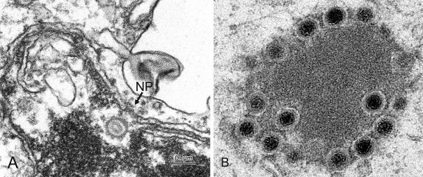
Many proteins that are involved in virus egress were discovered. However, the true function of many, if not all, is poorly understood. If the dual nuclear exit pathway is correct, the factors targeting capsids to the nuclear surface for budding and for exit via nuclear pores need to be investigated as demanded by Campadelli-Fiume and Roizman. Though most images show capsids lining the nuclear membrane at unspecific sites or at budding stages, there is clear evidence that capsids are transported towards nuclear pores (Fig. 1A), suggesting intranuclear sorting. Budding capsids acquire a dense envelope and tegument. Tegument consists of a compact intensely stained shell when imaged at a higher resolution. It is, however, poorly stained in routinely prepared specimens or in specimens prepared for immunolabeling, suggesting loss of substances. Nevertheless, tegument protein VP16 could be demonstrated on both perinuclear and extracellular virions, whereas others could be demonstrated only on extracellular virions (7, 10). It is a difficult task to prove that a given protein is absent from structures, especially if they occur only in low numbers. Thus, comparing very low intensities of immunolabeling of a few virions within the PNS with that of hundreds of extracellular virions in poorly preserved cells is not a reliable method. Furthermore, if many of the extracellular virions are not labeled (7), conclusions about viral origin cannot be drawn. To find a satisfying answer about the differences in tegument protein composition, immunolabeling must be performed on well-preserved cells, and labeling must be compared between virions within the PNS and virions that clearly originated by budding at Golgi membranes.

FIG. 1.

FIG. 1.

(A) Capsid of Epstein-Barr virus in front of the nuclear pore (NP) within the nucleus, showing folding of the nuclear membrane. Courtesy of Dr. V. Kushnaryov, Department of Microbiology and Molecular Genetics, Medical College of Wisconsin, Milwaukee. (B) Cytoplasmic capsid-tegument accumulation in gE-deleted bovine herpesvirus 1-infected cells.

US3, UL31, and UL34 were shown on nuclear membranes and on virions within the PNS and, in the case of US3, also on cytoplasmic and extracellular virions and on the plasma membrane (12). These proteins seem to be involved in nuclear envelopment. However, the conclusion that the perinuclear virions are de-enveloped is not valid because there are other possibilities for substances to be cleft from the viral periphery where these proteins seem to be located. gK is assumed to promote fusion of the primary envelope with the ONM because pseudorabies virus (PRV) deleted of gK was found to accumulate within the PNS (5). However, intact HSV-1 virions accumulate within the PNS (8, 15), and gK is not required in de-envelopment of HSV-1 (9). gK is also assumed to prevent PRV from reentry into cells (5), i.e., by preventing fusion. Astonishingly, the capsid membrane interactions assumed to represent fusion are identical to those assumed to represent budding in UL20-deleted PRV (2) and in intact HSV-1 (8) and human cytomegalovirus (4). gB and gD, two members of the quartet involved in cell entry (1), have been localized at nuclear membranes (3, 15). The question as to why they do not initiate de-envelopment arises. Many other proteins are suggested to be involved in envelopment on the basis of phenotypes found in cells infected with deletion mutants. The crux of using phenotypes as a indicator for the significance of a given protein is that identical phenotypes can occur in cells infected with intact virus or other deletion mutants. For example, simultaneous deletion of UL11 and gM is assumed to severely affect PRV envelopment, resulting in formation of capsid-tegument aggregates (6). Interestingly, identical aggregates occur in cells infected with gE-deleted bovine herpesvirus 1 (Fig. 1B).

There are many more questions. To understand capsid transportation, budding, fusion, and virion transportation, the signaling pathways and mechanisms need to be studied. To resolve confusing and controversial theories, factors playing a role in envelopment must be investigated with the aim to shed light onto their true significance. One powerful tool is electron microscopy employing techniques for improved retention of cellular material, which needs to be combined with high-level immunolabeling.

REFERENCES

  • 1.Avitabile, E., G. Lombardi, T. Gianni, M. Capri, and G. Campadelli-Fiume. 2004. Coexpression of UL20p and gK inhibits cell-cell fusion mediated by herpes simplex virus glycoproteins gD, gH-gL, and wild-type gB or an endocytosis-defective gB mutant and downmodulates their cell surface expression. J. Virol. 78:8015-8025. [DOI] [PMC free article] [PubMed] [Google Scholar]
  • 2.Fuchs, W., B. G. Klupp, H. Granzow, and T. C. Mettenleiter. 1997. The UL20 gene product of pseudorabies virus functions in virus egress. J. Virol. 71:5639-5646. [DOI] [PMC free article] [PubMed] [Google Scholar]
  • 3.Gilbert, R., and H. P. Ghosh. 1993. Immunoelectron microscopic localization of herpes simplex virus glycoprotein gB in the nuclear envelope of infected cells. Virus Res. 28:217-231. [DOI] [PubMed] [Google Scholar]
  • 4.Homman-Loudiyi, M., K. Hultenby, W. Britt, and C. Soderberg-Naucler. 2003. Envelopment of human cytomegalovirus occurs by budding into Golgi-derived vacuole compartments positive for gB, Rab 3, trans-golgi network 46, and mannosidase II. J. Virol. 77:3191-3203. (Erratum, 77:8179.) [DOI] [PMC free article] [PubMed] [Google Scholar]
  • 5.Klupp, B. G., J. Baumeister, P. Dietz, H. Granzow, and T. C. Mettenleiter. 1998. Pseudorabies virus glycoprotein gK is a virion structural component involved in virus release but is not required for entry. J. Virol. 72:1949-1958. [DOI] [PMC free article] [PubMed] [Google Scholar]
  • 6.Kopp, M., H. Granzow, W. Fuchs, B. Klupp, and T. C. Mettenleiter. 2004. Simultaneous deletion of pseudorabies virus tegument protein UL11 and glycoprotein M severely impairs secondary envelopment. J. Virol. 78:3024-3034. [DOI] [PMC free article] [PubMed] [Google Scholar]
  • 7.Kopp, M., B. G. Klupp, H. Granzow, W. Fuchs, and T. C. Mettenleiter. 2002. Identification and characterization of the pseudorabies virus tegument proteins UL46 and UL47: role for UL47 in virion morphogenesis in the cytoplasm. J. Virol. 76:8820-8833. [DOI] [PMC free article] [PubMed] [Google Scholar]
  • 8.Leuzinger, H., U. Ziegler, C. Fraefel, E. M. Schraner, D. Glauser, I. Held, M. Ackermann, M. Müller, and P. Wild. 2005. Herpes simplex virus 1 envelopment follows two diverse pathways. J. Virol. 79:13047-13059. [DOI] [PMC free article] [PubMed] [Google Scholar]
  • 9.Melancon, J. M., R. E. Luna, T. P. Foster, and K. G. Kousoulas. 2005. Herpes simplex virus type 1 gK is required for gB-mediated virus-induced cell fusion, while neither gB and gK nor gB and UL20p function redundantly in virion de-envelopment. J. Virol. 79:299-313. [DOI] [PMC free article] [PubMed] [Google Scholar]
  • 10.Naldinho-Souto, R., H. Browne, and T. Minson. 2006. Herpes simplex virus tegument protein VP16 is a component of primary enveloped virions. J. Virol. 80:2582-2584. [DOI] [PMC free article] [PubMed] [Google Scholar]
  • 11.Nii, S., C. Morgan, and H. M. Rose. 1968. Electron microscopy of herpes simplex virus. II. Sequence of development. J. Virol. 2:517-536. [DOI] [PMC free article] [PubMed] [Google Scholar]
  • 12.Reynolds, A. E., E. G. Wills, R. J. Roller, B. J. Ryckman, and J. D. Baines. 2002. Ultrastructural localization of the herpes simplex virus type 1 UL31, UL34, and US3 proteins suggests specific roles in primary envelopment and egress of nucleocapsids. J. Virol. 76:8939-8952. [DOI] [PMC free article] [PubMed] [Google Scholar]
  • 13.Schwartz, J., and B. Roizman. 1969. Concerning the egress of herpes simplex virus from infected cells: electron and light microscope observations. Virology 38:42-49. [DOI] [PubMed] [Google Scholar]
  • 14.Stackpole, C. W. 1969. Herpes-type virus of the frog renal adenocarcinoma. I. Virus development in tumor transplants maintained at low temperature. J. Virol. 4:75-93. [DOI] [PMC free article] [PubMed] [Google Scholar]
  • 15.Stannard, L. M., S. Himmelhoch, and S. Wynchank. 1996. Intra-nuclear localization of two envelope proteins, gB and gD, of herpes simplex virus. Arch. Virol. 141:505-524. [DOI] [PubMed] [Google Scholar]
J Virol. 2006 Jul;80(13):6716–6719.

Author's Reply 2

Thomas C Mettenleiter 1,2, Tony Minson 1,2,*

In their letter, Campadelli-Fiume and Roizman identify a number of key issues concerning the current debate over the route of herpesvirus egress from the nuclei of infected cells. We agree with much of their analysis and recognize the need for further work to resolve several unclarified issues. However, we believe that in presenting alternative models, it is important to consider the weight of evidence.

It is generally accepted that at least the majority of mature herpesvirus virions are enveloped in a cytoplasmic compartment (7). What is at issue is the source of cytoplasmic nucleocapsids. In our view these arise by a sequence of budding at the inner nuclear membrane, followed by fusion of the primary envelope with the outer nuclear membrane or the endoplasmic reticulum, a sequence that is consistent with electron microscopic observations. This process is independent of the four conserved herpesviral glycoproteins required for fusion of the mature envelope with the cellular plasma membrane during infectious entry but is enhanced in the presence of the US3 viral kinase (5). As Campadelli-Fiume and Roizman correctly point out, the mechanism of fusion during nuclear egress and the viral and/or cellular proteins involved have not yet been elucidated. However, this does not seem sufficient reason to relegate the perinuclear enveloped virion to a minor role in herpesvirus egress and to propose instead that transfer through enlarged nuclear pores provides the major source of cytoplasmic nucleocapsids and, hence, infectious progeny.

The envelopment-de-envelopment-reenvelopment model is supported by several lines of evidence and, in particular, is consistent with analysis of genetically well-defined mutants of the alphaherpesviruses pseudorabies virus and herpes simplex virus using several cell types (7). In contrast, the proposed egress via dilated nuclear pores is based on a limited number of morphological analyses without any biochemical or genetic support (6, 11). Our studies with herpes simplex virus and pseudorabies virus have not revealed impairment of and nucleocapsid exit through nuclear pores. In fact, the integrity of nuclear pores appears to be maintained until late in herpesvirus infection (3). No viral mutants that influence the size of nuclear pores have been identified, and there are no reports on the accumulation of nucleocapsids at nuclear pores during the productive phase of herpesvirus infection. In contrast, accumulation of nucleocapsids along the inner nuclear membrane is routinely seen in herpesvirus-infected cells, and enveloped virions within the perinuclear space are an accepted feature of herpesvirus infections, regardless of virus type, virus strain, or host cell. In addition, mutations that impair envelopment at the inner nuclear membrane have been identified, and the resulting reduction in perinuclear enveloped virions is associated with reduced infectious yield (1, 4, 9). These observations all imply a key role for the perinuclear virion. Since these primary virions contain tegument and envelope proteins which are absent from the mature virion, their integrity cannot be maintained during maturation, and a de-envelopment step must occur (1, 2, 8).

We believe, therefore, that the weight of evidence favors a pathway in which the nucleocapsid exits the nucleus by traversing the nuclear membrane following an envelopment-de-envelopment route (10), but we accept that several issues still need clarification. We agree with Campadelli-Fiume and Roizman that a complete understanding of herpesvirus egress at the molecular level is still lacking and that, in designing future experiments, we should keep our minds open.

REFERENCES

  • 1.Fuchs, W., B. G. Klupp, H. Granzow, N. Osterrieder, and T. C. Mettenleiter. 2002. The interacting UL31 and UL34 gene products of pseudorabies virus are involved in egress from the host cell nucleus and represent components of primary enveloped but not of mature virions. J. Virol. 76:364-378. [DOI] [PMC free article] [PubMed] [Google Scholar]
  • 2.Fuchs, W., H. Granzow, B. G. Klupp, M. Kopp, and T. C. Mettenleiter. 2002. The UL48 tegument protein of pseudorabies virus is critical for intracytoplasmic assembly of infectious virions. J. Virol. 76:6729-6742. [DOI] [PMC free article] [PubMed] [Google Scholar]
  • 3.Haines, H., and R. J. Baerwald. 1976. Nuclear membrane changes in herpes simplex virus-infected BHK-21 cells as seen by freeze-fracture. J. Virol. 17:1038-1042. [DOI] [PMC free article] [PubMed] [Google Scholar]
  • 4.Klupp, B. G., H. Granzow, and T. C. Mettenleiter. 2000. Primary envelopment of pseudorabies virus at the inner nuclear membrane requires the UL34 gene product. J. Virol. 74:10063-10073. [DOI] [PMC free article] [PubMed] [Google Scholar]
  • 5.Klupp, B. G., H. Granzow, and T. C. Mettenleiter. 2001. Effect of pseudorabies virus US3 protein on nuclear membrane localisation of the UL34 protein and virus egress from the nucleus. J. Gen. Virol. 82:2363-2371. [DOI] [PubMed] [Google Scholar]
  • 6.Leuzinger, H., U. Ziegler, E. M. Schraner, C. Fraefel, D. L. Glauser, I. Held, M. Ackermann, M. Mueller, and P. Wild. 2005. Herpes simplex virus type 1 envelopment follows two diverse pathways. J. Virol. 79:13047-13059. [DOI] [PMC free article] [PubMed] [Google Scholar]
  • 7.Mettenleiter, T. C. 2004. Budding events in herpesvirus morphogenesis. Virus Res. 106:167-180. [DOI] [PubMed] [Google Scholar]
  • 8.Naldinho-Souto, R., H. Browne, and T. Minson. 2006. Herpes simplex virus tegument protein VP16 is a component of primary enveloped virions. J. Virol. 80:2582-2584. [DOI] [PMC free article] [PubMed] [Google Scholar]
  • 9.Reynolds, A. E., B. J. Ryckman, J. D. Baines, Y. Zhou, L. Liang, and R. J. Roller. 2001. UL31 and UL34 proteins of herpes simplex virus type 1 form a complex that accumulates at the nuclear rim and is required for envelopment of nucleocapsids. J. Virol. 75:8803-8817. [DOI] [PMC free article] [PubMed] [Google Scholar]
  • 10.Skepper, J. N., A. Whiteley, H. Browne, and A. Minson. 2001. Herpes simplex virus nucleocapsids mature to progeny virions by an envelopment → deenvelopment → reenvelopment pathway. J. Virol. 75:5697-5702. [DOI] [PMC free article] [PubMed] [Google Scholar]
  • 11.Wild, P., M. Engels, C. Senn, K. Tobler, U. Ziegler, E. M. Schraner, E. Loepfe, M. Ackermann, M. Müller, and P. Walther. 2005. Impairment of nuclear pores in bovine herpesvirus 1-infected MDBK cells. J. Virol. 79:1071-1083. [DOI] [PMC free article] [PubMed] [Google Scholar]

Articles from Journal of Virology are provided here courtesy of American Society for Microbiology (ASM)

RESOURCES