Abstract
The C-type lectins DC-SIGN and DC-SIGNR (collectively referred to as DC-SIGN/R) bind to the ebolavirus glycoprotein (EBOV-GP) and augment viral infectivity. DC-SIGN/R strongly enhance infection driven by the GP of EBOV subspecies. Zaire (ZEBOV) but have a much less pronounced effect on infection mediated by the GP of EBOV subspecies. Sudan (SEBOV). For this study, we analyzed the determinants of the differential DC-SIGN/R interactions with ZEBOV- and SEBOV-GP. The efficiency of DC-SIGN engagement by ZEBOV-GP was dependent on the rate of GP incorporation into lentiviral particles, while appreciable virion incorporation of SEBOV-GP did not allow robust DC-SIGN/R usage. Forced incorporation of high-mannose carbohydrates into SEBOV-GP augmented the engagement of DC-SIGN/R to the levels observed with ZEBOV-GP, indicating that appropriate glycosylation of SEBOV-GP is sufficient for efficient DC-SIGN/R usage. However, neither signals for N-linked glycosylation unique to SEBOV- or ZEBOV-GP nor the highly variable and heavily glycosylated mucin-like domain modulated the interaction with DC-SIGN/R. In contrast, analysis of chimeric GPs identified the signal peptide as a determinant of DC-SIGN/R engagement. Thus, ZEBOV- but not SEBOV-GP was shown to harbor high-mannose carbohydrates, and GP modification with these glycans was controlled by the signal peptide. These results suggest that the signal peptide governs EBOV-GP interactions with DC-SIGN/R by modulating the incorporation of high-mannose carbohydrates into EBOV-GP. In summary, we identified the level of GP incorporation into virions and signal peptide-controlled glycosylation of GP as determinants of attachment factor engagement.
Ebolavirus (EBOV) and Marburgvirus constitute the Filoviridae, a family of negative-strand RNA viruses that cause hemorrhagic fever in humans. Four EBOV subspecies have been identified and are termed, according to the area of their emergence, Zaire ebolavirus (ZEBOV), Sudan ebolavirus (SEBOV), Ivory Coast ebolavirus, and Reston ebolavirus (REBOV) (13, 41). The EBOV subspecies exhibit differential pathogenicities in humans. For example, ZEBOV and SEBOV are highly pathogenic, while REBOV infection does not seem to be associated with disease (13, 41). The reasons for these differences are unknown. Several strategies of vaccination have proved successful in nonhuman primate models of EBOV infection (28, 53, 54), but no effective vaccines or antiviral therapies are currently approved to combat EBOV infection of humans.
The EBOV glycoprotein (GP) mediates viral entry into target cells (56, 64, 67). The globular extracellular unit, GP1, is thought to engage the receptor, while the transmembrane unit, GP2, harbors conserved domains required for fusion of the viral and cellular membranes (23, 61-63). The GP subunits are generated by cleavage of a precursor protein, GP0, by a cellular furin-like protease (59). However, cleavage is not required for GP-mediated infectious entry (24, 38, 65). A signal peptide at the GP N terminus targets the nascent polypeptide chain to the endoplasmatic reticulum (ER), where the protein is folded and modified by the addition of carbohydrates (14). EBOV-GP harbors various consensus signals for N-linked glycosylation, and its mucin-like domain is extensively modified by both O- and N-linked glycans (14, 47, 48).
It has recently been shown that the membrane fusion activity of EBOV-GP is activated by cathepsin-mediated GP cleavage in endosomal vesicles of target cells (10, 49). However, it is currently unknown which receptor is engaged by EBOV-GP for cellular entry and uptake of virions into the endosomal pathway. Folate receptor α has been implicated in EBOV entry (9), but these results could not be confirmed (51, 52). Notably, filovirus GP-driven infection can be strongly augmented by binding to cellular lectins (7, 21, 33, 55). Thus, we and others found that the C-type lectins DC-SIGN and DC-SIGNR (collectively referred to as DC-SIGN/R) enhance EBOV-GP-driven infection (1, 50). Both lectins recognize high-mannose carbohydrates (2, 22, 33) and bind to a variety of viral pathogens (58). The expression of DC-SIGN on dendritic cells (DCs) (17) and of DC-SIGNR on sinusoidal endothelial cells (5, 44), both important targets of EBOV infection (8, 18-20), suggests a role for these lectins in EBOV tropism (4). However, the GPs of the EBOV subspecies engage DC-SIGN/R differentially. Thus, ZEBOV-GP-driven infection is strongly enhanced by DC-SIGN/R, while these lectins have little impact on SEBOV-GP-mediated infection (50). The reasons for these differences are unclear, but differential incorporation of high-mannose carbohydrates has been suggested (33).
Here we report that the rate of EBOV-GP incorporation into virions can modulate interactions with DC-SIGN/R and that GP modification with high-mannose carbohydrates is sufficient for efficient DC-SIGN/R engagement. The latter function was found to be determined by the signal peptide of EBOV-GP, suggesting that processes during or right after the translocation of the nascent EBOV-GP into the ER impact GP modification with high-mannose carbohydrates and thus the interaction of mature GP with DC-SIGN/R.
MATERIALS AND METHODS
Plasmid construction.
Expression plasmids for the vesicular stomatitis virus G protein, murine leukemia virus (MLV) GP, ZEBOV-GP, SEBOV-GP, and a ZEBOV-GP-immunoglobulin fusion protein have been described previously (21, 50). Plasmids for the expression of EBOV-GP variants fused to the Fc portion of human immunoglobulin were generated as reported previously (21). ZEBOV- and SEBOV-GP sequences were subcloned via HindIII and XbaI into pcDNA3.1/Zeo(+) (Invitrogen, CA). Overlap extension PCR was used to generate chimeric GP sequences and ZEBOV-GP mutants N228A and ΔMU. All fragments were gel purified and cloned into pcDNA3.1/Zeo(+) vector (Invitrogen, CA), and the PCR-amplified sequences were confirmed by automated sequence analysis (ABI, Germany).
Cell culture, virus production, and infection with reporter viruses.
293T cells and 293 T-REx cells were maintained as described previously (50). DC-SIGN/R expression on T-REx cell lines was induced with doxycycline (Sigma, Germany) at a final concentration of 125 ng/ml. Simian immunodeficiency virus (SIV) pseudotypes were generated by cotransfection of 293T cells with EBOV-GP expression plasmids and a SIVmac239 Δenv Δnef Luc plasmid as described previously (32). Virus-like-particles (VLPs) were generated by cotransfection of 293T cells with EBOV-GP expression plasmids in combination with either p96ZM651gag-opt plasmid (16) (for the production of lentivirus-like particles [lVLP]), pCR 3.1 GFP EBOV-VP40, or pCR 3.1 myc EBOV-VP40 (36) (for the generation of filovirus-like particles [fVLP]). For the production of GPs harboring exclusively high-mannose carbohydrates, transfected 293T cells were incubated with deoxymannojirimycin (DMJ; Sigma, Germany) at a final concentration of 2.5 mM. All culture supernatants were harvested 48 h after transfection, passed through 0.4-μm-pore-size filters, aliquoted, and stored at −80°C. For infection experiments, target cells were seeded in 96-well plates at a density of 1 × 10⁴ per well and incubated with equal volumes of viral supernatants normalized for comparable luciferase production upon infection of control cells. Luciferase activities were determined 72 h after transduction with a commercially available kit (Promega, WI).
Western blotting and analysis of GP glycosylation.
Samples were diluted in Laemmli sodium dodecyl sulfate (SDS) buffer, boiled at 95°C for 15 min, separated by SDS-polyacrylamide gel electrophoresis (SDS-PAGE), transferred to nitrocellulose membranes (Schleicher & Schuell, Germany), and blocked overnight at 4°C in phosphate-buffered saline containing 5% skim milk and 0.1% Tween. EBOV-GP was detected with a polyclonal rabbit serum (1:1,000), mouse monoclonal antibody 1G12 or 3B11 (34) or monoclonal anti-V5 antibody (Invitrogen, CA) at a 1:2,000 dilution, or hybridoma supernatants containing anti-Myc antibody at a dilution of 1:50 and with appropriate peroxidase-coupled secondary antibodies. The expression of VP40 fusion protein was visualized with green fluorescent protein (GFP)-specific rabbit serum at a dilution of 1:5,000, and human immunodeficiency virus type 1 (HIV-1) Gag expression was detected with Gag-specific rabbit serum at a dilution of 1:1,000 and with appropriate peroxidase-coupled secondary antibodies. For enzymatic analysis of glycosylation, equal amounts of lVLPs were pelleted, and pellets were incubated for 10 min at 95°C, treated with endoglycosidase Hf (endo H) or peptide N-glycosidase F (PNGase F) (New England Biolabs, Germany), and analyzed by Western blotting. For lectin binding assays, lVLPs were concentrated through a cushion of 20% sucrose in TNE (0.01 M Tris-HCl, pH 7.4, 0.15 M NaCl, 2 mM EDTA) for 2 h at 20,000 rpm and 4°C. Pellets were resuspended in TNE, aliquoted, and stored at −20°C. lVLPs treated with endo H or PNGase F were subjected to 10% SDS-PAGE, blotted onto nitrocellulose membranes, and blocked overnight at 4°C in blocking reagent (Roche, Germany) in TBS (50 mM Tris-HCl, 0.15 M NaCl, pH 7.5). Lectin binding assays were performed according to the protocol for DIG Glycan differentiation kits (Roche, Germany). Lectin specificities and dilutions were as follows: Galanthus nivalis agglutinin (GNA), 1:1,000, specific for Manα(1-3)Man(α1-3 > α1-6 > α1-2); and Maackia amurensis agglutinin (MAA), 1:200, specific for NeuAcα(2-3)Gal.
Binding of filovirus-like particles and lentiviral pseudotypes to cellular DC-SIGN/R.
fVLPs were concentrated by centrifugation through a 20% sucrose cushion, resuspended in TNE buffer, and aliquoted, and GP incorporation was analyzed by Western blotting. Aliquots of fVLPs harboring comparable amounts of GP were added to 3 × 10⁵ B-THP cells stably expressing DC-SIGN/R or control B-THP cells (66) and incubated for 1 h at 4°C. Subsequently, cells were washed two times with fluorescence-activated cell sorting (FACS) buffer (phosphate-buffered saline with 3% fetal calf serum and 0.01% NaN3), fixed with 2% paraformaldehyde, and analyzed by FACS. Pseudotypes employed for binding studies were generated by cotransfection of pNL43 E−R−Luc (12) and EBOV-GP variants. Equal volumes of viral pseudotypes containing 25 ng of p24 capsid antigen (as determined by a p24-specific antigen-capture enzyme-linked immunosorbent assay [ELISA] [Murex, Germany]) were incubated with 5 × 10⁴ lectin-expressing T-REx cells or T-REx control cells for 1 hour at 4°C in order to prevent infection. Subsequently, the cells were washed and lysed in 1% Triton X-100, and the amount of bound p24 antigen was determined by ELISA.
In vitro transcription, translation, and translocation assays.
cDNAs encoding full-length EBOV-GPs were cloned into pTNT vector (Promega, WI). In vitro transcription was performed with SP6 RNA polymerase. Transcripts were translated with rabbit reticulocyte lysate in the presence or absence of canine pancreatic rough microsomal membrane. Translation and transcription reactions were carried out at 40°C and 26°C, respectively, for 90 min. ZEBOV-GP with N- and C-terminal antigenic tags was generated by in vitro transcription and translation, employing a commercially available kit (Promega).
Binding of soluble EBOV-GP to DC-SIGN/R.
Soluble EBOV-GP variants fused to the Fc portion of human immunoglobulin were transiently expressed in 293T cells and concentrated from cellular supernatants by employing Centricon Plus-20 centrifugal filters (Millipore). The concentrated supernatants were normalized for comparable EBOV-GP contents by Western blotting and incubated with DC-SIGN/R-expressing T-REx or control T-REx cells for 60 min on ice. Thereafter, the cells were washed and stained with Cyan5-conjugated anti-human immunoglobulin G (Jackson ImmunoResearch) for 60 min on ice. Cells were then washed, reconstituted in FACS buffer, and analyzed by flow cytometry using a FACSCalibur flow cytometer (Becton Dickinson).
RESULTS
The efficiency of GP incorporation into pseudovirions can impact DC-SIGN/R engagement.
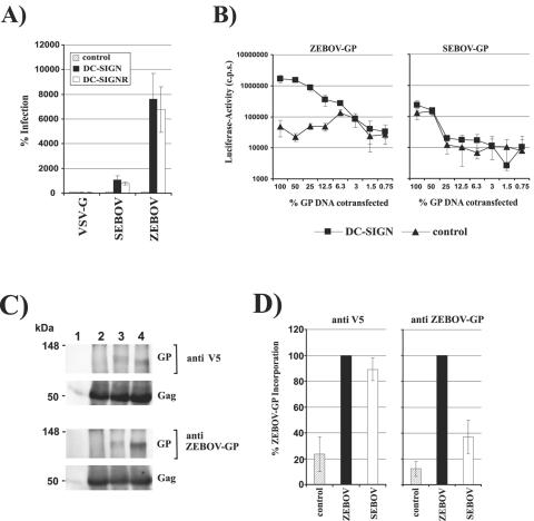
We and others previously found that DC-SIGN/R strongly augmented infections by HIV pseudovirions harboring ZEBOV-GP but had little impact on infections driven by SEBOV-GP (1, 50). Because of biosafety considerations, a SIV reporter genome with a large deletion in the envelope (env) gene (32) was chosen for the present study. Pseudovirions were generated as described previously (50), normalized for comparable luciferase activities upon infection of 293T cells, and employed for infection of 293 T-REx cells stably expressing the indicated lectins. DC-SIGN/R expression strongly enhanced infection by pseudotypes bearing ZEBOV-GP (∼70-fold increase compared to infection of control cells, which was set as 100%), whereas SEBOV-GP-driven infection was only moderately augmented (∼10-fold increase) (Fig. 1A). Infection mediated by vesicular stomatitis virus G was not modulated by DC-SIGN/R (Fig. 1A). These results confirm previous reports (1, 50) and indicate that SIV-based pseudovirions are adequate tools for analyzing EBOV-GP interactions with DC-SIGN/R.
FIG. 1.
Efficiency of ZEBOV-GP incorporation into pseudovirions modulates DC-SIGN/R engagement. (A) Pseudotypes harboring the indicated GPs were normalized for equal infection of 293T cells and used to infect T-REx cell lines expressing DC-SIGN/R or empty vector. Luciferase activities in cell lysates were determined 72 h after infection. The results are presented relative to infection of control cells, which was set as 100%. The averages of three independent experiments performed in triplicate are shown. Error bars indicate standard errors of the means (SEM). VSV-G, vesicular stomatitis virus G protein. (B) 293T cells were transiently cotransfected with constant amounts of plasmid carrying the SIV reporter genome and titrated amounts of EBOV-GP-encoding plasmids, and the supernatants were employed to infect T-REx DC-SIGN or T-REx control cells. Luciferase activities in cell lysates were determined 72 h after infection. The results of a representative experiment carried out in triplicate are shown, and similar results were obtained in an independent experiment. Error bars indicate standard deviations (SD). (C) lVLPs harboring ZEBOV- or SEBOV-GP with a C-terminal V5 tag were pelleted and lysed under nonreducing conditions, and Gag and GP were detected by Western blotting, using either an anti-V5 monoclonal antibody (top panels) or a rabbit serum raised against ZEBOV-GP (bottom panels) for detection of GP and a Gag-specific rabbit serum for visualization of Gag. Lane 1, control; lane 2, Gag; lane 3, Gag and SEBOV-GP; lane 4, Gag and ZEBOV-GP. (D) The incorporation of EBOV-GPs and Gag into lVLPs was analyzed as described for panel C and quantified by densitometry using AIDA image analyzer basic software (version 3.10). The incorporation of GP relative to Gag was determined, and the values obtained for ZEBOV-GP were set as 100%. The averages of three independent experiments with different lVLP preparations are shown. Error bars indicate SEM.
The amount of HIV and SIV Env incorporation into viral particles can affect infectivity, most likely by modulating the frequency of receptor binding events (3, 68). We asked if the level of ZEBOV- and SEBOV-GP incorporation into pseudovirions impacts DC-SIGN/R engagement. Viral pseudotypes were produced upon cotransfection of the SIV reporter plasmid with titrated amounts of ZEBOV- and SEBOV-GP and used for infections of DC-SIGN-expressing and control cells without prior normalization. With few exceptions, SEBOV-GP-bearing pseudotypes infected DC-SIGN-positive cells slightly more efficiently than control cells, and relative infections of both cell types were comparable, independent of the amount of GP transfected (Fig. 1B, right panel). In stark contrast, ZEBOV-GP-bearing pseudotypes infected DC-SIGN-expressing cells more efficiently than control cells when large amounts of GP were transfected, while infections of both cell types were comparable when small amounts of GP were cotransfected (Fig. 1B, left panel). Western blot analysis of virion-associated ZEBOV- and SEBOV-GP demonstrated that both GPs were incorporated into virions, with roughly comparable efficiencies (Fig. 1C and D), and that titrating out the amount of transfected GP indeed led to a gradual reduction in the GP:Gag ratio (data not shown). Thus, the rate of ZEBOV-GP incorporation can modulate lectin engagement, while SEBOV-GP incorporation, at least under the conditions tested, had no appreciable impact on interactions with DC-SIGN/R (Fig. 1B). Cotransfection of GP and SIV vector plasmids at a ratio of 3 to 1 did not increase DC-SIGN/R usage by SEBOV-GP-bearing pseudotypes (data not shown), further substantiating the observation that SEBOV-GP incorporation into virions does not limit DC-SIGN/R engagement.
ZEBOV- but not SEBOV-GP-bearing fVLPs bind efficiently to DC-SIGN/R.
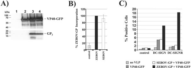
Retrovirus and filovirus particles differ in size and shape, which might impact attachment factor engagement. We therefore investigated if the differential DC-SIGN/R engagement of ZEBOV- and SEBOV-GP-bearing lentiviral pseudotypes reflects lectin binding by fVLPs bearing these GPs. fVLPs were generated by coexpression of GP and the EBOV matrix protein VP40 fused to GFP (36). In agreement with previous studies (6, 25, 40, 57, 60), an analysis of transfected cells by immunofluorescence revealed the generation of filamentous structures (data not shown), and Western blot analysis of fVLPs concentrated by centrifugation through a sucrose cushion demonstrated comparable virion incorporation of ZEBOV- and SEBOV-GP (Fig. 2A and B). Concentrated fVLPs bearing ZEBOV-GP bound efficiently to DC-SIGN/R-expressing cells but not to control cells (Fig. 2C). In contrast, binding of SEBOV-GP-harboring fVLPs to DC-SIGN/R-positive cells was inefficient and similar to that observed for control fVLPs harboring no GP. Thus, ZEBOV-GP and SEBOV-GP are incorporated into fVLPs to similar degrees, but only ZEBOV-GP-bearing particles bind to DC-SIGN/R with a high efficiency. These observations are in agreement with the results obtained with pseudotypes (Fig. 1A) and validate infection with lentiviral reporter viruses as a suitable system for studying attachment factor engagement by EBOV-GP.
FIG. 2.
ZEBOV- but not SEBOV-GP-bearing fVLPs bind to cellular DC-SIGN/R. (A) fVLPs were generated by cotransfection of VP40-GFP and the indicated GPs harboring a C-terminal V5 tag, concentrated by centrifugation through a sucrose cushion, and analyzed for VP40-GFP (top panel) and GP (bottom panel) expression by Western blotting with anti-GFP serum and an anti-V5 monoclonal antibody, respectively. The fVLPs analyzed were obtained upon transient transfection of 293T cells with plasmids encoding the following proteins: control (pcDNA3.1) (lane 1), VP40-GFP (lane 2), VP40-GFP and SEBOV-GP (lane 3), and VP40-GFP and ZEBOV-GP (lane 4). (B) The incorporation of EBOV-GP and VP40-GFP into fVLPs was analyzed as described for panel A and quantified by densitometry. The incorporation of GP relative to VP40-GFP was determined, and the values obtained for ZEBOV-GP were set as 100%. The averages of five independent experiments with different fVLP preparations are presented. Error bars indicate SEM. (C) Filovirus-like particles harboring comparable amounts of ZEBOV-GP and SEBOV-GP or no GP were incubated with B-THP DC-SIGN/R or control B-THP cells, and the percentage of GFP-positive cells was determined by FACS analysis. The results of a representative experiment performed in duplicate are shown, and comparable results were obtained in three independent experiments with two different fVLP preparations.
The glycosylation status of SEBOV-GP determines interaction with DC-SIGN/R.
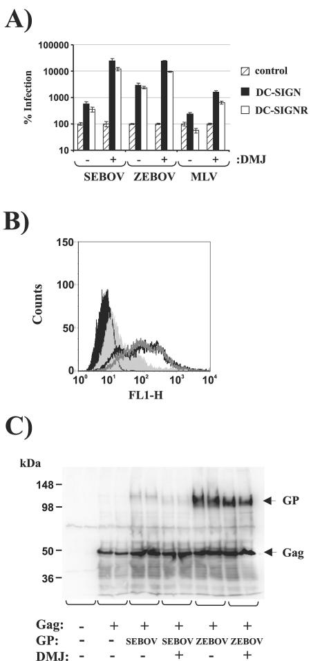
In order to address whether the type of glycosylation of EBOV-GP governs DC-SIGN/R engagement or if protein-protein interactions are involved, we asked if forced incorporation of high-mannose carbohydrates into SEBOV-GP is sufficient to confer efficient DC-SIGN/R utilization. Therefore, pseudotypes bearing ZEBOV-, SEBOV-, and MLV-GP were produced in the presence or absence of DMJ. DMJ blocks mannosidase I, resulting in the exclusive incorporation of high-mannose carbohydrates into glycoproteins. These viruses were normalized for comparable infections of control cells and employed for infections of lectin-expressing and lectin-negative control cells. The results are shown relative to infection of control cells, which was set as 100%. In agreement with previous results (33), DMJ treatment conferred functional DC-SIGN/R binding on MLV-GP (Fig. 3A), which normally does not interact with these lectins efficiently. DMJ treatment strongly increased DC-SIGN/R engagement by SEBOV-GP (80-fold) and slightly enhanced lectin engagement by ZEBOV-GP (10-fold). Importantly, the overall enhancements of infection by DC-SIGN/R was comparable for ZEBOV- and SEBOV-GP pseudotypes following incubation of producer cells with DMJ (Fig. 3A), and DMJ treatment did not enhance cell surface expression of GP (Fig. 3B) or GP incorporation into virions (Fig. 3C; note that the serum used for the detection of GP reacts preferentially with ZEBOV-GP, as shown in Fig. 1C and D), demonstrating that modification of ZEBOV- and SEBOV-GP with high-mannose carbohydrates is sufficient for robust DC-SIGN/R engagement.
FIG. 3.
EBOV-GP-bearing pseudotypes produced in the presence of DMJ use DC-SIGN/R for efficient enhancement of infection. (A) 293T cells were cotransfected with a SIV reporter genome and the indicated GP expression plasmids, the cells were cultivated in the absence or presence of DMJ, and supernatants were harvested. The supernatants were normalized for infectivity and employed for infection of T-REx cell lines expressing the indicated lectins. Luciferase activities in cell lysates were determined 72 h after infection. Results are presented relative to infection of control cells, which was set as 100%. The averages of three experiments are shown. Error bars indicate SEM. (B) 293T cells were transiently transfected with pcDNA3.1 (light gray shaded area) or ZEBOV-GP, the ZEBOV-GP-transfected cells were incubated in the presence (dark gray line) or absence (black line) of DMJ, and surface expression of GP was analyzed by FACS using a rabbit serum raised against ZEBOV-GP. The black shaded area indicates the background fluorescence of unstained cells. (C) Lentivirus-like particles bearing the indicated GPs were produced in the presence or absence of DMJ, and the GP and Gag contents were analyzed by Western blotting, using rabbit sera raised against ZEBOV-GP and Gag, respectively. Similar results were obtained in an independent experiment.
The mucin-like domain and glycosylation sites unique to ZEBOV- or SEBOV-GP do not modulate DC-SIGN/R engagement.
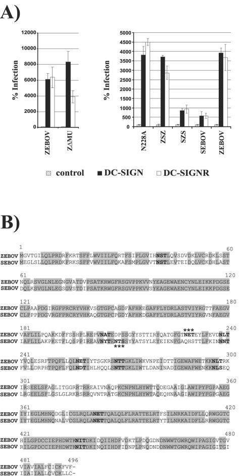
Since functional DC-SIGN/R binding was dependent on GP glycosylation, we first focused our analysis on sites of differential glycosylation in ZEBOV- and SEBOV-GP. The mucin-like domain of GP harbors several signals for N- and O-linked glycosylation and exhibits sequence variation between the EBOV subspecies, suggesting that it could modulate the interaction with DC-SIGN/R. However, the deletion of this domain in ZEBOV-GP (mutant ZΔMU; see Fig. 5A) did not appreciably impact the interaction with DC-SIGN/R and, in agreement with previous reports (26), did not diminish pseudotype infectivity (Fig. 4A, left panel; data not shown). Sequence alignment of ZEBOV- and SEBOV-GP without the mucin-like domains revealed that one signal for N-linked glycosylation out of nine was unique to each GP (Fig. 4B). In order to determine if these glycosylation signals are important for the interaction with DC-SIGN/R, a 27-amino-acid portion harboring both glycosylation signals was exchanged between ZEBOV- and SEBOV-GP (chimeras ZSZ and SZS; Fig. 5A). Additionally, the glycosylation signal unique to ZEBOV-GP was inactivated (mutant N228A; Fig. 5A). However, neither the exchange of the unique glycosylation signals between SEBOV- and ZEBOV-GP nor the inactivation of the glycosylation signal unique to ZEBOV-GP appreciably modulated the interaction with DC-SIGN/R (Fig. 4A, right panel), indicating that these motifs are not critical for DC-SIGN/R engagement.
FIG. 5.
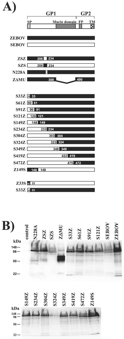
Expression of EBOV-GP mutants. (A) Schematic representation of the EBOV-GP mutants analyzed. SP, signal peptide; FP, fusion peptide; TM, transmembrane domain. (B) The indicated EBOV-GP variants were transiently expressed in 293T cells, and GP expression in cellular lysates was analyzed by Western blotting. Rabbit serum raised against ZEBOV-GP was used for detection. Similar results were obtained in two independent experiments. EBOV-GP variants were transiently expressed in 293T cells, and GP expression in cellular lysates was analyzed by Western blotting. Rabbit serum raised against ZEBOV-GP was used for detection. Similar results were obtained in two independent experiments.
FIG. 4.
The mucin-like domain and glycosylation signals unique to ZEBOV- or SEBOV-GP do not modulate DC-SIGN/R engagement. (A) T-REx cells expressing the indicated lectins were inoculated with infectivity-normalized pseudotypes bearing EBOV-GP variants in which the mucin-like domain was deleted (ZΔMU, left panel) or in which unique glycosylation sites were exchanged or inactivated (right panel). Luciferase activities were determined 72 h after infection. Results are presented relative to infection of control cells, which was set as 100%. The results of a representative experiment performed in triplicate are shown, and comparable results were obtained in two independent experiments. Error bars indicate SD. (B) Alignment of ZEBOV- and SEBOV-GP sequences without the variable mucin-like domain. Signals for N-linked glycosylation are indicated in bold, and unique glycosylation signals are marked with asterisks.
The signal peptide can determine GP interactions with DC-SIGN/R.
In order to map regions in ZEBOV-GP which contribute to DC-SIGN/R engagement, we analyzed a set of chimeric GPs in which amino acids 1 to 472 of ZEBOV-GP were replaced in a stepwise fashion by the corresponding sequences of SEBOV-GP (Fig. 5A). A subset of these chimeric GPs was generated previously, employing ZEBOV- and REBOV-GP, and was found to be functional in infection assays (26). In agreement with these observations, all chimeric GPs tested here were efficiently expressed (Fig. 5B) and mediated entry into target cells with efficiencies roughly comparable to those of the wild-type proteins (data not shown), indicating that the overall structure of the GPs was intact. The expression of GP variant S324Z and entry driven by variant Z149S were reduced compared to those of the wild-type proteins but were still readily detectable (Fig. 5B and data not shown).
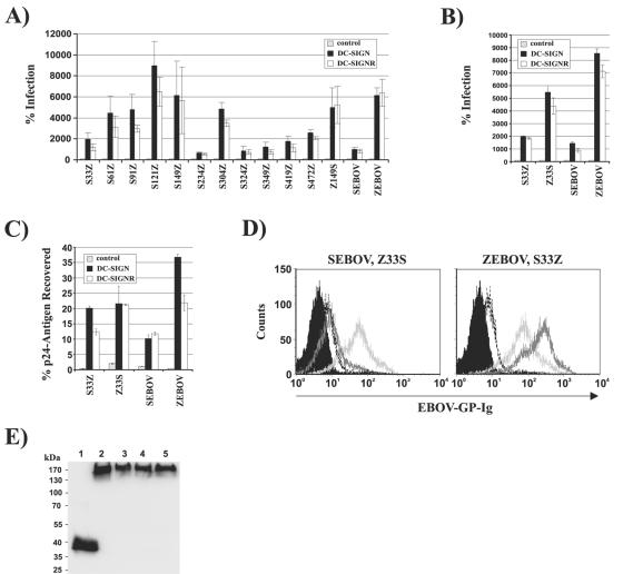
Infection of the indicated T-REx cell lines with infectivity-normalized pseudotypes revealed that several determinants in GP control DC-SIGN/R engagement. Thus, all chimeric GPs containing at least the N-terminal 324 amino acids of SEBOV-GP exhibited reduced DC-SIGN/R engagement, similar to that of wild-type SEBOV-GP, indicating that the N-terminal 324 amino acids in ZEBOV-GP control the interaction with DC-SIGN/R (Fig. 6A). However, several determinants of lectin engagement must be located within this sequence, since exchanges of S33Z and S234Z strongly reduced the DC-SIGN/R-mediated enhancement of infection, while all other exchanges in this region had only minor effects on the interaction with these lectins (Fig. 6A). Notably, the exchange of the first 32 amino acids, which correspond to the signal peptide, was sufficient to strongly diminish ZEBOV-GP-mediated engagement of DC-SIGN/R (Fig. 6A). This observation suggests that the signal peptide, which targets the nascent GP polypeptide chain for transport into the ER, is a determinant of DC-SIGN/R engagement. However, the signal peptide seems to exert its effect on lectin binding in conjunction with other regulatory sequences located between amino acids 33 and 324.
FIG. 6.
DC-SIGN/R usage by chimeric EBOV-GPs. (A) Infectivity-normalized pseudotypes bearing ZEBOV-GP variants in which the indicated portions were replaced by the corresponding sequences of SEBOV-GP were used to infect T-REx cell lines expressing the indicated lectins. Luciferase activities in cell lysates were determined 72 h after infection. Results are presented relative to infection of control cells, which was set as 100%. The averages of at least three independent experiments performed in triplicate are shown. Error bars indicate SEM. (B) Pseudotypes bearing ZEBOV- and SEBOV-GP variants in which the signal peptides were exchanged with the homologous sequences were used for infection of T-REx cells as described for panel A. The results of a representative experiment performed in triplicate are shown. Similar results were obtained in two independent experiments. Error bars indicate SD. (C) Pseudotypes bearing ZEBOV-, SEBOV-GP, or variants thereof in which the signal peptides were swapped were normalized for equal amounts of p24 antigen and incubated with the indicated T-REx cells. Incubation was performed at 4°C in order to prevent infection. Unbound virions were removed, the cells were lysed, and the amounts of p24 antigen in cellular lysates were determined. The percentages of input virus retained by the T-REx cell lines are indicated. The results of a representative experiment are shown and were confirmed in an independent experiment. Error bars indicate SD. (D) T-REx DC-SIGN cells were incubated with concentrated cellular supernatants containing wild-type EBOV-GP (dark gray line) or signal peptide variants (light gray line) fused to the Fc portion of human immunoglobulin or a control Fc protein (dotted black line). The cells were stained with Fc-specific antibody and analyzed by FACS. The results of a representative experiment are shown, and similar results were obtained in an independent experiment. The black shaded area indicates the fluorescence of cells stained with secondary antibody only. (E) The input proteins used for the binding experiment shown in panel D were analyzed by Western blotting employing an Fc-specific antibody. The samples were loaded in the following order: control Fc protein (lane 1), Z33S variant (lane 2), SEBOV-GP (lane 3), ZEBOV-GP (lane 4), and S33Z variant (lane 5).
To further investigate the importance of the signal peptide for lectin engagement, we also introduced the signal peptide of ZEBOV-GP into SEBOV-GP, resulting in GP variant Z33S (Fig. 5A). DC-SIGN/R engagement by the signal peptide variants was tested by infection of lectin-expressing T-REx lines with the indicated virus stocks normalized for equal luciferase production upon infection of control cells. The introduction of the SEBOV-GP signal peptide into ZEBOV-GP reduced the interaction with DC-SIGN/R, while the converse change enhanced lectin engagement by SEBOV-GP (Fig. 6B), confirming a role of the signal peptide in lectin interactions of ZEBOV- and SEBOV-GP.
We next asked if the differential capacities of the GP signal peptide variants to employ DC-SIGN/R for augmentation of infection are reflected by differential lectin binding levels of the respective GPs. To address this question, we first assessed binding of pseudotypes to lectin-expressing T-REx cells by employing a previously established binding assay (43, 50). In this assay format, SEBOV-GP-bearing pseudotypes bound less efficiently to DC-SIGN/R-expressing cells than did pseudotypes bearing ZEBOV-GP, while viruses harboring the S33Z and Z33S variants exhibited an intermediate phenotype (Fig. 6C). When DC-SIGN/R binding of soluble EBOV-GP variants was analyzed, strong binding was again observed with ZEBOV-GP but not with SEBOV-GP, and the signal peptide variants exhibited intermediate binding efficiencies (Fig. 6D and E). Thus, the signal peptide exchange between ZEBOV- and SEBOV-GP transfers the efficiencies of both lectin binding and lectin-mediated augmentation of infection from the wild-type proteins to the chimeric variants.
The signal sequence impacts EBOV-GP modification with high-mannose carbohydrates.
Modification of EBOV-GP with high-mannose carbohydrates is sufficient to confer efficient DC-SIGN/R engagement (Fig. 3). We therefore hypothesized that the signal peptide might modulate to what extent these carbohydrates are incorporated into EBOV-GP and that differential glycosylation might, at least in part, account for the differential DC-SIGN/R engagement by ZEBOV, the S33Z mutant, SEBOV, and the Z33S mutant. To address this question, we analyzed the glycosylation status of the respective GPs by enzymatic digestion. Digestion with PNGase F, which removes all N-linked glycans, increased the gel mobilities of all GPs tested (Fig. 7A), indicating the presence of N-linked glycosylation. However, only GPs from retroviral pseudovirions produced in the presence of DMJ were sensitive to endo H digestion (Fig. 7A), which removes N-linked high-mannose carbohydrates, suggesting that under normal conditions, incorporation of these carbohydrates into EBOV-GPs is inefficient.
FIG. 7.

Analysis of ZEBOV- and SEBOV-GP glycosylation. (A) lVLPs were produced by transient transfection of 293T cells in the presence or absence of DMJ, normalized for comparable Gag contents, digested with endo H and PNGase F for the indicated time periods, and analyzed by Western blotting. (B) lVLPs bearing the indicated GPs were concentrated by centrifugation through a sucrose cushion and analyzed for GP and Gag incorporation by Western blotting with antisera raised against ZEBOV-GP and Gag, respectively. lVLPs were obtained from cells expressing the following proteins: lane 1, Gag; lane 2, Gag and SEBOV-GP; lane 3, Gag and ZEBOV-GP; lane 4, Gag and S33Z variant; lane 5, Gag and Z33S variant. (C) lVLPs were analyzed for reactivity with lectins with known specificities. lVLPs were derived from cells expressing the following proteins: lane 1, Gag; lane 2, Gag and SEBOV-GP; lane 3, Gag and ZEBOV-GP; lane 4, Gag and S33Z variant; lane 5, Gag and Z33S variant. (D) Supernatants from pcDNA3 (lanes 1)-, SEBOV-GP (lanes 2)-, and Z33S (lanes 3)-transfected cells were concentrated using Centricon Plus columns and analyzed as described for panel C. Panels C and D show the results of representative experiments, and similar results were obtained in two independent experiments. cushion and analyzed for GP and Gag incorporation by Western blotting with antisera raised against ZEBOV-GP and Gag, respectively. lVLPs were obtained from cells expressing the following proteins: lane 1, Gag; lane 2, Gag and SEBOV-GP; lane 3, Gag and ZEBOV-GP; lane 4, Gag and S33Z variant; lane 5, Gag and Z33S variant. (C) lVLPs were analyzed for reactivity with lectins with known specificities. lVLPs were derived from cells expressing the following proteins: lane 1, Gag; lane 2, Gag and SEBOV-GP; lane 3, Gag and ZEBOV-GP; lane 4, Gag and S33Z variant; lane 5, Gag and Z33S variant. (D) Supernatants from pcDNA3 (lanes 1)-, SEBOV-GP (lanes 2)-, and Z33S (lanes 3)-transfected cells were concentrated using Centricon Plus columns and analyzed as described for panel C. Panels C and D show the results of representative experiments, and similar results were obtained in two independent experiments.
In order to detect low-level EBOV-GP modification with high-mannose carbohydrates, we employed a sensitive assay system based on labeled lectins with known carbohydrate specificities. Binding of these lectins to lVLP-associated GP was analyzed. Preparations of lVLPs containing ZEBOV-, S33Z-, SEBOV-, and Z33S-GP exhibited levels of incorporation of the signal peptide variants roughly comparable to those of the parental GPs (Fig. 7B), although somewhat lower signals were usually obtained for S33Z- and Z33S-GP than for ZEBOV- and SEBOV-GP, respectively. The stronger signal observed for ZEBOV-GP than for SEBOV-GP is due to preferential recognition of ZEBOV-GP by the antiserum employed for detection (Fig. 1C and D). Comparable signals were obtained for all GPs upon analysis of lVLPs with MAA, a lectin specific for sialic acid (Fig. 7C, right panel). In contrast, ZEBOV-GP- but not SEBOV-GP-harboring lVLPs consistently reacted with GNA, which is specific for high-mannose carbohydrates, indicating that some of the ZEBOV-GPs incorporated into virions are indeed modified with high-mannose carbohydrates (Fig. 7C, left panel). Importantly, introduction of the SEBOV-GP signal peptide into ZEBOV-GP repeatedly reduced the reactivity with GNA, while the converse exchange slightly enhanced the reactivity (Fig. 7C and D), suggesting that the signal peptide modulates GP glycosylation. Endo H and PNGase F digestion confirmed the specificity of lectin binding (Fig. 7C), and Western blot analysis revealed that the signals observed were indeed due to EBOV-GP (data not shown). Thus, the signal peptides can determine how efficiently high-mannose glycans are incorporated into EBOV-GP.
The signal peptide modulates introduction and/or glycosylation of GP in the secretory pathway and is not present in mature GP.
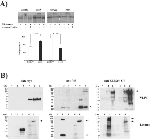
To further assess the role of the signal peptide in EBOV-GP expression, we next employed a cell-free system. The open reading frames for SEBOV-GP, Z33S-GP, ZEBOV-GP, and S33Z-GP were transcribed and translated in the presence or absence of ER-derived microsomes, which allow for glycosylation. Glycosylated proteins were detected thereafter due to their decreased gel mobilities (Fig. 8A, top panel, black arrows). A significantly larger fraction of the Z33S variant than of SEBOV-GP was found to be glycosylated, and similar results were obtained for ZEBOV-GP and the S33Z variant (Fig. 8A). These results indicate that the signal peptide variants might be differentially introduced and/or glycosylated within the secretory pathway and that these differences correlate with the efficiency of DC-SIGN/R engagement.
FIG. 8.
The signal peptide determines EBOV-GP interactions with the secretory pathway and is cleaved off during GP biogenesis. (A) The open reading frames encoding ZEBOV-GP, SEBOV-GP, Z33S-GP, and S33Z-GP were transcribed and translated in the presence and absence of ER-derived microsomes and an acceptor peptide, an inhibitor of glycosylation. The results of SDS-PAGE analysis of the reaction products from a representative experiment are shown in the top panel. The amounts of glycosylated reaction products were quantified by densitometry (bottom panel). The percent glycosylation represents the band intensity of the glycosylated fraction normalized to that of the precursor. The results of three independent experiments are shown. Error bars indicate SEM. (B) Western blot analysis of GP expression in cellular supernatants and purified fVLPs (top panels) or in in vitro transcription/translation reactions (bottom panels). The expression of ZEBOV- and SEBOV-GP variants harboring an N-terminal Myc and a C-terminal V5 antigenic tag was assessed by employing the indicated detection reagents. (Top panels) Supernatants of cells transfected with pcDNA3.1 (lane 1), SEBOV-GP (lane 2), or ZEBOV-GP (lane 3) or purified fVLPs produced upon cotransfection of Myc-tagged VP40 with pcDNA3.1 (lane 4), SEBOV-GP (lane 5), or ZEBOV-GP (lane 6). (Bottom panels) Cells transfected with pcDNA3.1 (lane 1), ZEBOV-GP (lane 2), Myc-tagged VP40 (lane 3), in vitro transcribed and translated pcDNA3.1 (lane 4), or in vitro transcribed and translated ZEBOV-GP (lane 5). Black arrows indicate bands corresponding to GP.
In order to test signal sequence cleavage of SEBOV-GP, ZEBOV-GP, and their respective chimeras, a competitive inhibitor of glycosylation (acceptor peptide) was added to the translation reaction. As expected, the glycosylated fraction of the GPs analyzed disappeared in the presence of the acceptor peptide (Fig. 8A, top panel). Under these conditions, a new species was generated with a lower molecular weight than the precursor present in the absence of microsomes, consistent with the estimated molecular weight of GP after the removal of the signal peptide (Fig. 8A, top panel, white arrows). Thus, these results suggest that both the wild-type GPs and the chimeric variants undergo signal sequence cleavage upon ER translocation in a cell-free assay.
To analyze signal sequence cleavage in cells, we introduced an N-terminal Myc and a C-terminal V5 antigenic tag into ZEBOV- and SEBOV-GP (the Myc tag was introduced at the N terminus of the signal peptide). The GP variants mediated pseudotype infections with high efficiencies (data not shown), indicating that the antigenic tags did not interfere with GP function. Expression of the tagged GPs was analyzed in supernatants of GP-transfected cells or in sucrose-purified fVLPs generated upon cotransfection of GP with Myc-tagged VP40. Staining with a rabbit serum raised against ZEBOV-GP revealed efficient GP incorporation into fVLPs, while less GP was detected in cellular supernatants (Fig. 8B, upper left panel). Similarly, staining with an anti-V5 antibody detected prominent bands corresponding to the GP2 subunit in fVLP lysates and, to a lesser degree, in supernatants of GP-transfected cells. In contrast, VP40, but not GP, was detected when the anti-Myc antibody was used for staining (Fig. 8B, upper left panel), suggesting that the signal peptide was cleaved off at the very early stages of GP synthesis in the ER. If the signal sequence is removed during ER import, the N-terminal tag should still be present in GP synthesized outside the ER. We therefore compared the expression of in vitro-translated ZEBOV-GP with that of GP produced in transfected 293T cells. Prominent GP signals in both cellular lysates and the in vitro translation reaction were obtained when a GP-specific or V5-specific antibody was used for staining (Fig. 8B, lower panels). However, anti-Myc antibody detected only in vitro-translated, not cellular, GP, strongly suggesting that the Myc-tagged signal peptide is present in GP generated outside the ER but is cleaved off upon transport of GP into the ER.
DISCUSSION
In this study, we analyzed the determinants of DC-SIGN/R engagement by the GPs of the EBOV subspecies Zaire and Sudan. In the context of filovirus and lentivirus particles, only ZEBOV-GP interacted with DC-SIGN/R efficiently, resulting in strongly enhanced infection by lentiviral pseudotypes bearing this GP. DC-SIGN/R engagement by ZEBOV-GP was dependent on the level of GP incorporation into virions, suggesting that multiple binding events are required for robust enhancement of infection. Modification of SEBOV-GP with high-mannose carbohydrates was sufficient to confer efficient DC-SIGN/R engagement, indicating that the DC-SIGN/R-EBOV-GP interaction is carbohydrate dependent. However, glycosylation sites unique to ZEBOV- or SEBOV-GP did not account for the differential DC-SIGN/R binding of these GPs. In contrast, the interactions of ZEBOV- and SEBOV-GP with DC-SIGN/R were controlled by the signal peptides of the respective GPs, which modulated GP modification with high-mannose glycans.
Filovirus entry into target cells is incompletely understood. Analyses of viral cell tropism both in cell culture (64) and in infected macaques (19) indicated that a broad range of cell types are permissive to EBOV infection, with lymphoid cells constituting an exception (19, 64). Therefore, the viral receptor(s) seems to be widely expressed. Extensive mutagenic analysis identified the N-terminal 150 amino acids of GP1 as critical for EBOV-GP-mediated entry into target cells, and these residues might constitute a receptor binding pocket (35). However, the cellular receptor(s) contacted by filovirus GPs for entry is unknown. Previous studies, including our own work, demonstrated that filovirus GPs interact with the cellular lectins DC-SIGN/R (1, 50), human macrophage C-type lectin specific for galactose and N-acetylglucosamine (hMGL) (55), asialoglycoprotein receptor (7, 33), and liver and lymph node sinusoidal endothelial cell C-type lectin (LSECtin) (21) and that these interactions can result in robust augmentation of GP-mediated cellular entry (4, 42). It is currently not clear if some or all of these lectins can function as true receptors or if these proteins simply augment viral attachment to cells and thereby increase entry via so far unknown cellular receptors (1, 50). Nevertheless, indirect evidence suggests that these lectins might play an important role in filovirus infection in vivo. Thus, despite the broad viral tropism observed in vitro (64) and at later stages of EBOV infection in vivo (19), the range of viral target cells and organs early in infection is quite narrow (19), and many of the early targets express lectins that augment filovirus entry. It is therefore conceivable that, e.g., DC-SIGN and hMGL focus EBOV infection on macrophages and DCs, which are early and sustained targets (19), while the asialoglycoprotein receptor might concentrate viral particles in the liver, a major target organ of filoviruses (19). Finally, DC-SIGN/R and LSECtin could facilitate infection of sinusoidal endothelial cells in the liver, which are the first type of endothelial cells infected in macaques experimentally inoculated with ZEBOV (19). The analysis of filovirus interactions with DC-SIGN/R and other cellular lectins might therefore reveal important insights into filovirus infection and might uncover novel targets for therapeutic intervention.
DC-SIGN/R-mediated enhancement of ZEBOV-GP-driven infection was dependent on the amount of GP incorporated into virions (Fig. 1B). Importantly, transduction of macrophages by DC-SIGN/R-encoding lentiviruses (50) or induced expression of DC-SIGN/R on cell lines (21) augments the susceptibility to infection by replication-competent ZEBOV, suggesting that EBOV incorporates sufficient GP to efficiently engage DC-SIGN/R. In this context, it is notable that decreasing the amount of GP incorporated into virions did not diminish ZEBOV-GP-driven infection of control cells and that SEBOV-GP-mediated infection was only moderately reduced (Fig. 1B). Thus, while the level of GP incorporation into virions can affect attachment factor engagement, even very small amounts of GP seem to be sufficient to allow efficient binding to the receptor and infection of target cells.
Forced incorporation of high-mannose carbohydrates into SEBOV-GP allowed efficient engagement of DC-SIGN/R without increasing GP cell surface expression or virion incorporation (Fig. 3), indicating that adequate carbohydrate modification of SEBOV-GP is sufficient to confer DC-SIGN/R binding. These data are in agreement with previous reports (22, 33) and suggest that specific glycosylation sites in ZEBOV- but not in SEBOV-GP might be modified with high-mannose carbohydrates and facilitate recognition by DC-SIGN/R. However, neither glycosylation signals unique to ZEBOV- or SEBOV-GP nor the mucin-like domain, which is quite divergent between the EBOV subspecies and harbors several signals for N- and O-linked glycosylation, was required for DC-SIGN/R engagement or infection of target cells (Fig. 4), with the latter being in agreement with published results (26). Linking DC-SIGN/R usage to the modification of specific glycosylation sites in EBOV-GP with high-mannose glycans will therefore be difficult.
In agreement with a pivotal role of high-mannose carbohydrates in EBOV-GP recognition by DC-SIGN/R, ZEBOV- but not SEBOV-GP was found to contain these glycans, as judged by its ability to bind to GNA (Fig. 7). By employing the same experimental system, similar results were previously obtained for GPs from replication-competent ZEBOV and SEBOV (14), indicating that lVLPs bearing EBOV-GP are a valid model for analyzing EBOV-GP glycosylation. These results suggest that appropriate virion incorporation and glycosylation of GP determine the interaction with DC-SIGN/R. Unexpectedly, an analysis of chimeric GPs revealed that the GP signal peptide modulated the latter function (Fig. 6) and therefore constitutes a determinant of DC-SIGN/R engagement. However, it is important that the signal peptide exerted its effects on DC-SIGN/R binding in conjunction with other sequences in GP, since several chimeric GPs which contained the SEBOV-GP signal peptide interacted with DC-SIGN/R efficiently (Fig. 6). Since the function of the signal peptide is context dependent, the introduction of a given signal peptide into a heterologous protein might not generally confer the glycosylation pattern of the parental protein on the chimeric one. Also, the introduction of the SEBOV-GP signal peptide into ZEBOV-GP had a more pronounced effect on DC-SIGN/R-mediated augmentation of infectivity than on binding to these lectins (Fig. 6B and C), raising the possibility that the signal peptide-induced defects in DC-SIGN/R usage are of a complex nature. Finally, it needs to be taken into account that glycosylation is cell type dependent, and it is currently unclear if GP glycosylation in 293 cells adequately mirrors GP glycosylation in primary EBOV target cells, such as macrophages. Detailed further analysis is therefore warranted to characterize the interplay between the signal peptide and sequences in mature GP and their potential interactions with cellular factors.
While signal peptides of a few viral glycoproteins have functions beyond targeting the nascent protein to the ER (37), a role of the signal peptide in GP glycosylation has not been documented. How does the EBOV-GP signal peptide, which is cleaved off after ER translocation (Fig. 8), modulate these functions? A large body of evidence suggests that signal sequences are multifunctional. Thus, the signal peptide can modulate interactions between the ribosome and translocon and can thereby determine to what extent the nascent chain is exposed to the cytoplasm and the ER lumen (15, 29, 30, 45). Also, the signal sequence can impact the topology of the mature protein (31). Finally, interactions between the signal peptide and the components of the translocon can determine when the signal peptide is cleaved off, which in turn controls how efficiently the protein is glycosylated (46).
The reported role of the signal peptide in protein glycosylation is in agreement with our observation that high-mannose carbohydrates are incorporated into ZEBOV- but not SEBOV-GP and that incorporation of these glycans depends on the signal sequence. It has been proposed that signal peptide cleavage modulates N-linked glycosylation by modifying the location and thus accessibility of N-terminal glycosylation signals to the oligosaccharyl transferase complex (11, 39) and/or by modulating the conformation of the N terminus and its accessibility to modifications (46). However, an analysis of translocation intermediates revealed that the signal peptides of ZEBOV-GP and a variant thereof harboring the SEBOV-GP signal were cleaved off at comparable stages of the translocation process (during translocation of amino acids 95 to 219 [data not shown]). Thus, either the time of signal peptide cleavage does not modulate EBOV-GP glycosylation or relatively subtle differences in signal peptide cleavage kinetics account for the differential modification of ZEBOV- and SEBOV-GP with high-mannose carbohydrates. In any event, further studies are required to elucidate the molecular basis for the present observations.
In summary, the EBOV-GP signal peptide impacts GP glycosylation and attachment factor engagement. The latter is thought to be an important determinant of EBOV tropism, especially during the early stages of infection, when DCs and macrophages are central targets (8, 19). Hence, the expression of DC-SIGN on DCs might contribute to their preferential infection. Detailed mutagenic analysis of the EBOV-GP signal peptide sequences will be required to determine which regions contribute to the incorporation of high-mannose carbohydrates into GP. Moreover, it will be interesting to determine if signal sequences of other viral GPs modulate glycosylation. Since carbohydrate modification of HIV Env is thought to play a crucial role in engagement of cellular membrane proteins and in protection against neutralizing antibodies (27), the analysis of this protein might be especially promising.
Acknowledgments
We thank B. Fleckenstein and K. von der Mark for constant support. We thank M. Geier and A. Wegele for expert technical assistance. We are especially indebted to S. Becker and P. Möller for discussions and antibody 1G12, to R. Grunow for antibody 3B11, to U. Schubert for antisera specific for p24 and GFP, to P. Bieniasz for VP40-GFP and VP40-myc expression plasmids, and to A. Sanchez for EBOV-GP expression plasmids. p96ZM651 gag.opt, deposited by B. H. Hahn, was obtained through the NIH AIDS Research and Reference Reagent Program, Division of AIDS, NIAID, NIH.
A.M., T.G., H.H., and S.P. were supported by grant SFB466, and G.S. was funded by NIH MARCE grant U54 AI057168.
REFERENCES
- 1.Alvarez, C. P., F. Lasala, J. Carrillo, O. Muniz, A. L. Corbi, and R. Delgado. 2002. C-type lectins DC-SIGN and L-SIGN mediate cellular entry by Ebola virus in cis and in trans. J. Virol. 76:6841-6844. [DOI] [PMC free article] [PubMed] [Google Scholar]
- 2.Appelmelk, B. J., I. van Die, S. J. Van Vliet, C. M. Vandenbroucke-Grauls, T. B. Geijtenbeek, and Y. van Kooyk. 2003. Cutting edge: carbohydrate profiling identifies new pathogens that interact with dendritic cell-specific ICAM-3-grabbing nonintegrin on dendritic cells. J. Immunol. 170:1635-1639. [DOI] [PubMed] [Google Scholar]
- 3.Bachrach, E., H. Dreja, Y. L. Lin, C. Mettling, V. Pinet, P. Corbeau, and M. Piechaczyk. 2005. Effects of virion surface gp120 density on infection by HIV-1 and viral production by infected cells. Virology 332:418-429. [DOI] [PubMed] [Google Scholar]
- 4.Baribaud, F., R. W. Doms, and S. Pöhlmann. 2002. The role of DC-SIGN and DC-SIGNR in HIV and Ebola virus infection: can potential therapeutics block virus transmission and dissemination? Expert Opin. Ther. Targets 6:423-431. [DOI] [PubMed] [Google Scholar]
- 5.Bashirova, A. A., T. B. Geijtenbeek, G. C. van Duijnhoven, S. J. Van Vliet, J. B. Eilering, M. P. Martin, L. Wu, T. D. Martin, N. Viebig, P. A. Knolle, V. N. KewalRamani, Y. van Kooyk, and M. Carrington. 2001. A dendritic cell-specific intercellular adhesion molecule 3-grabbing nonintegrin (DC-SIGN)-related protein is highly expressed on human liver sinusoidal endothelial cells and promotes HIV-1 infection. J. Exp. Med. 193:671-678. [DOI] [PMC free article] [PubMed] [Google Scholar]
- 6.Bavari, S., C. M. Bosio, E. Wiegand, G. Ruthel, A. B. Will, T. W. Geisbert, M. Hevey, C. Schmaljohn, A. Schmaljohn, and M. J. Aman. 2002. Lipid raft microdomains: a gateway for compartmentalized trafficking of Ebola and Marburg viruses. J. Exp. Med. 195:593-602. [DOI] [PMC free article] [PubMed] [Google Scholar]
- 7.Becker, S., M. Spiess, and H. D. Klenk. 1995. The asialoglycoprotein receptor is a potential liver-specific receptor for Marburg virus. J. Gen. Virol. 76:393-399. [DOI] [PubMed] [Google Scholar]
- 8.Bosio, C. M., M. J. Aman, C. Grogan, R. Hogan, G. Ruthel, D. Negley, M. Mohamadzadeh, S. Bavari, and A. Schmaljohn. 2003. Ebola and Marburg viruses replicate in monocyte-derived dendritic cells without inducing the production of cytokines and full maturation. J. Infect. Dis. 188:1630-1638. [DOI] [PubMed] [Google Scholar]
- 9.Chan, S. Y., C. J. Empig, F. J. Welte, R. F. Speck, A. Schmaljohn, J. F. Kreisberg, and M. A. Goldsmith. 2001. Folate receptor-alpha is a cofactor for cellular entry by Marburg and Ebola viruses. Cell 106:117-126. [DOI] [PubMed] [Google Scholar]
- 10.Chandran, K., N. J. Sullivan, U. Felbor, S. P. Whelan, and J. M. Cunningham. 2005. Endosomal proteolysis of the Ebola virus glycoprotein is necessary for infection. Science 308:1643-1645. [DOI] [PMC free article] [PubMed] [Google Scholar]
- 11.Chen, X., C. VanValkenburgh, H. Liang, H. Fang, and N. Green. 2001. Signal peptidase and oligosaccharyltransferase interact in a sequential and dependent manner within the endoplasmic reticulum. J. Biol. Chem. 276:2411-2416. [DOI] [PubMed] [Google Scholar]
- 12.Connor, R. I., B. K. Chen, S. Choe, and N. R. Landau. 1995. Vpr is required for efficient replication of human immunodeficiency virus type-1 in mononuclear phagocytes. Virology 206:935-944. [DOI] [PubMed] [Google Scholar]
- 13.Feldmann, H., S. Jones, H. D. Klenk, and H. J. Schnittler. 2003. Ebola virus: from discovery to vaccine. Nat. Rev. Immunol. 3:677-685. [DOI] [PubMed] [Google Scholar]
- 14.Feldmann, H., S. T. Nichol, H. D. Klenk, C. J. Peters, and A. Sanchez. 1994. Characterization of filoviruses based on differences in structure and antigenicity of the virion glycoprotein. Virology 199:469-473. [DOI] [PubMed] [Google Scholar]
- 15.Fons, R. D., B. A. Bogert, and R. S. Hegde. 2003. Substrate-specific function of the translocon-associated protein complex during translocation across the ER membrane. J. Cell Biol. 160:529-539. [DOI] [PMC free article] [PubMed] [Google Scholar]
- 16.Gao, F., Y. Li, J. M. Decker, F. W. Peyerl, F. Bibollet-Ruche, C. M. Rodenburg, Y. Chen, D. R. Shaw, S. Allen, R. Musonda, G. M. Shaw, A. J. Zajac, N. Letvin, and B. H. Hahn. 2003. Codon usage optimization of HIV type 1 subtype C gag, pol, env, and nef genes: in vitro expression and immune responses in DNA-vaccinated mice. AIDS Res. Hum. Retrovir. 19:817-823. [DOI] [PubMed] [Google Scholar]
- 17.Geijtenbeek, T. B., R. Torensma, S. J. Van Vliet, G. C. van Duijnhoven, G. J. Adema, Y. van Kooyk, and C. G. Figdor. 2000. Identification of DC-SIGN, a novel dendritic cell-specific ICAM-3 receptor that supports primary immune responses. Cell 100:575-585. [DOI] [PubMed] [Google Scholar]
- 18.Geisbert, T. W., and L. E. Hensley. 2004. Ebola virus: new insights into disease aetiopathology and possible therapeutic interventions. Expert Rev. Mol. Med. 6:1-24. [DOI] [PubMed] [Google Scholar]
- 19.Geisbert, T. W., L. E. Hensley, T. Larsen, H. A. Young, D. S. Reed, J. B. Geisbert, D. P. Scott, E. Kagan, P. B. Jahrling, and K. J. Davis. 2003. Pathogenesis of Ebola hemorrhagic fever in cynomolgus macaques: evidence that dendritic cells are early and sustained targets of infection. Am. J. Pathol. 163:2347-2370. [DOI] [PMC free article] [PubMed] [Google Scholar]
- 20.Geisbert, T. W., H. A. Young, P. B. Jahrling, K. J. Davis, T. Larsen, E. Kagan, and L. E. Hensley. 2003. Pathogenesis of Ebola hemorrhagic fever in primate models: evidence that hemorrhage is not a direct effect of virus-induced cytolysis of endothelial cells. Am. J. Pathol. 163:2371-2382. [DOI] [PMC free article] [PubMed] [Google Scholar]
- 21.Gramberg, T., H. Hofmann, P. Möller, P. F. Lalor, A. Marzi, M. Geier, M. Krumbiegel, T. Winkler, F. Kirchhoff, D. H. Adams, S. Becker, J. Münch, and S. Pöhlmann. 2005. LSECtin interacts with filovirus glycoproteins and the spike protein of SARS coronavirus. Virology 340:224-236. [DOI] [PMC free article] [PubMed] [Google Scholar]
- 22.Guo, Y., H. Feinberg, E. Conroy, D. A. Mitchell, R. Alvarez, O. Blixt, M. E. Taylor, W. I. Weis, and K. Drickamer. 2004. Structural basis for distinct ligand-binding and targeting properties of the receptors DC-SIGN and DC-SIGNR. Nat. Struct. Mol. Biol. 11:591-598. [DOI] [PubMed] [Google Scholar]
- 23.Ito, H., S. Watanabe, A. Sanchez, M. A. Whitt, and Y. Kawaoka. 1999. Mutational analysis of the putative fusion domain of Ebola virus glycoprotein. J. Virol. 73:8907-8912. [DOI] [PMC free article] [PubMed] [Google Scholar]
- 24.Ito, H., S. Watanabe, A. Takada, and Y. Kawaoka. 2001. Ebola virus glycoprotein: proteolytic processing, acylation, cell tropism, and detection of neutralizing antibodies. J. Virol. 75:1576-1580. [DOI] [PMC free article] [PubMed] [Google Scholar]
- 25.Jasenosky, L. D., G. Neumann, I. Lukashevich, and Y. Kawaoka. 2001. Ebola virus VP40-induced particle formation and association with the lipid bilayer. J. Virol. 75:5205-5214. [DOI] [PMC free article] [PubMed] [Google Scholar]
- 26.Jeffers, S. A., D. A. Sanders, and A. Sanchez. 2002. Covalent modifications of the Ebola virus glycoprotein. J. Virol. 76:12463-12472. [DOI] [PMC free article] [PubMed] [Google Scholar]
- 27.Johnson, W. E., and R. C. Desrosiers. 2002. Viral persistance: HIV's strategies of immune system evasion. Annu. Rev. Med. 53:499-518. [DOI] [PubMed] [Google Scholar]
- 28.Jones, S. M., H. Feldmann, U. Stroher, J. B. Geisbert, L. Fernando, A. Grolla, H. D. Klenk, N. J. Sullivan, V. E. Volchkov, E. A. Fritz, K. M. Daddario, L. E. Hensley, P. B. Jahrling, and T. W. Geisbert. 2005. Live attenuated recombinant vaccine protects nonhuman primates against Ebola and Marburg viruses. Nat. Med. 11:786-790. [DOI] [PubMed] [Google Scholar]
- 29.Kim, S. J., and R. S. Hegde. 2002. Cotranslational partitioning of nascent prion protein into multiple populations at the translocation channel. Mol. Biol. Cell 13:3775-3786. [DOI] [PMC free article] [PubMed] [Google Scholar]
- 30.Kim, S. J., D. Mitra, J. R. Salerno, and R. S. Hegde. 2002. Signal sequences control gating of the protein translocation channel in a substrate-specific manner. Dev. Cell 2:207-217. [DOI] [PubMed] [Google Scholar]
- 31.Kim, S. J., R. Rahbar, and R. S. Hegde. 2001. Combinatorial control of prion protein biogenesis by the signal sequence and transmembrane domain. J. Biol. Chem. 276:26132-26140. [DOI] [PubMed] [Google Scholar]
- 32.Kirchhoff, F., S. Pöhlmann, M. Hamacher, R. E. Means, T. Kraus, K. Uberla, and P. Di Marzio. 1997. Simian immunodeficiency virus variants with differential T-cell and macrophage tropism use CCR5 and an unidentified cofactor expressed in CEMx174 cells for efficient entry. J. Virol. 71:6509-6516. [DOI] [PMC free article] [PubMed] [Google Scholar]
- 33.Lin, G., G. Simmons, S. Pöhlmann, F. Baribaud, H. Ni, G. J. Leslie, B. S. Haggarty, P. Bates, D. Weissman, J. A. Hoxie, and R. W. Doms. 2003. Differential N-linked glycosylation of human immunodeficiency virus and Ebola virus envelope glycoproteins modulates interactions with DC-SIGN and DC-SIGNR. J. Virol. 77:1337-1346. [DOI] [PMC free article] [PubMed] [Google Scholar]
- 34.Lucht, A., R. Grunow, C. Otterbein, P. Moller, H. Feldmann, and S. Becker. 2003. Production of monoclonal antibodies and development of an antigen capture ELISA directed against the envelope glycoprotein GP of Ebola virus. Med. Microbiol. Immunol. (Berlin) 193:181-187. [DOI] [PubMed] [Google Scholar]
- 35.Manicassamy, B., J. Wang, H. Jiang, and L. Rong. 2005. Comprehensive analysis of Ebola virus GP1 in viral entry. J. Virol. 79:4793-4805. [DOI] [PMC free article] [PubMed] [Google Scholar]
- 36.Martin-Serrano, J., D. Perez-Caballero, and P. D. Bieniasz. 2004. Context-dependent effects of L domains and ubiquitination on viral budding. J. Virol. 78:5554-5563. [DOI] [PMC free article] [PubMed] [Google Scholar]
- 37.Martoglio, B., and B. Dobberstein. 1998. Signal sequences: more than just greasy peptides. Trends Cell Biol. 8:410-415. [DOI] [PubMed] [Google Scholar]
- 38.Neumann, G., H. Feldmann, S. Watanabe, I. Lukashevich, and Y. Kawaoka. 2002. Reverse genetics demonstrates that proteolytic processing of the Ebola virus glycoprotein is not essential for replication in cell culture. J. Virol. 76:406-410. [DOI] [PMC free article] [PubMed] [Google Scholar]
- 39.Nilsson, I. M., and G. von Heijne. 1993. Determination of the distance between the oligosaccharyltransferase active site and the endoplasmic reticulum membrane. J. Biol. Chem. 268:5798-5801. [PubMed] [Google Scholar]
- 40.Noda, T., H. Sagara, E. Suzuki, A. Takada, H. Kida, and Y. Kawaoka. 2002. Ebola virus VP40 drives the formation of virus-like filamentous particles along with GP. J. Virol. 76:4855-4865. [DOI] [PMC free article] [PubMed] [Google Scholar]
- 41.Peters, C. J., and J. W. LeDuc. 1999. An introduction to Ebola: the virus and the disease. J. Infect. Dis. 179(Suppl. 1):ix-xvi. [DOI] [PubMed] [Google Scholar]
- 42.Pöhlmann, S. 2006. Interaction of hemorrhagic fever viruses with dendritic cells, p. 829-854. In M. Lutz, N. Romani, and A. Steinkasserer (ed.), Handbook of dendritic cells: biology, diseases and therapies. Wiley-VCH, Weinheim, Germany.
- 43.Pöhlmann, S., F. Baribaud, B. Lee, G. J. Leslie, M. D. Sanchez, K. Hiebenthal-Millow, J. Munch, F. Kirchhoff, and R. W. Doms. 2001. DC-SIGN interactions with human immunodeficiency virus type 1 and 2 and simian immunodeficiency virus. J. Virol. 75:4664-4672. [DOI] [PMC free article] [PubMed] [Google Scholar]
- 44.Pöhlmann, S., E. J. Soilleux, F. Baribaud, G. J. Leslie, L. S. Morris, J. Trowsdale, B. Lee, N. Coleman, and R. W. Doms. 2001. DC-SIGNR, a DC-SIGN homologue expressed in endothelial cells, binds to human and simian immunodeficiency viruses and activates infection in trans. Proc. Natl. Acad. Sci. USA 98:2670-2675. [DOI] [PMC free article] [PubMed] [Google Scholar]
- 45.Rutkowski, D. T., V. R. Lingappa, and R. S. Hegde. 2001. Substrate-specific regulation of the ribosome-translocon junction by N-terminal signal sequences. Proc. Natl. Acad. Sci. USA 98:7823-7828. [DOI] [PMC free article] [PubMed] [Google Scholar]
- 46.Rutkowski, D. T., C. M. Ott, J. R. Polansky, and V. R. Lingappa. 2003. Signal sequences initiate the pathway of maturation in the endoplasmic reticulum lumen. J. Biol. Chem. 278:30365-30372. [DOI] [PubMed] [Google Scholar]
- 47.Sanchez, A., S. G. Trappier, B. W. Mahy, C. J. Peters, and S. T. Nichol. 1996. The virion glycoproteins of Ebola viruses are encoded in two reading frames and are expressed through transcriptional editing. Proc. Natl. Acad. Sci. USA 93:3602-3607. [DOI] [PMC free article] [PubMed] [Google Scholar]
- 48.Sanchez, A., Z. Y. Yang, L. Xu, G. J. Nabel, T. Crews, and C. J. Peters. 1998. Biochemical analysis of the secreted and virion glycoproteins of Ebola virus. J. Virol. 72:6442-6447. [DOI] [PMC free article] [PubMed] [Google Scholar]
- 49.Schornberg, E., S. Matsuyama, K. Kabsch, S. Delos, A. Bouton, and J. White. 2006. Role of endosomal cathepsins in entry mediated by the Ebola virus glycoprotein. J. Virol. 80:4174-4178. [DOI] [PMC free article] [PubMed] [Google Scholar]
- 50.Simmons, G., J. D. Reeves, C. C. Grogan, L. H. Vandenberghe, F. Baribaud, J. C. Whitbeck, E. Burke, M. J. Buchmeier, E. J. Soilleux, J. L. Riley, R. W. Doms, P. Bates, and S. Pöhlmann. 2003. DC-SIGN and DC-SIGNR bind Ebola glycoproteins and enhance infection of macrophages and endothelial cells. Virology 305:115-123. [DOI] [PubMed] [Google Scholar]
- 51.Simmons, G., A. J. Rennekamp, N. Chai, L. H. Vandenberghe, J. L. Riley, and P. Bates. 2003. Folate receptor alpha and caveolae are not required for Ebola virus glycoprotein-mediated viral infection. J. Virol. 77:13433-13438. [DOI] [PMC free article] [PubMed] [Google Scholar]
- 52.Sinn, P. L., M. A. Hickey, P. D. Staber, D. E. Dylla, S. A. Jeffers, B. L. Davidson, D. A. Sanders, and P. B. McCray, Jr. 2003. Lentivirus vectors pseudotyped with filoviral envelope glycoproteins transduce airway epithelia from the apical surface independently of folate receptor alpha. J. Virol. 77:5902-5910. [DOI] [PMC free article] [PubMed] [Google Scholar]
- 53.Sullivan, N. J., T. W. Geisbert, J. B. Geisbert, L. Xu, Z. Y. Yang, M. Roederer, R. A. Koup, P. B. Jahrling, and G. J. Nabel. 2003. Accelerated vaccination for Ebola virus haemorrhagic fever in non-human primates. Nature 424:681-684. [DOI] [PMC free article] [PubMed] [Google Scholar]
- 54.Sullivan, N. J., A. Sanchez, P. E. Rollin, Z. Y. Yang, and G. J. Nabel. 2000. Development of a preventive vaccine for Ebola virus infection in primates. Nature 408:605-609. [DOI] [PubMed] [Google Scholar]
- 55.Takada, A., K. Fujioka, M. Tsuiji, A. Morikawa, N. Higashi, H. Ebihara, D. Kobasa, H. Feldmann, T. Irimura, and Y. Kawaoka. 2004. Human macrophage C-type lectin specific for galactose and N-acetylgalactosamine promotes filovirus entry. J. Virol. 78:2943-2947. [DOI] [PMC free article] [PubMed] [Google Scholar]
- 56.Takada, A., C. Robison, H. Goto, A. Sanchez, K. G. Murti, M. A. Whitt, and Y. Kawaoka. 1997. A system for functional analysis of Ebola virus glycoprotein. Proc. Natl. Acad. Sci. USA 94:14764-14769. [DOI] [PMC free article] [PubMed] [Google Scholar]
- 57.Timmins, J., S. Scianimanico, G. Schoehn, and W. Weissenhorn. 2001. Vesicular release of Ebola virus matrix protein VP40. Virology 283:1-6. [DOI] [PubMed] [Google Scholar]
- 58.van Kooyk, Y., and T. B. Geijtenbeek. 2003. DC-SIGN: escape mechanism for pathogens. Nat. Rev. Immunol. 3:697-709. [DOI] [PubMed] [Google Scholar]
- 59.Volchkov, V. E., H. Feldmann, V. A. Volchkova, and H. D. Klenk. 1998. Processing of the Ebola virus glycoprotein by the proprotein convertase furin. Proc. Natl. Acad. Sci. USA 95:5762-5767. [DOI] [PMC free article] [PubMed] [Google Scholar]
- 60.Warfield, K. L., C. M. Bosio, B. C. Welcher, E. M. Deal, M. Mohamadzadeh, A. Schmaljohn, M. J. Aman, and S. Bavari. 2003. Ebola virus-like particles protect from lethal Ebola virus infection. Proc. Natl. Acad. Sci. USA 100:15889-15894. [DOI] [PMC free article] [PubMed] [Google Scholar]
- 61.Watanabe, S., A. Takada, T. Watanabe, H. Ito, H. Kida, and Y. Kawaoka. 2000. Functional importance of the coiled coil of the Ebola virus glycoprotein. J. Virol. 74:10194-10201. [DOI] [PMC free article] [PubMed] [Google Scholar]
- 62.Weissenhorn, W., L. J. Calder, S. A. Wharton, J. J. Skehel, and D. C. Wiley. 1998. The central structural feature of the membrane fusion protein subunit from the Ebola virus glycoprotein is a long triple-stranded coiled coil. Proc. Natl. Acad. Sci. USA 95:6032-6036. [DOI] [PMC free article] [PubMed] [Google Scholar]
- 63.Weissenhorn, W., A. Carfi, K. H. Lee, J. J. Skehel, and D. C. Wiley. 1998. Crystal structure of the Ebola virus membrane fusion subunit, GP2, from the envelope glycoprotein ectodomain. Mol. Cell 2:605-616. [DOI] [PubMed] [Google Scholar]
- 64.Wool-Lewis, R. J., and P. Bates. 1998. Characterization of Ebola virus entry by using pseudotyped viruses: identification of receptor-deficient cell lines. J. Virol. 72:3155-3160. [DOI] [PMC free article] [PubMed] [Google Scholar]
- 65.Wool-Lewis, R. J., and P. Bates. 1999. Endoproteolytic processing of the Ebola virus envelope glycoprotein: cleavage is not required for function. J. Virol. 73:1419-1426. [DOI] [PMC free article] [PubMed] [Google Scholar]
- 66.Wu, L., T. D. Martin, M. Carrington, and V. N. KewalRamani. 2004. Raji B cells, misidentified as THP-1 cells, stimulate DC-SIGN-mediated HIV transmission. Virology 318:17-23. [DOI] [PubMed] [Google Scholar]
- 67.Yang, Z., R. Delgado, L. Xu, R. F. Todd, E. G. Nabel, A. Sanchez, and G. J. Nabel. 1998. Distinct cellular interactions of secreted and transmembrane Ebola virus glycoproteins. Science 279:1034-1037. [DOI] [PubMed] [Google Scholar]
- 68.Yuste, E., J. D. Reeves, R. W. Doms, and R. C. Desrosiers. 2004. Modulation of Env content in virions of simian immunodeficiency virus: correlation with cell surface expression and virion infectivity. J. Virol. 78:6775-6785. [DOI] [PMC free article] [PubMed] [Google Scholar]


