Abstract
We report the first full-length infectious clone of strain JX/CHA/97 of rabbit hemorrhagic disease virus (RHDV). The transcripts from the full-length cDNA clones were infectious when they were directly injected into rabbits. The sequence of the virus recovered from the rabbits was identical to that of the injected RNA transcripts. The cDNA clone was engineered to contain one silent nucleotide change to create an EcoRV site (A to T at nucleotide 2908). The genetic marker was retained in the recovered progeny virus. The transfection of RNA transcripts into RK-13 cells resulted in the synthesis of viral antigens, indicating that the cDNA clones were replication competent. This stable infectious molecular clone should be an important tool for developing a better understanding of the molecular biology and pathogenesis of RHDV.
Rabbit hemorrhagic disease virus (RHDV) represents the causative agent of a highly contagious disease in rabbits often associated with liver necrosis, hemorrhages, and high mortality. Outbreaks of rabbit hemorrhagic disease were first reported from China (7) and later from other areas of Asia, different European countries, Mexico, and elsewhere (5, 13). In 1995, the virus reached the mainland of Australia after escaping from an island where it had been kept for experimental purposes (8). Two years later, rabbit hemorrhagic disease was also observed in New Zealand (14). The etiological agent was identified as a calicivirus (10), a positive-sense, single-stranded RNA virus which is antigenically related to European brown hare syndrome virus (12, 16). The complete genome of the virus has been elucidated for the German isolate (9) and shown to comprise a single-stranded, positive-sense RNA genome of 7,437 nucleotides (nt). The genome of RHDV contains two open reading frames (ORFs), the long ORF of 2,344 codons (ORF1), which codes for a polyprotein containing nonstructural proteins as well as the virion coat protein, is at the C terminus; the short ORF of 118 codons (ORF2), which codes for a minor structural protein (VP10), is at the N terminus. The genome also has a virus-encoded protein, VPg, attached covalently to the 5′ end (4) and is polyadenylated at the 3′ end. Viral particles also encapsidate an abundant VPg-linked polyadenylated subgenomic RNA of about 2.2 kb (11). Progress on the replication of RHDV has been negligible compared with the advances made with other RNA viruses, such as foot-and-mouth disease virus, avian influenza virus, poliovirus, etc., because of the lack of a suitable cell culture system for RHDV (13). The mechanisms involved in RHDV virulence and pathogenicity are not fully understood. A reverse genetic system has been crucial for the production and characterization of viral proteins. In this article, we describe the construction of the first infectious cDNA clone of RHDV. RNA transcripts of the cDNA were infectious when inoculated into the bodies of rabbits, and the sequence of the rescued virus was identical to that of the parental virus. The model offers a novel approach to elucidate the mechanisms involved in viral pathogenesis.
MATERIALS AND METHODS
Virus stock and cell lines.
Rabbits were inoculated with RHDV by intramuscular injection of a filtered liver homogenate obtained from infected rabbits. Livers from infected animals were homogenized and virus particles purified from the clarified suspension as previously described (2). Rabbit kidney cells (RK-13, ATCC CCL37) were grown in monolayers in Dulbecco modified Eagle medium-Glutamax (Life Technologies) supplemented with 10% fetal calf serum, 100 IU of penicillin per ml, and 100 μg of streptomycin per ml. Cells (5 × 10⁵ cells/well) were seeded in six-well plates (Nunc and Life Technologies) and grown to 80% confluence.
RNA extraction and RT-PCR.
Genomic RNA was extracted from the viral suspension with RNeasy (QIAGEN, Germany) and used immediately for cDNA synthesis. cDNA synthesis was performed with SuperScript II reverse transcriptase (RT) (Invitrogen) and specific RT primers (Table 1). A total of three fragments, covering the complete JX/CHA/97 genome, were subsequently PCR amplified with Pfu Turbo DNA polymerase according to the manufacturer's protocol (Stratagene).
TABLE 1.
Oligonucleotides used for PCR amplification of strain JX/CHA/97 of RHDV
| Oligonucleotide designation | Oligonucleotide sequence (5′→3′)a and restriction site(s) |
|---|---|
| DF | CGGGGTACCCATACGATTTAGGTGACACTATAGGTGAAAATTATGGCGGCTATG (KpnI) |
| DR | CATGAAGATGAGGCAGCCAGCA |
| CF | CTCTGTCCCCACGGGCCCAAC |
| CR | TTTGCGCACAGGTGTGATATCACC (EcoRV) |
| ABF | GGTGATATCACACCTGTGCGCAAA (EcoRV) |
| ABR | AGCTCTAGAGCGGCCGCTCGCGATTTTTTTTTTTTTTTTT (XbaI, NotI, NruI) |
RHDV sequences are shown in lightface type, non-RHDV-specific sequences are shown in boldface type, and restriction sites used for cDNA cloning are underlined. The core sequence of the Sp6 promoter in primer DF is shown in italics. The EcoRV site was engineered into the cDNA clone and was the genetic marker to distinguish recombinant progeny virus from the corresponding parental virus.
PCR was done using specific primers at several positions along the template RNA based on the published Czech strain (U54983) sequence (Table 1). The cDNA fragment from the 3′ end of the viral genome was amplified by the rapid amplification of cDNA ends method.
Construction of the full-length cDNA clone.
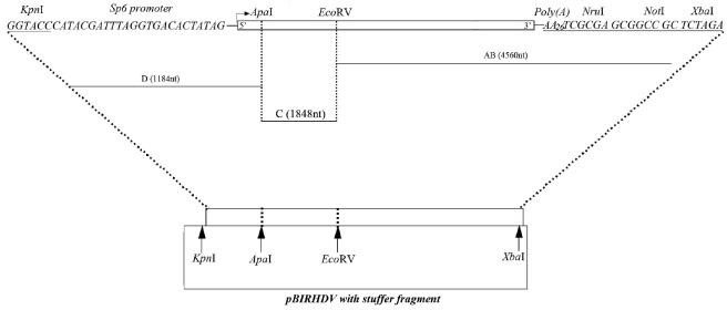
By following the multistep strategy illustrated in Fig. 1, we assembled a full-length cDNA clone of RHDV. First, pBluescript II SK+ plasmid was digested with XbaI and EcoRV and ligated to the AB fragment, which was digested with XbaI and EcoRV. The ligation yielded a recombinant plasmid, pBLABf, which was confirmed by sequence analysis to contain 27 A's. Fragments C and D, covering the remainder of the genome of RHDV JX/CHA/97, were assembled into pBLABf with unique restriction sites naturally found in the viral sequence. After transformation of competent Escherichia coli JM109 cells, ampicillin-resistant colonies were screened for recombinant plasmid. Positive clones were characterized by restriction endonuclease analysis and determination of the nucleotide sequence at the ends of the inserted fragment. The recombinant vector containing the full-length cDNA was named pBlRHDV.
FIG. 1.
Schematic diagram of steps used in the construction of a full-length cDNA clone of RHDV. At the 5′ end of the genome, the KpnI restriction site and an Sp6 RNA promoter were fused to the genome. The arrow indicates the transcription start site of Sp6 RNA polymerase. Downstream of the 3′ untranslated region, a poly(A) tail of 27 A's and the restriction sites NruI, NotI, and XbaI were inserted. The complete viral genome was divided into three fragments flanked by unique restriction sites, represented by the horizontal lines labeled AB, C, and D. The length of each fragment is indicated in parentheses. As shown at the bottom of the figure, fragments AB, C, and D were cloned into the pBluescript II SK+ vector in the order AB to D.
In vitro transcription.
The plasmid pBlRHDV was linearized with NruI, treated with proteinase K, purified by phenol-chloroform extraction and ethanol precipitation, and resuspended in 20 μl of RNase-free water. The linearized plasmid DNA was used for in vitro transcription with a transcription kit (Promega) according to the manufacturer's instructions, including treatment of the RNA with DNase to remove input plasmid. The RNA was purified by acid phenol-chloroform followed by isopropanol precipitation and redissolved in Tris-EDTA buffer by being heated at 70°C. To check the size and quality of the in vitro-transcribed RNA, a sample was denatured in urea-based RNA sample buffer (New England Biolabs) and electrophoresed on a 1% native agarose gel in Tris-borate-EDTA buffer with 1 μg of ethidium bromide per ml.
IFA.
Indirect immunofluorescence assays (IFA) were used to detect viral protein expression in RHDV RNA-transfected RK-13 cells. After transfection, approximately 105 transfected cells were spotted onto 10-mm glass coverslips. Cells on coverslips were analyzed by IFA at various times posttransfection for viral protein synthesis. Cells were fixed in 3.7% paraformaldehyde with phosphate-buffered saline (PBS), pH 7.5, at room temperature for 30 min followed by incubation in methanol at −20°C for 30 min. The fixed cells were washed with PBS, incubated at room temperature for 45 min in RHDV immune mouse ascites fluid (1:100 dilution), and further reacted with goat anti-mouse immunoglobulin G conjugated with fluorescein isothiocyanate at room temperature for 30 min (1:100 dilution). The coverslips were washed with PBS, mounted to a slide by use of fluorescent mounting medium (KPL), and observed under a fluorescence microscope equipped with a video documentation system.
Inoculation in rabbits.
In order to evaluate their infectivity in vivo, transcripts from pBlRHDV were inoculated into rabbits. Twelve 8-week-old rabbits from a specific-pathogen-free and RHDV-seronegative herd were divided into three groups, each consisting of four animals. The first group was inoculated intraperitoneally with 100 μl of different amounts of the viral RNAs described above, diluted in PBS containing 20 μg of Lipofectin (Gibco BRL, Rockville, Md.), the second group was inoculated by intrahepatic injection with the same dose of RNAs as the first group, and the third group was mock inoculated with PBS. The animals were kept in separate rooms throughout the experiment and observed daily for clinical signs of disease.
Electron microscopy.
Supernatants of a filtered liver homogenate obtained from infected rabbits were preclarified by low-speed centrifugation (6,000 rpm for 30 min at 4°C), virus was subsequently pelleted through a 25% sucrose cushion in NTE buffer (NaCl, 100 mM; Tris, 10 mM; EDTA, 1 mM [pH 7.4]) by centrifugation at 28,000 rpm for 120 min in an SW41 rotor at 4°C, and the purified virions were resuspended in NTE buffer. Anti-RHDV virion mouse serum at a dilution of 1:100 in PBS was added to the virions, and they were then incubated for 2 h at 37°C. Then, the complex was precipitated by low-speed centrifugation (3,000 rpm for 20 min at 4°C). After being washed with PBS, the precipitate was resuspended again with PBS. Negative staining of virus was performed using the immune complex resuspended in PBS. The immune complex was allowed to adhere to carbon-Formvar grids (Electron Microscopy Sciences) for 3 to 5 min, washed once with PBS, and stained for 15 s with 1% ammonium phosphotungstate (Sigma), pH 7.0. Specimens were viewed with a JEM-1200EX transmission electron microscope, and three representative photographs per virus were taken.
Genetic marker analysis of the recombinant and parental viruses.
Recombinant virus and parental virus were subjected to RNA extraction with RNeasy (QIAGEN). A 443-bp fragment including the genetic marker was amplified by RT-PCR from RNA extracted from either recombinant or parental virus with primers 2729R (5′-CCAACTGCACAATTCAAATCC-3′) and 3151F (5′-TGAACATGACGGAGTCCTGGT-3′). The RT-PCR products were digested with EcoRV and analyzed on a 2% agarose gel.
Quantification of RHDV RNA.
A comparative analysis of an increase in genome copies between transfected RNA and that isolated from liver tissue was carried out. PCR primers were designed to amplify a conserved region proximal to the 5′ end of the genome of RHDV. The forward primer was from nt 153 to 174 (5′-AGGACAAAACGAGAATGAAGGA-3′), and the reverse primer was from nt 274 to 295 (5′-GCTGGGCTATGGAACACAAAC-3′). PCR amplification yielded a 122-bp product. Briefly, 4 mg RNA was transfected into specific-pathogen-free rabbits (no. 1 to 6). RNA was extracted from liver samples prepared after euthanasia of all of these rabbits and reverse transcribed into cDNA by using a strand-specific reverse primer, as described above. The cDNA was amplified by real-time PCR using SYBR green PCR mix (Applied Biosystems). The reaction was carried out with 2× SYBR green PCR mastermix in a 25-ml volume. The samples were aliquoted into a MicroAmp Optical 96-well reaction plate (Perkin Elmer Applied Biosystems) and sealed. PCR amplification was performed using a program of 10 min at 95°C followed by 40 cycles of 1 min at 94°C, 1 min at 60°C, and 1 min at 72°C. Each reaction was done in triplicate with a Perkin Elmer ABI Prism 7700 sequence detection system (TaKaRa). Standards to establish genome equivalents were synthetic RNAs transcribed from a clone of the full-length cDNA of the RHDV. RNA was quantified by absorbance, and 10-fold serial dilutions from 106 to 10 copies were prepared. These standards were run in duplicate for all assays in order to calculate genome equivalents in the experimental samples. The calibration curves from one preparation of synthetic RNA to the next were essentially identical and yielded values comparable to those from commercially available assays.
Sequence analysis.
The sequences of the rescued virus and the parental virus were determined from the double-stranded plasmid by the dye chain termination method. Nucleotide sequences were analyzed with an IBM compatible personal computer by using program Lasergene99 (DNASTAR).
RESULTS
Construction and sequencing of the full-length RHDV cDNA clone.
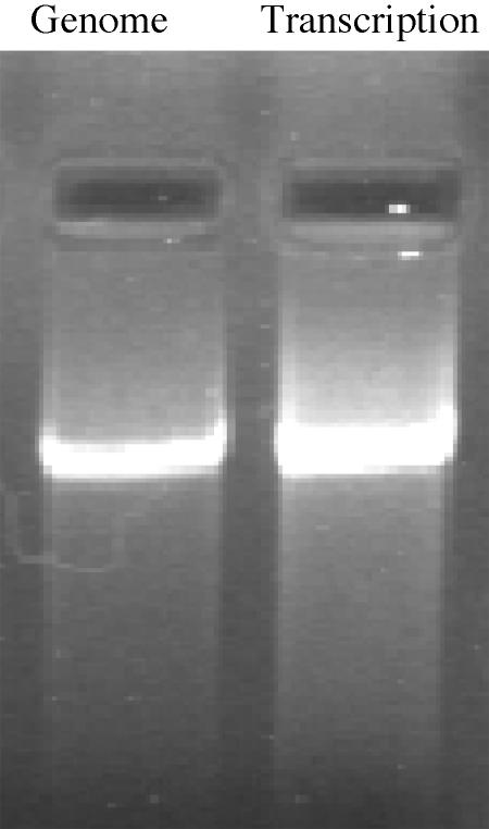
The cDNA fragments were assembled into a single clone from three overlapping cDNA fragments by use of the restriction sites XbaI, EcoRV, ApaI, and KpnI. The primers used to generate individual fragments are listed in Table 1. The full-length cDNA clone was constructed in three steps. First, the AB fragment was amplified by primers ABF and ABR and cloned into pBluescript II SK+ at their respective sites (Fig. 1), yielding plasmid pBlAB. In order to distinguish the recombinant virus from the parental virus, an EcoRV site was designed in primer ABF containing silent mutations of nucleotide A to T at position 2908. Second, the C fragment, covering ApaI to EcoRV, was amplified by primers CF and CR and inserted into plasmid pBlAB to generate clone pSpe-Xba pBlABC. Finally, the D fragment, covering KpnI to ApaI, was amplified by primers DF and DR (Table 1) and cloned into plasmid pBlABC. The resulting plasmid, pBlRHDV, contained the complete RHDV cDNA under the Sp6 promoter for in vitro transcription. For RNA synthesis, the full-length cDNA plasmid was linearized by NruI. RNA that was in vitro transcribed from this DNA template in the absence of a cap analog had three redundant residues (TCG) at the 3′ end of the RHDV genomic RNA and a 5′ nonviral G nucleotide derived from the Sp6 promoter. Analysis of the RNA transcript on a formaldehyde-denaturing agarose gel showed a single band with a mobility identical to that of genomic RNA extracted from RHDV (Fig. 2).
FIG. 2.
Transcription of RHDV RNA. Formaldehyde-denaturing 1.0% agarose electrophoresis of RNA transcript together with genomic RNA purified from RHDV.
Results of IFA.
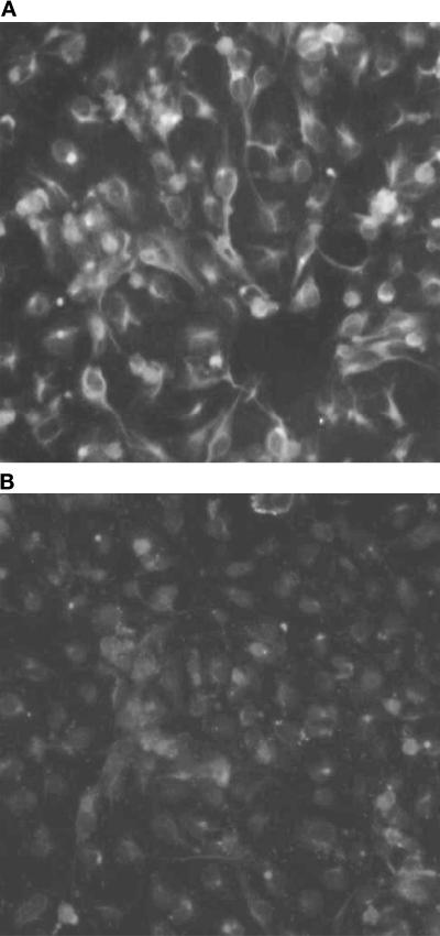
To determine if viral protein was expressed in RHDV RNA-transfected RK-13 cells, an RHDV-positive RK-13 cell line was analyzed by IFA. The normal RK-13 cell line was also analyzed by IFA as a negative control. The results showed that there was viral protein expression in the transfected cells (Fig. 3).
FIG. 3.
IFA of viral protein expression in cells transfected with full-length RHDV RNA transcript. (Top) RK-13 cells transfected with in vitro transcripts. (Bottom) Normal RK-13 cells.
Virulence of RHDV RNAs after direct inoculation of rabbits.
In order to evaluate their infectivity in vivo, RNA transcripts of cDNA clones were inoculated into rabbits. All transcripts were generated by using Sp6 RNA polymerase (Promega, Madison, Wis.), and plasmids were linearized with NruI (New England Biolabs, Beverly, Mass.) as a template. The transcript mixture was next injected into rabbits. Group 1 and group 2 rabbits showed typical symptoms (fever, anepithymia, screaming, hemorrhage, and opisthotonos) similar to those observed prior to death in animals inoculated with infectious virus. Forty-eight to 72 h later, all rabbits were killed. None of the rabbits inoculated with PBS died in the experiment, suggesting that viral RNAs were the only cause of death. To confirm that infectious virus had indeed been generated following RNA inoculation in rabbits, crude extracts from these animals were assayed for infectivity in rabbits. Dilution of the homogenates in PBS up to 10−2 was inoculated into rabbits by the same procedure described for RNA primary inoculation. All animals inoculated with these homogenates were found dead at day 3 postinoculation. The ability to cause infection and death by using the homogenates of rabbit livers strongly suggested the presence of infectious viral particles.
Electron microscopy.
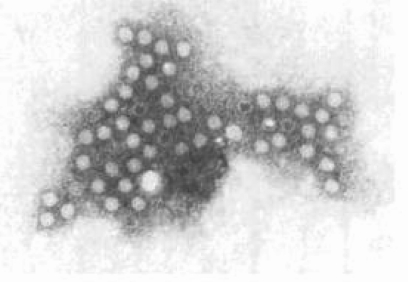
The morphology of RHDV particles negatively stained with phosphotungstate is shown in Fig. 4. Immune electron microscopic observation of the virus particles revealed that the particles were rotund, with a diameter of 35 nm (Fig. 4).
FIG. 4.
Immune electron micrograph of negatively stained RHDV particles.
Discrimination between cloned virus and parental virus.
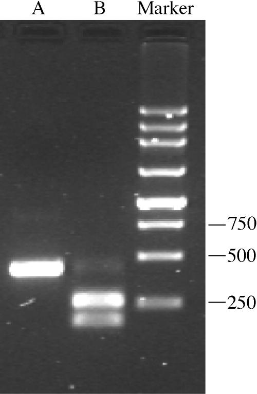
To exclude the possibility that the virus recovered from the transfected cells was due to contamination by the parental virus, we assayed the cloned virus for the presence of an EcoRV restriction site. As expected, the RT-PCR fragment derived from the cloned virus was cleaved by EcoRV, generating two fragments, one of 180 bp and one of 262 bp. In contrast, the PCR fragment derived from the parental isolate was not cleaved by EcoRV (Fig. 5).
FIG. 5.
Differentiation between cloned virus and parental JX/CHA/97 strain. An EcoRV restriction site was introduced in the full-length cDNA clone of JX/CHA/97 to allow discrimination between cloned virus (tagged with the EcoRV site) and parental virus (lacking an EcoRV site). RNA was extracted from the viral suspension of rabbits inoculated with either the cloned virus or the parental JX/CHA/97 isolate (lane A), and a 443-bp fragment was amplified by RT-PCR as described in Materials and Methods (lane B). The amplicons were digested with EcoRV and analyzed on a 2.0% agarose gel. The presence of an EcoRV restriction site resulted in fragments of 263 bp and 180 bp. As expected, the restriction site was found in the cloned virus but not in the parental JX/CHA/97 virus isolate. Molecular size markers (in base pairs) are noted at the right of blots.
Detection of full-length RHDV replication.
To confirm that the transfected RNA was replicating, levels of RHDV RNA were quantified by real-time RT-PCR in rabbits transfected with in vitro-transcribed RNA from the full-length cDNA clone of RHDV after 6 to 72 h. Virus titer quantification obtained by quantitative RT-PCR (qRT-PCR), expressed as genome equivalents per mg of total RNA, is reported in Table 2 and shows a correlation between time of infection and virus titer.
TABLE 2.
Detection of RHDV RNA in the livers of infected rabbits
| Rabbit no. | Source of infection | Phase of infection (hours postinoculation) | qRT-PCR titer (genome equivalent/mg total RNA) |
|---|---|---|---|
| 1 | RNA | 6 | 4.40 × 10⁶ |
| 2 | RNA | 18 | 3.49 × 10⁷ |
| 3 | RNA | 38 | 1.52 × 10¹⁰ |
| 4 | RNA | 48 | 5.90 × 10¹⁰ |
| 5 | RNA | 56 | 1.59 × 10⁸ |
| 6 | RNA | 72 | 7.45 × 10⁷ |
Sequence analysis.
The sequences of the rescued virus and the parental virus were determined by direct sequencing of PCR products obtained by RT-PCR. The results of sequence analyzing showed that the viruses shared very high homology in nucleotide sequence (99.9%), and the sequence of the rescued virus contained six mutations, including the artificial EcoRV cleavage site. However, the coding products were not altered (data not shown). Therefore, RNA transcripts of this clone of RHDV were infectious. Our data also showed that the length of poly(A) contained at least 30 A's, which was somewhat longer than the transcripts. Whether the length of poly(A) is related to the infection of RHDV will be investigated in another paper.
DISCUSSION
RHDV causes an economically important disease of rabbits. The mechanisms involved in RHDV virulence and pathogenicity are not well known. Full-length cDNA clones from RHDV strains allowing the recovery of infectious virus would be an invaluable tool for the determination of virulence factors and also for the elucidation of mechanisms involved in viral pathogenesis. Furthermore, the RHDV infectious cDNA clone could provide precious information in the study of genetic expression and replication of caliciviruses. Unfortunately, such an infectious clone of RHDV has not been constructed.
In the present study, we report the construction of the first full-length cDNA clone of the strain JX/CHA/97 of RHDV. RNA transcripts transcribed from the cDNA clone were highly infectious by direct injection into rabbits. The identification of genetic markers engineered into the clone confirmed that the progeny virus was derived from the cDNA clone and thus was not a contaminant. Sequence analysis demonstrated that the recombinant virus recovered from the rabbits shared the highest homology in nucleotide sequence with its parental virus. In addition, our results show that the infectivity of the RNA is independent of the presence of a 5′-end cap structure, which is different from the infectious cDNA clone of feline calicivirus, another member of the family Caliciviridae (15).
The results of qRT-PCR showed that the genome copies of the engineering virus increased along with the course of infection, arrived at the highest level at 48 h (5.9 × 10¹⁰) (and then the curve declined), and arrived at the lowest level (7.45 × 10⁷) at 72 h, when the experimental rabbits died.
Whether a full-length cDNA clone is infectious depends on two points. One point is that the cDNA clone should ideally be genetically completely identical to the parental virus. Another is that we should have proper rescue systems. As for our infectious clone, because the core sequence of the Sp6 promoter was positioned immediately before the authentic G of the 5′-untranslated-region sequence, the RNA transcripts should include the exact 5′ end of RHDV. Furthermore, after plasmid linearization with NruI, the template DNA and therefore the RNA transcripts should end exactly at the 3′ end. However, for RHDV, hepatitis C virus, hepatitis E virus, etc., due to the lack of reliable in vitro propagation systems we could not perform screening for infectivity in cell cultures. However, infectious hepatitis C virus and foot-and-mouth disease virus have been rescued by injecting transcription mixtures into animals (1, 17). In the present study, our results showed that RNA transcribed from full-length cDNA clones is infectious after direct transfection of rabbits. The model offers a novel approach to access the virulence of RHDV RNAs in vivo. The absence of a robust cell culture model of RHDV infection has severely limited analysis of the RHDV life cycle and the development of effective antivirals and vaccines. Based on the infectious clone of RHDV, we are considering constructing a highly efficient in vitro infection system of RHDV.
In conclusion, we have constructed a genetically stable infectious clone of RHDV. The approach used to engineer this clone should be applicable to the construction of infectious cDNA of other RHDVs, as well as a number of recently discovered related viruses. Moreover, it will be possible to target RHDV to multiple species by simple replacement of the VP60 gene (3, 15), allowing for the development of RHDV as a vaccine vector in domesticated animals. Furthermore, the availability of an infectious RHDV clone makes it possible to study in detail the mechanisms of viral replication and pathogenesis. For example, by using the reverse genetic system, we analyzed the functional role of the 3′ untranslated region and the poly(A) tail. The results showed that these regions are essential for the infectivity of RHDV; however, the replicon still retains RHDV RNA synthesis ability (to be reported elsewhere). In addition, by using the replicon system, we would analyze the role of some RNA elements at the 3′ untranslated region during translation and RNA synthesis.
Acknowledgments
This study was supported by grants from the Zhejiang Natural Sciences Foundation (Y305047).
We thank Jinqing Chen for his support and Xuelong Wu and Tao Zheng for their technical assistance.
REFERENCES
- 1.Baranowski, E., N. Molina, J. I. Núñez, F. Sobrino, and M. Sáiz. 2003. Recovery of infectious foot-and-mouth disease virus from suckling mice after direct inoculation with in vitro-transcribed RNA. J. Virol. 77:11290-11295. [DOI] [PMC free article] [PubMed] [Google Scholar]
- 2.Collins, B. J., J. R. White, C. Lenghaus, V. Boyd, and H. A. Westbury. 1995. A competition ELISA for the detection of antibodies to rabbit haemorrhagic disease virus. Vet. Microbiol. 43:85-96. [DOI] [PubMed] [Google Scholar]
- 3.El Mehdaoui, S., A. Touze, S. Laurent, P. Y. Sizaret, D. Rasschaert, and P. Coursaget. 2000. Gene transfer using recombinant rabbit hemorrhagic disease virus capsids with genetically modified DNA encapsidation capacity by addition of packaging sequences from the L1 or L2 protein of human papillomavirus type 16. J. Virol. 74:10332-10340. [DOI] [PMC free article] [PubMed] [Google Scholar]
- 4.Gould, A. R., J. A. Kattenbelt, C. Lenghaus, C. Morrissy, T. Chamberlain, B. J. Collings, and H. A. Westbury. 1997. The complete nucleotide sequence of rabbit haemorrhagic disease virus (Czech strain V351): use of the polymerase chain reaction to detect replication in Australian vertebrates and analysis of viral population sequence variation. Virus Res. 47:7-17. [DOI] [PubMed] [Google Scholar]
- 5.Gregg, D. A., C. House, R. Meyer, and M. Berninger. 1991. Viral haemorrhagic disease of rabbits in Mexico: epidemiology and viral characterization. Rev. Sci. Tech. 10:435-451. [DOI] [PubMed] [Google Scholar]
- 6.Laurent, S., J.-F. Vautherot, M.-F. Madelaine, G. Le Gall, and D. Rasschaert. 1994. Recombinant rabbit hemorrhagic disease virus capsid protein expressed in baculovirus self-assembles into virus-like particles and induces protection. J. Virol. 68:6794-6798. [DOI] [PMC free article] [PubMed] [Google Scholar]
- 7.Liu, S. J., H. P. Xue, B. Q. Pu, and S. H. Qian. 1984. A new viral disease in rabbits. Anim. Husb. Vet. Med. 16:253-255. [Google Scholar]
- 8.Meyers, G., C. Wirblich, and H.-J. Thiel. 1991. Rabbit hemorrhagic disease virus molecular cloning and nucleotide sequencing of a calicivirus genome. Virology 184:664-676. [DOI] [PubMed] [Google Scholar]
- 9.Meyers, G., C. Wirblich, and H.-J. Thiel. 1991. Genomic and subgenomic RNAs of rabbit hemorrhagic disease virus are both protein linked and packaged into particles. Virology 184:677-686. [DOI] [PMC free article] [PubMed] [Google Scholar]
- 10.Mitro, S., and H. Krauss. 1993. Rabbit hemorrhagic disease: a review with special reference to its epizootiology. Eur. J. Epidemiol. 9:70-78. [DOI] [PubMed] [Google Scholar]
- 11.Morales, M., J. Barcena, M. A. Ramirez, J. A. Boga, F. Parra, and J. M. Torres. 2004. Synthesis in vitro of rabbit hemorrhagic disease virus subgenomic RNA by internal initiation on (−)sense genomic RNA: mapping of a subgenomic promoter. J. Biol. Chem. 279:17013-17018. [DOI] [PubMed] [Google Scholar]
- 12.Mutze, G., B. Cooke, and P. Alexander. 1998. The initial impact of rabbit hemorrhagic disease on European rabbit populations in South Australia. J. Wildl. Dis. 34:221-227. [DOI] [PubMed] [Google Scholar]
- 13.Nowotny, N., C. R. Bascunana, A. Ballagi-Pordany, D. Gavier-Widen, M. Uhlen, and S. Belak. 1997. Phylogenetic analysis of rabbit haemorrhagic disease and European brown hare syndrome viruses by comparison of sequences from the capsid protein gene. Arch. Virol. 142:657-673. [DOI] [PubMed] [Google Scholar]
- 14.Ohlinger, V. F., B. Haas, G. Meyers, F. Weiland, and H.-J. Thiel. 1990. Identification and characterization of the virus causing rabbit hemorrhagic disease. J. Virol. 64:3331-3336. [DOI] [PMC free article] [PubMed] [Google Scholar]
- 15.Thumfart, J. O., and G. Meyers. 2002. Feline calicivirus: recovery of wild-type and recombinant viruses after transfection of cRNA or cDNA constructs. J. Virol. 76:6398-6407. [DOI] [PMC free article] [PubMed] [Google Scholar]
- 16.Wirblich, C., G. Meyers, V. F. Ohlinger, L. Capucci, U. Eskens, B. Haas, and H.-J. Thiel. 1994. European brown hare syndrome virus: relationship to rabbit hemorrhagic disease virus and other caliciviruses. J. Virol. 68:5164-5173. [DOI] [PMC free article] [PubMed] [Google Scholar]
- 17.Yanagi, M., R. H. Purcell, S. U. Emerson, and J. Bukh. 1997. Transcripts from a single full-length cDNA clone of hepatitis C virus are infectious when directly transfected into the liver of a chimpanzee. Proc. Natl. Acad. Sci. USA 94:8738-8743. [DOI] [PMC free article] [PubMed] [Google Scholar]


