Abstract
The leptin receptor, LRb, and other cytokine receptors are devoid of intrinsic enzymatic activity and rely upon the activity of constitutively associated Jak family tyrosine kinases to mediate intracellular signaling. In order to clarify mechanisms by which Jak2, the cognate LRb-associated Jak kinase, is regulated and mediates downstream signaling, we employed tandem mass spectroscopic analysis to identify phosphorylation sites on Jak2. We identified Ser523 as the first-described site of Jak2 serine phosphorylation and demonstrated that this site is phosphorylated on Jak2 from intact cells and mouse spleen. Ser523 was highly phosphorylated in HEK293 cells independently of LRb-Jak2 activation, suggesting a potential role for the phosphorylation of Ser523 in the regulation of LRb by other pathways. Indeed, mutation of Ser523 sensitized and prolonged signaling by Jak2 following activation by the intracellular domain of LRb. The effect of Ser523 on Jak2 function was independent of Tyr570-mediated inhibition. Thus, the phosphorylation of Jak2 on Ser523 inhibits Jak2 activity and represents a novel mechanism for the regulation of Jak2-dependent cytokine signaling.
Type I cytokines mediate a plethora of physiologic processes, ranging from hematopoietic and immune functions (such as those mediated by erythropoietin [Epo] and the interleukins) to growth and neuroendocrine responses (such as those mediated by growth hormone and leptin) (11, 15, 16, 23, 31). These actions are mediated by the activation of cytokine receptor proteins found on the surface of target cells. Cytokine receptors each contain an extracellular domain that recognizes its specific cytokine ligand, a single transmembrane domain, and an intracellular domain that, although devoid of enzymatic activity, transmits intracellular signals by means of an associated Jak family tyrosine kinase. Ligand binding activates the associated intracellular Jak kinase, resulting in Jak kinase autophosphorylation and activation and the subsequent tyrosine phosphorylation of the intracellular domain of the cytokine receptor. These tyrosine phosphorylation events mediate downstream signaling by the LRb/Jak2 complex (11, 16, 19, 23).
The Jak kinase family contains four members: Jak1 to Jak3 and Tyk2 (11, 16). Of these, Jak1 and -2 and Tyk2 are ubiquitously expressed, while Jak3 is found predominantly in immune and hematopoietic tissues. Jak kinases are composed of four conserved domains. The NH2-terminal FERM domain is required for interaction with cytokine receptors (32, 35), while the adjacent SH2-like fold has no known function. The COOH-terminal portion of Jak kinases contains a kinase-like JH2 domain that is devoid of enzymatic activity but that regulates the activity of the COOH-terminal JH1 tyrosine kinase domain (9, 21, 28, 33, 34).
Our laboratory studies signaling by the long form of the leptin receptor (LRb), which regulates feeding, neuroendocrine, and immune function in response to leptin, which is in turn regulated by nutritional cues (8, 10, 23, 31). Stimulation of LRb mediates the activation and tyrosine phosphorylation of the LRb-associated Jak2, resulting in the phosphorylation of tyrosine residues on the intracellular tail of LRb and on Jak2 (2, 17, 23). Tyrosine phosphorylation sites on LRb mediate signaling by STAT proteins and SHP-2, as well as binding the suppressor of cytokine signaling 3 (SOCS3) to attenuate LRb signaling (2, 3, 23). Several sites of Jak2 tyrosine phosphorylation have been identified, and functions for a few of these sites are known: phosphorylation of Tyr1007 and Tyr1008 within the kinase domain participates in kinase activation (7), phosphorylated Tyr813 mediates binding of SH2-B to increase Jak2 signaling (19), and phosphorylation of Tyr570 within the JH2 domain inhibits Jak2 kinase activity (1, 6).
During our continuing analysis of Jak2 by liquid chromatography-tandem mass spectroscopy (LC-MS/MS), we identified the first-described site of serine phosphorylation (Ser523) on Jak2 protein from intact cells. Here, we report that phosphorylation of Ser523 on Jak2 inhibits Jak2-mediated signaling; this and the high-stoichiometry phosphorylation of this site in intact cells suggest an important role for this phosphorylation event in the regulation of cytokine signaling.
MATERIALS AND METHODS
Antibodies, growth factors, and reagents.
Rabbit anti-Jak2 (758) [αJak2(758)] antiserum has been described previously (2, 6). Antigen affinity-purified phosphorylation-state-specific antibodies to phosphorylated Tyr1007 and Tyr1008 [αJak2(PY1007,8)] and Tyr570 [αJak2(PY570)] have also been described previously (6). Antisera for immunoblotting of Jak2 [αJak2(NT)] were prepared in rabbits by injection of a keyhole limpet hemocyanin-coupled synthetic peptide corresponding to the NH2-terminal 12 amino acids of Jak2. Antibodies recognizing phosphorylated Ser523 of Jak2 were raised in rabbits by injection of a keyhole limpet hemocyanin-coupled synthetic 11-amino-acid phosphorylated peptide centered on Ser523. All site- and phosphospecific antisera were affinity purified on the antigen peptide coupled to a mixture of Affigel-10 and -15 (Bio-Rad), followed by passage over Affigel coupled to irrelevant phosphopeptides and nonphosphorylated antigen peptide to remove antibodies directed against other sites of phosphorylation and to the nonphosphorylated form of the site. The independent preparation of αJak2(PS523) by the Carter-Su laboratory is described in reference 21a. Synthetic peptides were purchased from Boston Biomolecules (Woburn, MA). 9-Fluorenylmethoxy carbonyl-[13C, 15N]-Ile was purchased from Cambridge Isotope Laboratories (Cambridge, MA) for the synthesis of mass-labeled peptides by Boston Biomolecules. Recombinant murine interleukin-3 (IL-3) was obtained from Pierce Endogen (Rockford, IL); monoclonal 4G10 was used for antiphosphotyrosine (αPY) immunoblotting (Upstate Biotechnology). Antibodies directed against the phosphorylated (activated) forms of extracellular signal-regulated kinase (ERK) and STAT3(PY705) were purchased from Cell Signaling Technology (Beverly, MA). Recombinant human Epo was purchased from Amgen. Bovine serum albumin fraction V was purchased from Sigma. Protein A-Sepharose 6MB and horseradish peroxidase-protein A were from Amersham Pharmacia Biotech (Piscataway, NJ), and secondary antibodies for immunoblotting were from Santa Cruz Biotechnology (Santa Cruz, CA). Dimethylpimelimidate was from Pierce Endogen (Rockford, IL).
Generation of mutant Jak2 cDNAs.
pcDNA3Jak2 (17) and pcDNA3Jak2Y570F (6) were used as templates for mutagenesis using the QuickChange kit (Stratagene) to replace Ser523 with Ala or Glu individually (to generate pcDNA3Jak2S523A and pcDNA3Jak2S523E, respectively) or in combination with replacement of Tyr570 by Phe (pcDNA3Jak2S523A/Y570F). The presence of the desired mutations and the absence of adventitious mutations were confirmed by DNA sequencing. Other Jak2 mutants have been described previously (6).
Cell lines.
All cells were maintained in a humidified atmosphere containing 5% CO2 and 95% air at 37°C. 32D cells were grown in RPMI 1640 medium supplemented with 10% fetal bovine serum and 5% WEHI-3 conditioned medium (a source of IL-3) (2). HEK293 cells were grown in Dulbecco's modified Eagle's medium supplemented with 10% fetal bovine serum. ELR constructs in pcDNA3 were transiently cotransfected with pcDNA3 alone or with the appropriate pcDNA3Jak2 isoform into subconfluent HEK293 cells using Lipofectamine (Invitrogen) as described previously (6).
Preparation of cell lysates for immunoprecipitation.
Prior to each experiment, subconfluent cells plated in 15-cm dishes were made quiescent by incubation in Dulbecco's modified Eagle's medium containing 0.5% bovine serum albumin (32D cells, 4 h; 293 cells, overnight) before stimulation with Epo or IL-3 at 37°C. Cells were lysed in 20 mM Tris, pH 7.4, containing 137 mM NaCl, 2 mM EDTA, 10% glycerol, 50 mM β-glycerophosphate, 50 mM NaF, 1% Nonidet P-40, 2 mM phenylmethylsulfonyl fluoride, and 2 mM sodium orthovanadate (lysis buffer). Insoluble material was removed by centrifugation at 16,000 × g at 4°C for 20 min. Protein concentrations of the resulting lysates were determined using the bicinchoninic acid protein assay kit (Pierce) and bovine serum albumin standards, and equivalent amounts of protein were added to the appropriate antibodies for immunoprecipitation or denatured in Laemmli buffer for direct resolution by 10% sodium dodecyl sulfate-polyacrylamide gel electrophoresis (SDS-PAGE). For immunoprecipitates, lysates were incubated with antibody at 4°C overnight followed by incubation with protein A-Sepharose for 60 min. All Jak2 immunoprecipitations were performed with αJak2(758). Immune complexes were collected by centrifugation and washed three times in lysis buffer before denaturation in Laemmli buffer and separation by 8% SDS-PAGE. Immunoblotting was performed as previously described (6). Quantification of immunoblots was accomplished by scanning densitometry of film using QuantityOne software (Bio-Rad).
For stripping blots, the membrane was stripped with Re-blot Plus (Chemicon) according to the manufacturer's instructions. Stripped membranes were blocked overnight in block buffer and reprobed as described elsewhere.
Analysis of Jak2 from spleen was accomplished by lysing freshly isolated spleen in a Dounce homogenizer; lysates were clarified and protein concentration determined as above. Lysates containing 4.5 mg of protein each were immunoprecipitated with affinity-purified αJak2(758) covalently coupled to protein A-Sepharose by the dimethylpimelimidate method (24). Immunoblotting of control immunoprecipitates (no tissue lysate) using this coupled antibody revealed no reactivity with either αJak2(PS523) or αJak2(NT) (data not shown). Mice were C57BL/6 wild-type and obese, leptin-deficient ob/ob mice from our in-house breeding program at the University of Michigan. Mice had ad libitum access to food and water, and all experimental procedures were approved by the University Committee on the Use and Care of Animals.
Analysis of SOCS3 mRNA expression.
HEK293 cells were transfected in triplicate with the ELR and the appropriate Jak2 constructs and made quiescent overnight before stimulation with vehicle or various concentrations of Epo for an additional 2 h. Cells were lysed and RNA purified using Trizol reagent (Invitrogen). RNA was subjected to reverse transcription with the Superscript first-strand synthesis system (Invitrogen) and subjected to quantitative PCR analysis for SOCS3 and glyceraldehyde-3-phosphate dehydrogenase using 6-carboxytetramethylrhodamine probes and primers from Applied Biosystems. Relative amounts of SOCS3 RNA were determined by the 2−ΔΔCt method (20).
Analysis of STAT3 reporter activity.
HEK293 cells were transfected in triplicate with the ELR and the appropriate Jak2 constructs plus STAT3-responsive gamma interferon activated sequence (GAS)-Luc and control Renilla luciferase plasmids and made quiescent overnight before stimulation with vehicle or various concentrations of Epo for an additional 12 h. Cells were lysed and assayed for firefly and Renilla luciferase using the dual-luciferase reporter assay system (Promega) on a Victor3 instrument (Perkin-Elmer). Firefly luciferase activity was normalized to Renilla luciferase activity and plotted.
LC-MS/MS analysis.
For preparation of protein for LC-MS/MS analysis, material was immunoprecipitated from 5 to 10 15-cm dishes of HEK293 cells and resolved on a single lane of a 7% SDS-polyacrylamide gel. Jak2 protein was visualized by staining with Coomassie brilliant blue G-250 (Bio-Rad) and destaining overnight in 10% methanol, 10% glacial acetic acid. Gel slices containing Jak2 were digested with 5 ng/μl sequencing-grade modified trypsin (Promega) in 25 mM ammonium bicarbonate containing 0.01% n-octylglucoside for 18 h at 37°C. Peptides were eluted from the gel slices with 80% acetonitrile, 1% formic acid. Tryptic digests were separated by capillary high-pressure liquid chromatography (C18, 75-μm-inside-diameter Picofrit column; New Objective) using a flow rate of 100 nl/min over a 3-h reverse-phase gradient and analyzed using an LTQ two-dimensional linear ion trap mass spectrometer (ThermoFinnigan). Resultant MS/MS spectra were matched against mouse Jak2 sequence using TurboSequest (BioWorks 3.1) with a fragment ion tolerance of <0.5 and amino acid modification variables including phosphorylation (80 Da) of Ser, Thr, and Tyr; oxidation (16 Da) of Met; and methylation (14 Da) of Lys. Synthetic phosphopeptides, corresponding to tryptic sequences derived from Jak2, were obtained (Boston Biomolecules, Inc.) and analyzed using the LC-MS/MS protocol described above. MS/MS spectra from these synthetic peptide controls and Jak2-derived tryptic peptides were compared for correlation of fragmentation ion m/z and abundance. Peptides were quantified using peak areas generated by liquid chromatography-selective reaction monitoring (LC-SRM; 2+ precursor ion → 1+ fragment ion) for Jak2-derived tryptic peptides TNGISDVQIS(p)PTLQR (854.9 → 928.5 m/z) and HDFVHGWIK (570.31 → 886.5 m/z) using internal standards TNGISDVQ(13C/15N-I)S(p)PTLQR(858.7 → 935.5 m/z) and HDFVHGW(13C/15N-I)K (573.47 → 893.6 m/z). Mass spectroscopic analysis was performed using a sequence of MS (370 to 2,000 m/z) and data-dependent MS2 followed by the four SRM events described above. Precursor and fragment ion isolation widths were 2 and 3 m/z, respectively.
Phosphatase treatment.
Washed immunoprecipitated protein complexes immobilized on protein A-Sepharose were prepared as described above, washed three times in the absence of phosphatase inhibitors, and either resuspended in sample buffer immediately or resuspended in phosphatase buffer in the absence or presence of 200 U of λ-phosphatase (New England Biolabs) and incubated for 30 min at 30°C before denaturation in sample buffer. Samples were resolved by SDS-PAGE and analyzed by immunoblotting as described above.
RESULTS
Phosphorylation of Ser523 on Jak2.
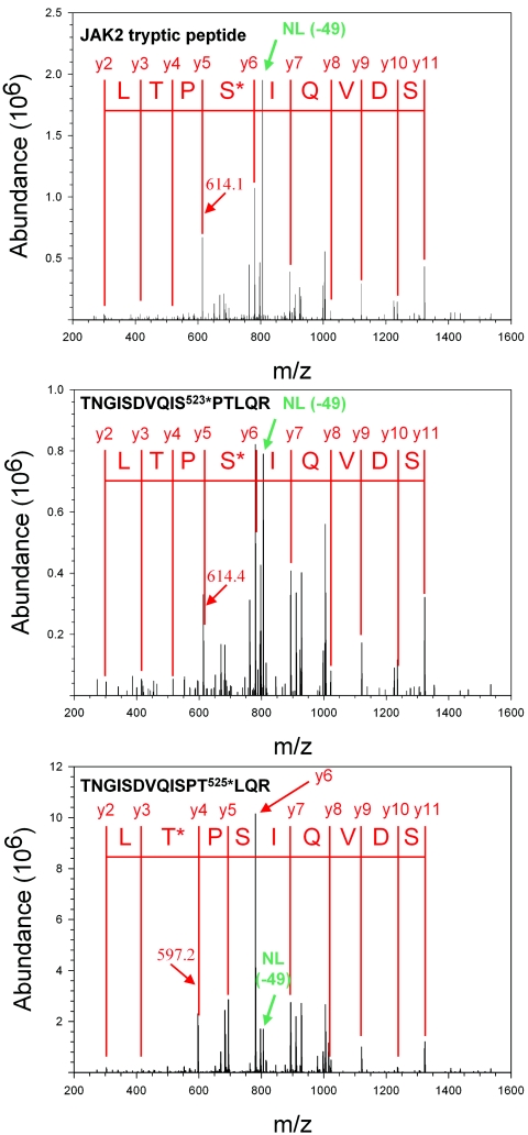
Since we are interested in mechanisms by which Jak2 contributes to LRb signaling, we undertook to identify sites of Jak2 phosphorylation that may modulate the activity or signaling of LRb-associated Jak2. We prepared Jak2 protein by αJak2 immunoprecipitation from HEK293 cells transfected with Jak2 and an Epo receptor/LRb chimera (ELR) that places the intracellular domain of LRb under the control of Epo. We employ this chimeric receptor in place of native LRb since ELR is expressed at much higher levels, thus facilitating the study of signaling by the intracellular domain of LRb in transfected cells (2). Jak2 protein purified by immunoprecipitation from cells that had been incubated in the absence or presence of Epo for 15 min was resolved by SDS-PAGE and visualized by staining with Coomassie brilliant blue (data not shown). This Jak2 protein was subjected to tryptic proteolysis and extracted from the gel, and the resulting peptides were subjected to LC-MS/MS analysis. TurboSequest analysis of MS/MS spectra from Jak2-derived material identified a predicted Jak2 tryptic peptide containing phosphoserine, TNGISDVQIS(p)PTLQR, with Xcorr scores of >3.0 for 2+ charged precursor (Fig. 1, top panel). The spectra displayed ions consistent with the neutral ion loss of 49 m/z resulting from the loss of HPO3 from doubly charged phosphoserine-containing peptides. MS/MS spectra corresponding to peptides containing phosphorylated Ser523 from Jak2 were detected from numerous independent analyses from both stimulated and unstimulated cells, suggesting that this residue represents a major site of phosphorylation in intact cells. In order to confirm the assignment of this phosphorylation site, phosphopeptides corresponding to the candidate tryptic Jak2 peptide containing phosphorylated Ser523 and the closest potential alternative phosphorylation site (Thr525) were synthesized and subjected to LC-MS/MS analysis (Fig. 1, middle and lower panels). The appearance and relative abundance of the y4 ion at 614.1 m/z and the neutral loss ion at −49 m/z correlated with the synthetic peptide containing phosphorylated Ser523, but not the peptide containing phosphorylated Thr525, confirming the identification of Jak2 phosphorylation at Ser523.
FIG. 1.
LC-MS/MS identification of Ser523 phosphorylation. Shown are MS/MS spectra for the Jak2 tryptic peptide phosphorylated at Ser523 (top) and synthetic peptides TNGISDVQIS(p)PTLQR (middle) and TNGISDVQISPT(p)LQR (bottom). Sequest assignments of y+ ions are shown in red. Fragment ions corresponding to the neutral loss of 49 m/z from these doubly charged precursors are shown in green.
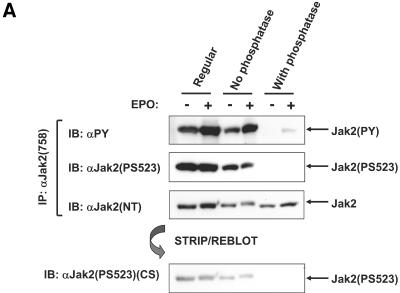
In order to facilitate the study of the phosphorylation status of these residues and to study their regulation in intact cells, we generated and affinity purified an antibody specific for the phosphorylated form of Ser523 [αJak2(PS523)]. We initially tested this antibody for phosphorylation-dependent recognition of Jak2 by examining reactivity with exogenously expressed Jak2 from 293 cells and the effect of dephosphorylation on this recognition (Fig. 2A). Cells were transfected with ELR and Jak2, made quiescent, and incubated in the absence or presence of Epo for 15 min before immunoprecipitation. Cells were lysed, and Jak2 was immunoprecipitated; washed immunoprecipitates were then either directly denatured or incubated in the presence or absence of alkaline phosphatase before denaturation. Denatured proteins were resolved by SDS-PAGE for immunoblotting with αPY, αJak2(PS523), or αJak2(NT), as indicated. The αJak2(NT) immunoblots demonstrated that, while some Jak2 protein was lost during the incubation, the presence of the phosphatase during the incubation did not alter the amount of protein retained. Immunoblotting with αPY demonstrated the tyrosine phosphorylation of Jak2 and confirmed the expected increase in Jak2 tyrosine phosphorylation upon ligand stimulation. Incubation of the immunoprecipitated Jak2 with alkaline phosphatase almost entirely abrogated the αPY reactivity of Jak2, suggesting that this treatment effectively dephosphorylated Jak2. Immunoblotting with αJak2(PS523) revealed similar levels of immunoreactivity in the absence and in the presence of Epo stimulation and showed that phosphatase treatment abrogated immunoreactivity, demonstrating the phosphospecificity of αJak2(PS523) reactivity. Stripping of the αJak2(NT) membrane and reprobing with αJak2(PS523)(CS) antiserum provided by the Carter-Su laboratory (see reference 21a) demonstrated similar phosphospecificity of this antiserum, as well.
FIG. 2.
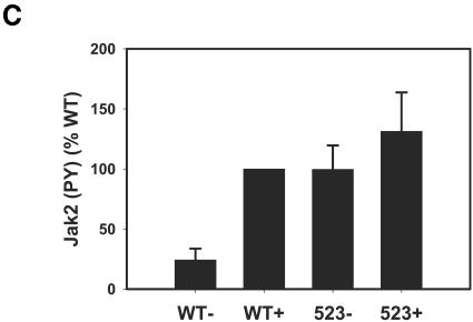
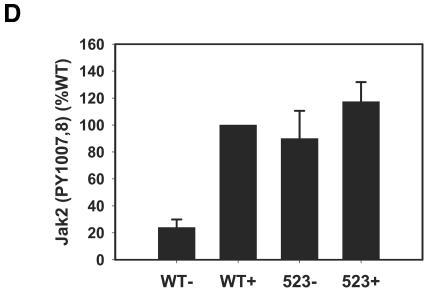
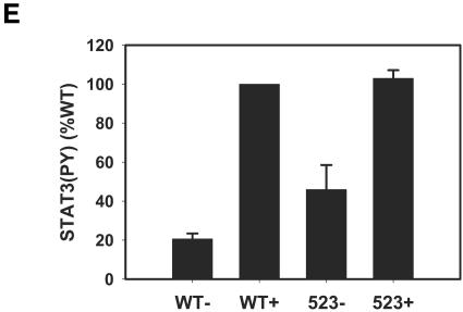
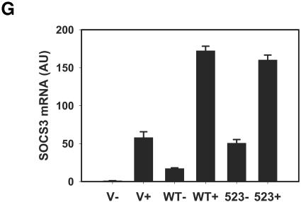
Specificity of αJak2(PS523) and increased signaling activity by Jak2S523A. A. HEK293 cells were cotransfected with ELR plus Jak2, made quiescent, and incubated for 15 min in the absence (−) or presence (+) of Epo (10 units/ml). Cells were lysed and immunoprecipitated with αJak2(758). Washed immunoprecipitates were either directly denatured (Regular) or were incubated with (With phosphatase) or without (No phosphatase) phosphatase for 30 min at 30°C. All proteins were resolved by SDS-PAGE and transferred to a nitrocellulose membrane for immunoblotting with the indicated antibody. Lower panel: the αJak2(NT) immunoblot was stripped and reprobed with an independently prepared αJak2(PS523)(CS). B. HEK293 cells were cotransfected with ELR plus control plasmid (pcDNA3) or the indicated Jak2 isoforms (Jak2 and Jak2S523A), made quiescent, incubated in the absence (−) or presence (+) of Epo (10 units/ml) for 15 min, and lysed. Lysates or αJak2(758) immunoprecipitates were resolved by SDS-PAGE, transferred to a nitrocellulose membrane, and immunoblotted with the indicated antibodies. The migration of detected proteins is noted to the right of the panels. C to F. Films corresponding to three independent experiments similar to that shown in panel B were quantified and the results plotted (means ± standard errors of the means). C and D. Tyrosine-phosphorylated Jak2 and Jak2 phosphorylated on Tyr1007,1008, respectively, normalized to total Jak2. E and F. Tyrosine-phosphorylated STAT3 and phosphorylated ERK, respectively. G. HEK293 cells were cotransfected with ELR plus vector (V) or Jak2, made quiescent, and incubated for 2 h in the absence (−) or presence (+) of Epo (10 units/ml). Cells were lysed, and RNA was prepared and converted to cDNA for quantification of SOCS3 mRNA relative to glyceraldehyde-3-phosphate dehydrogenase mRNA. Relative expression was calculated by the 2−ΔΔCt method; data are expressed as means ± standard errors of the means (n = 3). WT, wild type.
In order to further test the specificity of αJak2(PS523) and to probe the potential function of Ser523 phosphorylation, we also generated a phosphorylation site-defective mutant of Jak2 in which Ser523 was replaced by Ala (Jak2S523A). We transfected HEK293 cells with ELR in combination with control plasmid or plasmids encoding Jak2 or Jak2S523A (Fig. 2B). Cells were incubated in the absence or presence of Epo and lysed, and lysates were immunoprecipitated with αJak2(758) or directly resolved for the determination of downstream ELR signaling. αJak2(PS523) immunoblotting of αJak2-precipitable material again demonstrated ligand-independent immunoreactivity; this reactivity was absent in Jak2S523A. Similar results were obtained with αJak2(PS523) prepared independently by the Carter-Su laboratory. Thus, αJak2(PS523) reactivity is specific for phosphorylation of Ser523; these data also suggest either that the basal phosphorylation of Ser523 in HEK293 cells is very high or that brief stimulation of Jak2 activity is insufficient to increase the phosphorylation of this site.
The analysis of αPY immunoreactivity again demonstrated the expected ligand-stimulated tyrosine phosphorylation of Jak2; the tyrosine phosphorylation of Jak2S523A was increased compared to that of wild-type Jak2 in the absence of ligand stimulation and only slightly increased over this high baseline level upon Epo treatment (Fig. 2B). In order to examine whether the increased overall tyrosine phosphorylation of Jak2S523A was likely to reflect increased activation of Jak2S523A, we performed immunoblot analysis of Jak2 and Jak2S523A using αJak2(PY1007,8), which detects the phosphorylation of the activation loop within the kinase domain of Jak2 and thus reflects the activation of the Jak2 tyrosine kinase. This analysis demonstrated increased phosphorylation of Tyr1007,8 on Jak2S523A compared to Jak2 in the basal state as well as the expected Epo-stimulated increase in phosphorylation. Overexpression of Jak2 with ELR also increased the ligand-dependent and -independent phosphorylation of the downstream STAT3 and ERK molecules compared to the levels observed in cells expressing ELR alone (which mediated some activation of these signals via endogenous Jak2 in the absence of overexpressed Jak2 isoforms). The presence of Jak2S523A increased the phosphorylation of these molecules compared to that observed with Jak2. While quantification demonstrated that the increase in these downstream signaling events in cells expressing Jak2S523A was not statistically significant compared to cells expressing Jak2 (perhaps due to the maximal amplitude of these signals in cells expressing Jak2), the increase in basal activity was increased by two- to threefold (P < 0.05) by Jak2S523A compared to Jak2 (Fig. 2C to F).
Furthermore, the expression of endogenous SOCS3 mRNA (a reflection of ELR-mediated STAT3 activation [27]) was similarly increased in unliganded cells expressing Jak2S523A compared to those expressing Jak2 (P < 0.05). Again, the failure to detect increased signaling following ligand stimulation may reflect the maximal nature of the signal generated by overexpressed Jak2 in the presence of ligand especially at short times of incubation (see below and Fig. 6D). In aggregate, these data demonstrated that mutation of Ser523 increased the tyrosine phosphorylation and activity of Jak2, suggesting that the phosphorylation of Ser523 may inhibit Jak2.
FIG. 6.
Role of Jak2 kinase activity in the phosphorylation of Ser523 and independent roles for Ser523 and Tyr570 in Jak2 regulation. A. HEK293 cells were cotransfected with ELR plus Jak2, Jak2S523A, or Jak2Y1007F; made quiescent; incubated in the absence (−) or presence (+) of Epo (10 units/ml) for 15 min; and lysed. Lysates were immunoprecipitated with αJak2(758). Lysates or immunoprecipitated proteins were resolved by SDS-PAGE, transferred to nitrocellulose membranes, and immunoblotted with the indicated antibodies. The migration of detected signaling proteins is noted to the right of the panels. B and C. HEK293 cells were cotransfected with ELR plus pcDNA3, Jak2 (WT), Jak2S523A (523), Jak2Y570F (570), or Jak2S523A/Y570F (A/F); made quiescent; incubated in the absence (−) or presence (+) of Epo (10 units/ml) for 15 min; and lysed. Lysates were immunoprecipitated with αJak2(758). Lysates or immunoprecipitated proteins were resolved by SDS-PAGE, transferred to a nitrocellulose membrane, and immunoblotted with the indicated antibodies. The migration of detected signaling proteins is noted to the right of the panels. Each panel is representative of two independent experiments. The phosphorylation of each of these Jak2 isoforms on Tyr1007,8 was quantified in each of these experiments and is plotted in panel C. D. HEK293 cells were cotransfected with ELR, GAS-luciferase, and control plasmids, plus pcDNA3, Jak2 (WT), Jak2S523A (523), Jak2Y570F (570), or Jak2S523A/Y570F (A/F); made quiescent; incubated in the indicated concentration of Epo for 12 h; and lysed for assay of luciferase activity. Mean luciferase activity normalized to control is plotted (± standard errors of the means) (n = 3). WT, wild type.
Phosphorylation of endogenous Jak2 on Ser523.
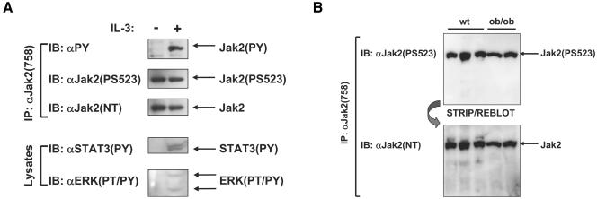
In order to examine the regulation of Ser523 phosphorylation at endogenous levels of receptor and Jak2, we employed the IL-3-dependent 32D myeloid progenitor cell line, which expresses endogenous IL-3 receptor and Jak2. Untransfected 32D cells were made quiescent, incubated for 15 min in the absence or presence of IL-3, and lysed. Lysates were immunoprecipitated with αJak2(758) or directly resolved by SDS-PAGE (Fig. 3). Immunoprecipitated Jak2 protein was immunoblotted with αPY, αJak2(PS523), and αJak2(NT), and total lysates were probed with αSTAT3(PY) and αERK(PT/PY). As expected, IL-3 stimulation resulted in increased phosphorylation of Jak2, STAT3, and ERK, consistent with the known effects of IL-3 in these cells. Immunoblotting of αJak2(758) immunoprecipitates with αJak2(PS523) demonstrated the phosphorylation of endogenous Jak2 on Ser523 in these cells in the absence or presence of IL-3 stimulation. Thus, Ser523 of Jak2 is phosphorylated in multiple cell types, and the regulation of Ser523 phosphorylation is not affected by acute leptin or IL-3 stimulation in HEK293 or 32D cells, respectively.
FIG. 3.
Phosphorylation of Ser523 on endogenous Jak2. A. Quiescent 32D cells were incubated in the absence (−) or presence (+) of IL-3 (10 ng/ml) for 15 min and lysed. Lysates were immunoprecipitated with αJak2(758). B. Spleens were removed from mice of the indicated genotypes, lysed, and subjected to immunoprecipitation with αJak2(758) as described in Materials and Methods. Lysates or immunoprecipitated proteins were resolved by SDS-PAGE, transferred to a nitrocellulose membrane, and immunoblotted with the indicated antibodies. The migration of detected signaling proteins is noted to the right of the panels. The upper panel in panel B was stripped and reprobed with αJak2(NT) to determine the amount of Jak2 protein present. Each figure is representative of two similar independent experiments. wt, wild type.
We also assayed the phosphorylation of Ser523 on Jak2 derived from mouse spleen (Fig. 3B) (we chose to examine spleen since Jak2 is highly expressed in this tissue, facilitating the detection of phosphorylated Ser523). Immunoblotting of Jak2 protein from mouse spleen with αJak2(PS523) demonstrated that this residue was phosphorylated in mouse tissue, although its phosphorylation was not detectably altered by leptin deficiency or obesity in obese, leptin-deficient ob/ob mice. Thus, Ser523 is phosphorylated on endogenous Jak2 in cultured cells and tissues. The failure of absent leptin action to appreciably alter phosphorylation of this site in spleen suggests that the phosphorylation of Ser523 of Jak2 is regulated by signaling pathways whose activity converges upon the Jak2 pathway, at least in this tissue.
Replacement of Ser523 by Glu activates Jak2 in HEK293 cells.
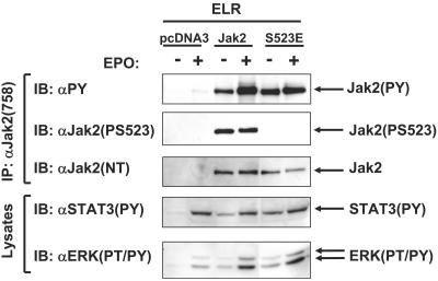
Replacement of amino acids with acidic side chains for Ser or Thr phosphorylation sites on enzymes (such as kinases) often modestly mimics phosphorylation (4). We therefore replaced Ser523 with Glu on Jak2 (Jak2S523E) in order to determine whether this mutation might mimic phosphorylation of Ser523. We transfected HEK293 cells with ELR plus control plasmid, Jak2, or Jak2S523E and rendered the cells quiescent before incubating them in the absence or presence of Epo for 15 min and lysing them. Lysates were directly resolved by SDS-PAGE for immunoblotting with αSTAT3(PY) and αERK(PT/PY) or immunoprecipitated with αJak2(758) for immunoblotting with αPY, αJAK2(PS523), and αJak2(NT) (Fig. 4). We found that basal Jak2S523E tyrosine phosphorylation was increased compared to that of Jak2, although the Epo-stimulated tyrosine phosphorylation of Jak2S523E was similar to that of wild-type Jak2. Quantification of tyrosine-phosphorylated Jak2 relative to Jak2 expression for two independent experiments demonstrated an approximately twofold increase in the basal tyrosine phosphorylation of Jak2S523E for each experiment (data not shown); the tyrosine phosphorylation of ligand-stimulated Jak2S523E was also modestly increased compared to Jak2 in each experiment. Furthermore, the basal phosphorylation of STAT3 and that of ERK were also increased in cells expressing Jak2S523E compared to cells expressing Jak2 in each experiment (data not shown). These data suggest that the substitution of Glu for Ser523 results in an activated/disinhibited phenotype similar to that for the substitution of Ala for Ser523, at least in the unstimulated state.
FIG. 4.
Replacement of Ser523 by Glu increases Jak2 phosphorylation and signaling. HEK293 cells were cotransfected with ELR plus pcDNA3 or the indicated Jak2 isoforms (Jak2 and Jak2S523E), made quiescent, incubated in the absence (−) or presence (+) of Epo (10 units/ml) for 15 min, and lysed. Lysates were immunoprecipitated with αJak2(758). Lysate or immunoprecipitated proteins were resolved by SDS-PAGE, transferred to a nitrocellulose membrane, and immunoblotted with the indicated antibodies. The migration of detected signaling proteins is noted to the right of the panels. The figure shown is representative of two similar independent experiments.
Given that the substitution of an acidic residue in place of a phosphorylation site usually mimics only a minority of the full phosphorylation-dependent function of the site and that the acidic substitution phenotype is generally observed against the backdrop of negligible baseline phosphorylation (see, for example, reference 4), one reasonable explanation for the increased basal activity of Jak2S523E would be the virtually stoichiometric phosphorylation of Ser523 in HEK293 cells. Under these conditions, the presence of Glu in place of Ser523 would be expected to inhibit Jak2 less well than the presence of phosphorylated Ser523 on virtually every Jak2 molecule. This interpretation is also consistent with the apparently high phosphorylation of Ser523 on Jak2 in the baseline and stimulated states in HEK293 and 32D cells, as high-stoichiometry phosphorylation in the baseline state would render it impossible to observe increased phosphorylation following ligand stimulation.
High-stoichiometry phosphorylation of Ser523 in HEK293 cells.
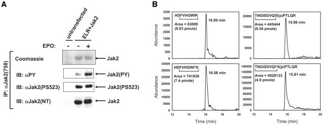
We thus employed LC-SRM using mass-tagged synthetic peptide standards (13) in order to examine the stoichiometry of Ser523 phosphorylation on Jak2 from HEK293 cells. Jak2 was isolated from unstimulated and Epo-stimulated HEK293 cells, as described for Fig. 1 (Fig. 5A). Six picomoles of TNGISDVQ(13C/15N-I)S(p)PTLQRand HDFVHGW(13C/15N-I)Kwere added to both Jak2 tryptic digests, and LC-SRM was performed as described in Materials and Methods. The mass-tagged peptides and corresponding Jak2-derived peptides eluted with identical retention times (Fig. 5B). Area under the curve was calculated for the SRM peak, and peptide amounts were calculated from standard curves of mass-tagged peptides. The concentration of HDFVHGWIK was used to estimate the amount of total Jak2 present and compared with the amount of TNGISDVQIS(p)PTLQR in each sample. Multiple LC-SRM experiments revealed that approximately 80 to 95% of Jak2 molecules from HEK293 cells are phosphorylated on Ser523 under the conditions employed for this analysis, with no detectable difference between stimulated and unstimulated HEK293 cells (Fig. 5B). The completeness of Ser523 phosphorylation in cultured cells suggests its importance in the regulation of Jak2 function.
FIG. 5.
Stoichiometry of phosphorylation of Ser523 from HEK293 cells. A. Untransfected HEK293 cells or cells transfected with ELR plus Jak2 were made quiescent, incubated in the absence (−) or presence (+) of Epo (10 units/ml) for 15 min, and lysed. Lysates were immunoprecipitated with αJak2(758), and immunoprecipitates were resolved by SDS-PAGE for immunoblotting with the indicated antibodies or for detection of proteins by staining with Coomassie brilliant blue. The migration of proteins is indicated to the right of the panels. B. Coomassie blue-stained Jak2 protein from panel A was digested with trypsin, mixed with synthetic mass-labeled internal standards, and subjected to LC-SRM analysis for the quantitative determination of total and Ser523-phosphorylated Jak2. A representative LC-SRM analysis (from two separate analyses) of tryptic peptides derived from unstimulated Jak2 (top panels) and internal peptide standards (bottom panels) is shown. Fragment ions that were monitored are underlined. I* indicates location of mass label (six 13C and one 15N). Peptide amounts were calculated from peak areas and standard curves.
Phosphorylation of Ser523 independently of Jak2 kinase activation in HEK293 cells.
Tyr1007 lies in the activation loop of Jak2 and is required for the activation of the catalytic activity of Jak2 (7); thus, the replacement of Tyr1007 by Phe abrogates kinase activity in Jak2Y1007F. In order to examine whether Jak2 kinase activation was required to promote the phosphorylation of Ser523 in HEK293 cells, we transfected these cells with the cDNAs encoding ELR plus Jak2, Jak2S523A, or Jak2Y1007F (Fig. 6A). Cells were made quiescent and incubated in the absence or presence of Epo for 15 min before lysis. Lysates were immunoprecipitated with αJak2(758) for immunoblotting with αPY, αJak2(PS523), and αJak2(NT) or directly resolved by SDS-PAGE for immunoblotting with αSTAT3(PY) and αERK(PT/PY). As expected, the tyrosine phosphorylation of Jak2 and the phosphorylation of STAT3 and ERK were increased in cells expressing Jak2S523A compared to cells expressing Jak2, while the tyrosine phosphorylation of Jak2Y1007F was undetectable. The phosphorylation of STAT3 and ERK in cells expressing Jak2Y1007F was also decreased compared to cells expressing Jak2 (the remaining signaling was presumably due to the activity of endogenous Jak2 in HEK293 cells, as seen in Fig. 2 and 4, above). The phosphorylation of Ser523 in Jak2Y1007F was detected at similar levels both in the absence and in the presence of ligand stimulation and at levels comparable to those observed in Jak2. These data suggest that the kinase activity of Jak2 is not required for the phosphorylation of Ser523 in HEK293 cells and that converging signaling pathways that can be activated independently of Jak2 are thus responsible for the phosphorylation of Ser523 in these cells.
Additive functions of Ser523 and Tyr570 in the regulation of Jak2 activity.
We and others have previously demonstrated that the phosphorylation of Tyr570 on Jak2 inhibits Jak2-dependent signaling (1, 6). Given the relative proximity and similar functions of Ser523 and Tyr570, we generated a Jak2 molecule containing nonphosphorylatable amino acid substitutions at each of these sites (Jak2S523A/Y570F) in order to examine the possibility of interaction between these two sites (Fig. 6B to D). HEK293 cells transfected with ELR alone or in combination with Jak2, Jak2S523A, Jak2Y570F, or Jak2S523A/Y570F were made quiescent overnight, incubated in the absence or presence of Epo for 15 min, and lysed. In this case, we decreased the amount of Jak2 isoform DNA that was transfected in each case by a factor of 3.3 compared to that used in preceding figures in order to decrease the amplitude of the effects of the mutations and thus permit the more facile determination of differences between these Jak2 isoforms. Lysates were immunoprecipitated with αJak2(758) for immunoblotting with αJak2(PY1007,8), αJak2(PS523), and αJak2(NT) (Fig. 6B). The baseline activation of each of the single point mutants was increased compared to Jak2, as judged by the activation-loop-specific tyrosine phosphorylation; the phosphorylation of the double Jak2S523A/Y570F mutant was increased beyond that observed with the single point mutants (quantification of phosphorylation normalized to Jak2 protein levels in two independent experiments is shown in Fig. 6C). As expected, phosphorylation of Ser523 was absent in Jak2S523A and Jak2S523A/Y570F, and phosphorylation of Tyr570 was absent in Jak2Y570F and Jak2S523A/Y570F.
In order to quantitatively examine the ligand sensitivity of these mutants, we cotransfected them with ELR plus a STAT3-responsive GAS-luciferase reporter plasmid and examined the effect of these Jak2 mutants on STAT3-mediated transcription in response to various submaximal doses of Epo (Fig. 6D). This analysis demonstrated that each Jak2 mutant mediated increased STAT3-mediated transcription compared to wild-type Jak2 at each dose of ligand tested (P < 0.001) and demonstrated that the double mutant mediated increased reporter activity compared to the single mutants at baseline and the lowest dose of Epo (0.1 units/ml) tested (P < 0.05). Overall, these data demonstrate independent and additive effects of Ser523 and Tyr570 in the inhibition of Jak2 activity, as well as demonstrating increased ligand sensitivity of Jak2 molecules defective for phosphorylation of either or both of these sites.
Increased and prolonged signaling by Jak2S523A.
The observed increase in ELR signaling in the presence of Jak2S523A compared to Jak2, above, suggested that mutation of the Ser523 phosphorylation site might increase the amplitude and/or duration of signaling by the intracellular tail of LRb. We thus examined the possibility that Ser523 regulates the attenuation of Jak2-dependent signaling by studying the temporal regulation of Jak2 phosphorylation in HEK293 cells transfected with ELR and control plasmid, Jak2, or Jak2S523A (Fig. 7). Since the dephosphorylation and attenuation of STAT3 signaling are modest over the first 24 h of stimulation (5) and since the first phase of ERK signaling attenuates very rapidly, we did not analyze the phosphorylation of these proteins in this assay but rather focused upon the tyrosine phosphorylation of Jak2 and Jak2S523A. Cells were incubated with maximal amounts (10 units/ml) of Epo for various times up to 8 h and lysed. Immunoprecipitated Jak2 protein was resolved by SDS-PAGE and immunoblotted with αPY, αJak2(PS523), and αJak2(NT) (Fig. 7A and B). In two independent experiments, immunoblotting with αPY demonstrated the expected rapid Epo-stimulated increase in tyrosine phosphorylation of Jak2 and the expected decrease in tyrosine phosphorylation of Jak2 after approximately 30 min of stimulation, as previously reported (5, 6). While Epo stimulation also increased the tyrosine phosphorylation of Jak2S523A, the tyrosine phosphorylation of Jak2S523A again reached much higher levels of activation than Jak2 and did not attenuate appreciably over the 8 h of stimulation. Furthermore, differences in tyrosine phosphorylation between Jak2 and Jak2S523A were more pronounced after 30 to 60 min of stimulation, suggesting that the differences between Jak2 and Jak2S523A at 15 min of stimulation in the preceding figures underestimate the magnitude in the differences between these two molecules. In order to facilitate the comparison of signal activation and attenuation between Jak2 and Jak2S523A, we quantified and graphed the tyrosine phosphorylation of each Jak2 isoform relative to Jak2 expression for each of these molecules in the experiments shown (Fig. 7C), demonstrating the increased duration of signaling in cells expressing Jak2S523A following a maximal stimulus. These data suggest that Ser523 is required for the normal attenuation of Jak2 phosphorylation following ligand stimulation. Interestingly, in each of these experiments, in which the transfected HEK293 cells were serum deprived for longer than in the preceding experiments, some increased phosphorylation of Ser523 on Jak2 was detected at brief times of stimulation. This result is consistent with the idea that high baseline activation of a variety of signaling pathways in HEK293 cells may result in high baseline phosphorylation of Ser523 but that increased times of serum starvation may decrease this activity to yield somewhat lower stoichiometry of Ser523 phosphorylation at baseline, such that activation of ELR-Jak2 signaling results in increased Ser523 phosphorylation.
FIG. 7.
Increased and prolonged signaling by Jak2S523A. HEK293 cells were cotransfected with ELR plus pcDNA3 or the indicated Jak2 isoforms (Jak2 and Jak2S523A) and made quiescent. Cells were incubated with Epo (10 units/ml) for the indicated time (0 to 8 h) before lysis. Lysates were immunoprecipitated with αJak2(758). Immunoprecipitated proteins were resolved by SDS-PAGE and transferred to a nitrocellulose membrane before immunoblotting with the indicated antibodies. The migration of detected signaling proteins is noted to the right of the panels. A and B. Two representative independent experiments are shown. C. Quantification of Jak2 tyrosine phosphorylation normalized for Jak2 expression is shown for the experiments from panels A and B. Means ± standard errors of the means are plotted. WT, wild type.
DISCUSSION
Using LC-MS/MS analysis of Jak2 protein purified from HEK293 cells, we have identified Ser523 as the first-described site of serine phosphorylation on Jak2. This site is phosphorylated in multiple cell types, including on endogenous Jak2 protein in the IL-3-dependent 32D myeloid progenitor cell line and in mouse spleen. Taken together, these data demonstrate the physiologic phosphorylation and likely relevance of Ser523 in the regulation of Jak2 function. Indeed, our analysis suggests that the phosphorylation of Ser523 inhibits Jak2-dependent signaling, as Jak2 molecules containing nonphosphorylatable substitution mutants for Ser523 exhibited enhanced signaling: cells expressing Jak2 substitution mutants for Ser523 consistently demonstrated increased phosphorylation of Jak2 (overall tyrosine phosphorylation and on the activating Tyr1007,8), STAT3, and ERK. For each of these molecules, increased phosphorylation is tantamount to increased signaling activity, as suggested by increased STAT3 transcriptional activity on a reporter plasmid and on the endogenous SOCS3 promoter. The increased activation of STAT3 (the activation of which is initiated predominantly via the phosphorylation of Tyr1138 on the intracellular tail of LRb) (2, 23) in cells expressing mutants for Ser523 suggests that the increased activity of Jak2S523A occurs in association with cytokine receptors (as well as potentially in the absence of cytokine receptor association). Indeed, Jak2S523A not only demonstrates increased signaling in the unstimulated condition but also increases the ligand sensitivity of signaling by the intracellular domain of LRb and impairs the normal attenuation of Jak2 phosphorylation during prolonged ligand stimulation.
Overall, our data suggest that the phosphorylation of Ser523 inhibits Jak2 signaling. This is consistent with the known effects of serine phosphorylation in the inhibition of a variety of other tyrosine kinases and their downstream signaling pathways and previous reports suggesting an inhibitory effect of increased overall Ser/Thr phosphorylation on Jak2 (12, 18, 26, 29, 30). While the exact mechanism of this inhibition is not clear, the findings of increased basal activity and increased duration of signaling during cytokine receptor activation in Jak2S523A suggest that phosphorylation of Ser523 may act to inhibit the enzymatic activity of the Jak2 tyrosine kinase, rather than altering the transmission of the activating signal from the ligand/receptor complex to the associated Jak2 molecule.
Ser523 is not conserved among other Jak kinase family members. Thus, this regulatory phosphorylation event is likely to be unique to Jak2. This residue lies within a stretch of approximately 15 amino acids within the region between the SH2-like domain and JH2 domains of Jak2 and is poorly conserved in other Jak kinases. In one predicted model of the overall structure of Jak2 (14), this region forms a loop that directly overlies the interface between the SH2-like and JH2 domains, and this loop projects Ser523 directly toward a closely apposed tyrosine kinase domain. Thus, phosphorylated Ser523 could theoretically regulate the activity of the Jak2 tyrosine kinase by modifying the overall conformation of the JH2/SH2-like domain interface and thus the tertiary structure of Jak2, or it could directly impinge upon the kinase domain to regulate its activity.
Given the proximity of Ser523 to the JH2 domain and the previously reported role for the JH2 tyrosine phosphorylation site (Tyr570) in inhibiting Jak2 activity, we explored the possibility that these sites might operate in an interdependent manner. This analysis demonstrated that the phosphorylation of Ser523 was independent of Tyr570 and that mutation of Ser523 further activated a Jak2 molecule with a substitution mutation at Tyr570, demonstrating that the effect of Ser523 is independent of Tyr570 and suggesting that the phosphorylation of each site represents an independent mechanism for Jak2 inhibition. This independence of Ser523- and Tyr570-mediated inhibition of Jak2 activity is consistent with the likely requirement for the regulation of Jak2 activity by numerous pathways within a given cell and is also consistent with the different mechanisms by which these two sites are regulated. Not only is the phosphorylation of these sites mediated via different kinases (Tyr570 by Jak2-mediated autophosphorylation and Ser523 by an unknown serine kinase), but the phosphorylation of Ser523 is also apparently high in unstimulated cells and not dependent upon the activity of Jak2 in HEK293 cells.
We have found it difficult to detect regulation of Ser523 phosphorylation in HEK293 or 32D cells. Based upon the finding that mutation of Ser523 to Ala or Glu activates Jak2, we hypothesized that the phosphorylation of Ser523 may be close to stoichiometric in unstimulated HEK293 cells under our usual conditions. Under these conditions, the presence of Glu in place of Ser523 might be expected to inhibit Jak2 to some extent compared to the Ala mutant but would inhibit much less well than the presence of phosphorylated Ser523 on virtually every Jak2 molecule. Furthermore, high levels of Ser523 phosphorylation in unstimulated cells would render it difficult to stimulate further phosphorylation of the residue. Indeed, our LC-SRM analysis with mass-labeled peptide standards demonstrated that the stoichiometry of Ser523 phosphorylation approached unity in unstimulated and Epo-stimulated HEK293 cells expressing exogenous ELR and Jak2. The finding of phosphorylated Ser523 on endogenous Jak2 from 32D cells and mouse spleen coupled with the observed high stoichiometry of Ser523 phosphorylation in HEK293 cells suggests the potential physiologic importance of Ser523 phosphorylation in the regulation of Jak2 function. Furthermore, the finding that neither leptin (in spleen) nor Jak2 activity (in transfected HEK293 cells) is required for the phosphorylation of Ser523 suggests the importance of converging signaling pathways for the regulation of LRb-Jak2 signaling by this phosphorylation event.
Furthermore, while a Pro lies directly COOH terminal to Ser523, 30 to 60 min of inhibition of Pro-directed Ser/Thr kinases (e.g., ERK) in HEK293 cells failed to alter the phosphorylation of Ser523 (see Fig. S1 in the supplemental material). It is important to note that in the presence of very high baseline phosphorylation of Ser523, however, the failure of this inhibitor to block phosphorylation could reflect a low rate of dephosphorylation, rather than the irrelevance of these kinases. Indeed, the Carter-Su laboratory, which independently identified and studied the phosphorylation of Ser523, describes in reference 21a the MEK-dependent regulation of Ser523 phosphorylation by growth hormone in 3T3-F442A preadipocytes. We do not attribute this discrepancy in our findings to differences in antibody preparation, as we have used a highly purified antibody (see Materials and Methods) and have recapitulated our results from HEK293 cells with αJak2(PS523) antisera provided by the Carter-Su laboratory (Fig. 2). Rather, we hypothesize that the observed differences in the regulation of Ser523 phosphorylation reflect differences in cell systems, in which 3T3-F442A cells are more fully quiescent than HEK293 cells (which contain high levels of tyrosine-phosphorylated IRS-4 even following prolonged starvation) or 32D cells (which can be deprived of growth factor for only a few hours prior to study lest they become apoptotic), such that the endogenous Jak2 protein from 3T3-F442A cells may contain significantly less phosphorylated Ser523 on a per-mole basis than that from HEK293 or 32D cells. Indeed, not only is the phosphorylation of Ser523 detectable at baseline in 3T3-F442A cells but prolonged serum starvation of HEK293 cells appears to lower baseline phosphorylation of Ser523, enabling the detection of regulation of Ser523 phosphorylation during signaling by the intracellular domain of LRb. Thus, in aggregate, the phosphorylation of Ser523 can likely function to mediate feedback inhibition of Jak2 activity during cytokine stimulation as well as inhibiting Jak2 signaling downstream of other, Jak2-independent, pathways within the cell.
While determining the exact mechanism by which phosphorylation of Ser523 inhibits Jak2 signaling will require further study, it is clear that the phosphorylation of this site represents an important mechanism by which Jak2-dependent signaling is regulated. Enhancing the phosphorylation of Ser523 would necessarily decrease the strength of Jak2-dependent signals and could underlie impaired signaling by Jak2-dependent cytokines, such as leptin in the leptin resistance that accompanies common forms of obesity (8, 10, 22). On the other hand, impairment of Ser523 phosphorylation or Ser523-mediated inhibition would be expected to increase cytokine action, as in autoimmunity or tumor promotion (25). Further investigation will be required to determine the role of Jak2 inhibition via Ser523 phosphorylation in these and other disease processes.
Supplementary Material
Acknowledgments
This work was supported by NIH DK56731 and grants from the American Diabetes Association (to M.G.M.), the Cellular and Molecular Biology Core of the Michigan Diabetes Research and Training Center (P60 DK20572), and Joslin's Diabetes and Endocrinology Research Center DK36836 (Proteomics Core laboratory).
We are grateful to Diane Fingar for a critical reading of the manuscript. Thanks go to the laboratory of Christin Carter-Su for sharing their preparation of αJak2(PS523).
Footnotes
Supplemental material for this article may be found at http://mcb.asm.org/.
REFERENCES
- 1.Argetsinger, L. S., J. L. Kouadio, H. Steen, A. Stensballe, O. N. Jensen, and C. Carter-Su. 2004. Autophosphorylation of JAK2 on tyrosines 221 and 570 regulates its activity. Mol. Cell. Biol. 24:4955-4967. [DOI] [PMC free article] [PubMed] [Google Scholar]
- 2.Banks, A. S., S. M. Davis, S. H. Bates, and M. G. Myers, Jr. 2000. Activation of downstream signals by the long form of the leptin receptor. J. Biol. Chem. 275:14563-14572. [DOI] [PubMed] [Google Scholar]
- 3.Bjorbaek, C., H. J. Lavery, S. H. Bates, R. K. Olson, S. M. Davis, J. S. Flier, and M. G. Myers, Jr. 2000. SOCS3 mediates feedback inhibition of the leptin receptor via Tyr985. J. Biol. Chem. 275:40649-40657. [DOI] [PubMed] [Google Scholar]
- 4.Dennis, P. B., N. Pullen, S. C. Kozma, and G. Thomas. 1996. The principal rapamycin-sensitive p70s6k phosphorylation sites, T-229 and T-389, are differentially regulated by rapamycin-insensitive kinase kinases. Mol. Cell. Biol. 16:6242-6251. [DOI] [PMC free article] [PubMed] [Google Scholar]
- 5.Dunn, S. L., M. Bjornholm, S. H. Bates, Z. Chen, M. Seifert, and M. G. Myers, Jr. 2005. Feedback inhibition of leptin receptor/Jak2 signaling via Tyr1138 of the leptin receptor and suppressor of cytokine signaling 3. Mol. Endocrinol. 19:925-938. [DOI] [PubMed] [Google Scholar]
- 6.Feener, E. P., F. Rosario, S. L. Dunn, Z. Stancheva, and M. G. Myers, Jr. 2004. Tyrosine phosphorylation of Jak2 in the JH2 domain inhibits cytokine signaling. Mol. Cell. Biol. 24:4968-4978. [DOI] [PMC free article] [PubMed] [Google Scholar]
- 7.Feng, J., B. A. Witthuhn, T. Matsuda, F. Kohlhuber, I. M. Kerr, and J. N. Ihle. 1997. Activation of Jak2 catalytic activity requires phosphorylation of Y1007 in the kinase activation loop. Mol. Cell. Biol. 17:2497-2501. [DOI] [PMC free article] [PubMed] [Google Scholar]
- 8.Flier, J. S., and E. Maratos-Flier. 1998. Obesity and the hypothalamus: novel peptides for new pathways. Cell 92:437-440. [DOI] [PubMed] [Google Scholar]
- 9.Frank, S. J., W. Yi, Y. Zhao, J. F. Goldsmith, G. Gilliland, J. Jiang, I. Sakai, and A. S. Kraft. 1995. Regions of the Jak2 tyrosine kinase required for coupling to the growth hormone receptor. J. Biol. Chem. 270:14776-14785. [DOI] [PubMed] [Google Scholar]
- 10.Friedman, J. M. 2002. The function of leptin in nutrition, weight, and physiology. Nutr. Rev. 60:S1-S14. [DOI] [PubMed] [Google Scholar]
- 11.Gadina, M., D. Hilton, J. A. Johnston, A. Morinobu, A. Lighvani, Y. J. Zhou, R. Visconti, and J. J. O'Shea. 2001. Signaling by type I and II cytokine receptors: ten years after. Curr. Opin. Immunol. 13:363-373. [DOI] [PubMed] [Google Scholar]
- 12.Gandino, L., M. F. Di Renzo, S. Giordano, F. Bussolino, and P. M. Comoglio. 1990. Protein kinase-c activation inhibits tyrosine phosphorylation of the c-met protein. Oncogene 5:721-725. [PubMed] [Google Scholar]
- 13.Gerber, S. A., J. Rush, O. Stemman, M. W. Kirschner, and S. P. Gygi. 2003. Absolute quantification of proteins and phosphoproteins from cell lysates by tandem MS. Proc. Natl. Acad. Sci. USA 100:6940-6945. [DOI] [PMC free article] [PubMed] [Google Scholar]
- 14.Giordanetto, F., and R. T. Kroemer. 2002. Prediction of the structure of human Janus kinase 2 (JAK2) comprising JAK homology domains 1 through 7. Protein Eng. 15:727-737. [DOI] [PubMed] [Google Scholar]
- 15.Herrington, J., and C. Carter-Su. 2001. Signaling pathways activated by the growth hormone receptor. Trends Endocrinol. Metab. 12:252-257. [DOI] [PubMed] [Google Scholar]
- 16.Ihle, J. N., W. Thierfelder, S. Teglund, D. Stravapodis, D. Wang, J. Feng, and E. Parganas. 1998. Signaling by the cytokine receptor superfamily. Ann. N. Y. Acad. Sci. 865:1-9. [DOI] [PubMed] [Google Scholar]
- 17.Kloek, C., A. K. Haq, S. L. Dunn, H. J. Lavery, A. S. Banks, and M. G. Myers, Jr. 2002. Regulation of Jak kinases by intracellular leptin receptor sequences. J. Biol. Chem. 277:41547-41555. [DOI] [PubMed] [Google Scholar]
- 18.Kovanen, P. E., I. Junttila, K. Takaluoma, P. Saharinen, L. Valmu, W. Li, and O. Silvennoinen. 2000. Regulation of Jak2 tyrosine kinase by protein kinase C during macrophage differentiation of IL-3-dependent myeloid progenitor cells. Blood 95:1626-1632. [PubMed] [Google Scholar]
- 19.Kurzer, J. H., L. S. Argetsinger, Y.-J. Zhou, J.-L. Kouadio, J. J. O'Shea, and C. Carter-Su. 2004. Tyrosine 813 is a site of JAK2 autophosphorylation critical for activation of JAK2 by SH2-Bβ. Mol. Cell. Biol. 24:4557-4570. [DOI] [PMC free article] [PubMed] [Google Scholar]
- 20.Livak, K. J., and T. D. Schmittgen. 2001. Analysis of relative gene expression data using real-time quantitative PCR and the 2(−ΔΔC(T)) method. Methods 25:402-408. [DOI] [PubMed] [Google Scholar]
- 21.Luo, H., P. Rose, D. Barber, W. P. Hanratty, S. Lee, T. M. Roberts, A. D. D'Andrea, and C. R. Dearolf. 1997. Mutation in the Jak kinase JH2 domain hyperactivates Drosophila and mammalian Jak-Stat pathways. Mol. Cell. Biol. 17:1562-1571. [DOI] [PMC free article] [PubMed] [Google Scholar]
- 21a.Mazurkiewicz-Munoz, A. M., L. S. Argetsinger, J.-L. K. Kouadio, A. Stensballe, O. N. Jensen, J. M. Cline, and C. Carter-Su. 2006. Phosphorylation of JAK2 at serine 523: a negative regulator of JAK2 that is stimulated by growth hormone and epidermal growth factor. Mol. Cell. Biol. 26:4052-4062. [DOI] [PMC free article] [PubMed] [Google Scholar]
- 22.Munzberg, H., and M. G. Myers, Jr. 2005. Molecular and anatomical determinants of central leptin resistance. Nat. Neurosci. 8:566-570. [DOI] [PubMed] [Google Scholar]
- 23.Myers, M. G., Jr. 2004. Leptin receptor signaling and the regulation of mammalian physiology. Recent Prog. Horm. Res. 59:287-304. [DOI] [PubMed] [Google Scholar]
- 24.Myers, M. G., Jr., L. M. Wang, X. J. Sun, Y. Zhang, L. Yenush, J. Schlessinger, J. H. Pierce, and M. F. White. 1994. The role of IRS-1/GRB2 complexes in insulin signaling. Mol. Cell. Biol. 14:3577-3587. [DOI] [PMC free article] [PubMed] [Google Scholar]
- 25.O'Shea, J. J., A. Ma, and P. Lipsky. 2002. Cytokines and autoimmunity. Nat. Rev. Immunol. 2:37-45. [DOI] [PubMed] [Google Scholar]
- 26.Rui, L., V. Aguirre, J. K. Kim, G. I. Shulman, A. Lee, A. Corbould, A. Dunaif, and M. F. White. 2001. Insulin/IGF-1 and TNF-alpha stimulate phosphorylation of IRS-1 at inhibitory Ser307 via distinct pathways. J. Clin. Investig. 107:181-189. [DOI] [PMC free article] [PubMed] [Google Scholar]
- 27.Saatcioglu, F., G. Lopez, B. L. West, E. Zandi, W. Feng, H. Lu, A. Esmaili, J. W. Apriletti, P. J. Kushner, J. D. Baxter, and M. Karin. 1997. Mutations in the conserved C-terminal sequence in thyroid hormone receptor dissociate hormone-dependent activation from interference with AP-1 activity. Mol. Cell. Biol. 17:4687-4695. [DOI] [PMC free article] [PubMed] [Google Scholar]
- 28.Saharinen, P., and O. Silvennoinen. 2002. The pseudokinase domain is required for suppression of basal activity of Jak2 and Jak3 tyrosine kinases and for cytokine-inducible activation of signal transduction. J. Biol. Chem. 277:47954-47963. [DOI] [PubMed] [Google Scholar]
- 29.Takayama, S., M. F. White, and C. R. Kahn. 1988. Phorbol ester-induced serine phosphorylation of the insulin receptor decreases its tyrosine kinase activity. J. Biol. Chem. 263:3440-3447. [PubMed] [Google Scholar]
- 30.Tanti, J. F., T. Gremeaux, E. Van Obberghen, and Y. Le Marchand-Brustel. 1994. Serine/threonine phosphorylation of insulin receptor substrate 1 modulates insulin receptor signaling. J. Biol. Chem. 269:6051-6057. [PubMed] [Google Scholar]
- 31.Tartaglia, L. A. 1997. The leptin receptor. J. Biol. Chem. 272:6093-6096. [DOI] [PubMed] [Google Scholar]
- 32.Velazquez, L., K. E. Mogensen, G. Barbieri, M. Fellous, G. Uze, and S. Pellegrini. 1995. Distinct domains of the protein tyrosine kinase tyk2 required for binding of interferon-α/β and for signal transduction. J. Biol. Chem. 270:3327-3334. [DOI] [PubMed] [Google Scholar]
- 33.Yeh, T. C., E. Dondi, G. Uze, and S. Pellegrini. 2000. A dual role for the kinase-like domain of the tyrosine kinase Tyk2 in interferon-alpha signaling. Proc. Natl. Acad. Sci. USA 97:8991-8996. [DOI] [PMC free article] [PubMed] [Google Scholar]
- 34.Zhao, Y., F. Wagner, S. J. Frank, and A. S. Kraft. 1995. The amino-terminal portion of the JAK2 protein kinase is necessary for binding and phosphorylation of the granulocyte-macrophage colony-stimulating factor receptor beta c chain. J. Biol. Chem. 270:13814-13818. [DOI] [PubMed] [Google Scholar]
- 35.Zhou, Y. J., M. Chen, N. A. Cusack, L. H. Kimmel, K. S. Magnuson, J. G. Boyd, W. Lin, J. L. Roberts, A. Lengi, R. H. Buckley, R. L. Geahlen, F. Candotti, M. Gadina, P. S. Changelian, and J. J. O'Shea. 2001. Unexpected effects of FERM domain mutations on catalytic activity of Jak3: structural implication for Janus kinases. Mol. Cell 8:959-969. [DOI] [PubMed] [Google Scholar]
Associated Data
This section collects any data citations, data availability statements, or supplementary materials included in this article.


