Abstract
The objective of this research was to optimize sampling parameters for increased recovery and detection of airborne porcine reproductive and respiratory syndrome virus (PRRSV) and swine influenza virus (SIV). Collection media containing antifoams, activated carbons, protectants, and ethylene glycol were evaluated for direct effects on factors impacting the detection of PRRSV and SIV, including virus infectivity, viability of continuous cell lines used for the isolation of these viruses, and performance of reverse transcriptase PCR assays. The results showed that specific compounds influenced the likelihood of detecting PRRSV and SIV in collection medium. A subsequent study evaluated the effects of collection medium, impinger model, and sampling time on the recovery of aerosolized PRRSV using a method for making direct comparisons of up to six treatments simultaneously. The results demonstrated that various components in air-sampling systems, including collection medium, impinger model, and sampling time, independently influenced the recovery and detection of PRRSV and/or SIV. Interestingly, it was demonstrated that a 20% solution of ethylene glycol collected the greatest quantity of aerosolized PRRSV, which suggests the possibility of sampling at temperatures below freezing. Based on the results of these experiments, it is recommended that air-sampling systems be optimized for the target pathogen(s) and that recovery/detection results should be interpreted in the context of the actual performance of the system.
Airborne pathogens are detected by recovering the target microorganism in a collection medium (liquid, semisolid, or solid substrate) and then assaying the substrate for the presence of the target pathogen by using an appropriate microbiological assay. Various air-sampling devices are available, but “impingers” are generally used to collect airborne viruses. Impingers direct a converged stream of environmental air onto a liquid collection medium to recover airborne viral particles in the liquid phase of the collection system (1, 11, 12, 14, 16, 17). Impingers are generally considered more effective than filters, bubblers, or impactors for capturing airborne viruses (18, 19, 20).
A number of variables are known to affect impinger collection efficiency. These include impinger design (5, 13, 24), sampling time (28), and the composition of collection medium (31). In addition, specific compounds are sometimes added to impinger collection medium to preserve virus infectivity during the collection process (37, 38, 40).
This research focused on specific aspects of optimizing the collection and detection of aerosolized porcine reproductive and respiratory syndrome virus (PRRSV) and swine influenza virus (SIV) in air-sampling systems. The first study (experiment 1) focused on virus detection. Compounds added to collection media to enhance collection efficiency (i.e., antifoams, bovine serum albumin, gelatin, mucin, activated carbon, and ethylene glycol) were evaluated for direct effects on virus infectivity, on the viability of continuous cell lines used for the isolation of these viruses, and on the performance of reverse transcriptase PCR (RT-PCR) assays. The second study (experiment 2) focused on optimizing sampling parameters, including medium composition, impinger model, and sampling time, for the collection of PRRSV from aerosols.
MATERIALS AND METHODS
Porcine reproductive respiratory syndrome virus.
The North American prototype PRRSV ATCC VR-2332 (American Type Culture Collection, Manassas, VA) was used in this study. The complete virus genomic sequence has been published previously (GenBank accession no. PRU87392). The virus was propagated on MARC-145 cells, which are clones of the African monkey kidney cell line MA-104 that is considered highly permissive to PRRSV (27).
Swine influenza virus.
A field isolate of SIV designated A/Swine/Iowa/73 (H1N1) (National Veterinary Service Laboratories, Ames, IA) was used in this study. The virus was propagated on Madin-Darby canine kidney (MDCK) cells.
Cell lines.
Virus propagation, microinfectivity assays, and neutral red (NR) cell viability assays were performed on MARC-145 and MDCK (American Type Culture Collection, Manassas, VA) continuous cell lines. Cells were propagated and maintained in 75-cm2 flasks (catalog no. 3150; Corning, Corning, NY). Growth medium for both cell lines consisted of minimal essential medium (MEM) (catalog no. M4655; Sigma Chemical Co., St. Louis, MO) supplemented with 10% fetal bovine serum (catalog no. F4922; Sigma, St. Louis, MO), 50 μg of gentamicin (catalog no. G1272; Sigma, St. Louis, MO) per ml, 0.25 μg of amphotericin B (Fungizone A2942; Sigma, St. Louis, MO) per ml, and 100 μg of penicillin-streptomycin (catalog no. P0781, Sigma; St. Louis, MO) per ml.
Virus titration.
Samples were titrated following the protocols described below. Virus titers were calculated using the Spearman-Kärber method (22) and expressed as 50% tissue culture infective dose (TCID50) per milliliter.
(i) PRRSV.
For PRRSV, 200 μl of MARC-145 cells suspended in MEM growth medium at a concentration of 4 × 10⁵ cells per ml were added to each well of a 96-well plate (catalog no. 3596; Corning, Corning, NY). Plates were incubated at 37°C in a humidified 5% CO2 incubator until the cell monolayer was confluent. Samples were serially 10-fold diluted (100 to 10−5) in MEM. Growth medium was discarded, and four wells were inoculated with 100 μl of sample at each dilution. After incubating for 2 h, the inoculum was discarded and 200 μl of growth medium with reduced fetal bovine serum (5%) was added to each well. Plates were incubated at 37°C in a humidified 5% CO2 incubator for 48 h. Following incubation, cells were fixed with aqueous 80% acetone solution and stained with a fluorescein isothiocyanate-conjugated monoclonal antibody specific for PRRSV (SDOW17; Rural Technologies, Inc., Brookings, SD). Virus titers were calculated on the basis of the number of wells showing a PRRSV-specific fluorescence reaction at each dilution. Each sample was run in duplicate, and the titers were averaged.
(ii) Swine influenza virus.
For SIV, 200 μl of MDCK cells suspended in MEM growth medium at a concentration of 4 × 10⁵ cells per ml were added to each well of a 96-well plate. Plates were incubated at 37°C in a humidified 5% CO2 incubator until cell monolayers were confluent. Treatment samples were serially 10-fold diluted (100 to 10−5) in inoculation medium. Inoculation medium consisted of MEM supplemented with 5% (vol/vol) bovine serum albumin (BSA) (catalog no. 15260-037; Invitrogen, Carlsbad, CA), 50 μg of gentamicin per ml, 0.25 μg of amphotericin B per ml, 100 μl per ml of 200 mM l-glutamine, and tosylsulfonyl phenylalanyl chloromethyl ketone (TPCK)-treated trypsin (catalog no. LS003740; Worthington Biochemical, Lakewood, NJ) at 4 μg per ml. Growth medium was discarded, and cells were rinsed three times with MEM containing TPCK-treated trypsin at 4 μg per ml. Four wells were inoculated with 100 μl of sample at each dilution, and then plates were incubated at 37°C in a humidified 5% CO2 incubator. After incubating for 2 h, the inoculum was discarded and 200 μl of the inoculation medium was added to each well. Plates were then incubated at 37°C in a humidified 5% CO2 incubator for 3 to 6 days. Cells were examined for cytopathic effects (CPE) daily. At day 6 or after the appearance of CPE, plates were fixed and stained as previously described by Clavijo et al. (8). The presence of SIV in cells was confirmed by an immunoperoxidase assay using a monoclonal antibody specific for influenza A virus nucleoprotein. Virus titers were determined by the number of wells at each dilution showing CPE and/or a positive reaction. Each sample was run in duplicate, and the titers averaged.
PCR.
PRRSV and SIV RNA for real-time RT-PCR amplification was extracted from 0.14 ml of sample with a QIAamp viral RNA mini kit (catalog no. 210210; QIAGEN Inc., Valencia, CA), following the protocols recommended by the manufacturer. Real-time RT-PCR quantification was performed using an ABI Prism 7900 HT sequence detection system (Applied Biosystems, Foster City, CA). Primers specific for ORF7 (PRRSV) and neuraminidase segment (SIV) were synthesized by Integrated DNA Technologies (Coralville, IA), and minor groove binder probes were synthesized by Applied Biosystems (Foster City, CA). The thermal profile for amplification of both PRRSV and SIV viral RNA was a reverse transcription at 50°C for 30 min, followed by enzyme activation at 95°C for 15 min, then 40 cycles of denaturation at 94°C for 15 s and a combined annealing/extension step at 60°C for 60 s. Fluorescence data capture occurred at the combined annealing/extension stage. For each assay, a standard curve was generated using standards (101 to 106 TCID50 equivalents per ml) and positive and negative control samples were tested with the unknowns. The unit expression for RT-PCR for PRRSV is TCID50/ml, which represents quantity of total viral RNA in samples relative to standards in which the amount of measurable infectious viruses was quantified using microtitration infectivity assays. Quantitative RT-PCR values are estimates of total viral RNA present in samples including both infectious and inactivated virus.
Experiment 1: effects of compounds on cell viability, virus infectivity, and PCR performance.
Specific compounds sometimes added to air-sampling collection media to improve collection efficiency were evaluated for direct effects on cell viability, virus infectivity, and quantitative RT-PCR assays specific for PRRSV and SIV.
Medium compounds.
Compounds tested included antifoams, protectants (BSA, gelatin, and mucin), sorbents (activated carbon), and ethylene glycol. Effects on cell viability were assessed by exposing MARC-145 and MDCK cells to these compounds for 2 h and then measuring differences between treated and untreated cells using a neutral red assay. Effects on virus infectivity were measured by exposing PRRSV and SIV to these compounds for 6 h at 37°C and then comparing pre- and postexposure titers (TCID50) to those of nonexposed controls. Effects on the diagnostic performance of quantitative RT-PCR assays were evaluated by comparing the PCR results from virus samples with and without these compounds. Compounds exhibiting no deleterious effects on cells, virus, or PCR assays were selected for further evaluation in experiment 2.
(i) Antifoams.
The process of impingement produces extensive foaming when the liquid collection medium contains proteins and/or carbohydrates. Antifoams are added to the collection medium to eliminate this problem (26, 37). Based on these reports, six antifoams (Sigma, St. Louis, MO), i.e., antifoam 204 (A6426), antifoam A emulsion (A5758), antifoam B emulsion (A5757), antifoam C emulsion (A8011), antifoam O-30 (A8082), and antifoam SE-15 (A8582), were evaluated. Although their exact compositions are proprietary information, this selection included both organic (antifoam 204 and antifoam O-30) and silicone-based (antifoam A, emulsion, antifoam B emulsion, antifoam C emulsion, and antifoam SE-15) antifoams. Antifoams were diluted in phosphate-buffered saline (PBS) (1×) to 0.01% (vol/vol) and tested for their effects on cell viability (neutral red assay), virus infectivity (TCID50), and diagnostic performance (RT-PCR).
(ii) Protectants.
The addition of BSA (38), mucin (35), and/or gelatin (38) to collection or suspension medium has been shown to reduce the rate of virus inactivation and preserve virus infectivity during the process of impingement or aerosolization. Based on these reports, solutions of BSA (A9418), gelatin (G1890), and mucin (M1788) (Sigma, St. Louis, MO) at a concentration of 1.0% (wt/vol) in PBS (1×) were examined for effects on cell viability (neutral red assay), virus infectivity (TCID50), and diagnostic performance (RT-PCR).
(iii) Sorbents.
Activated carbon adsorbs viruses (3, 9) and may enhance infectivity (7). Based on these reports, one peat-based (catalog no. C9157; Sigma, St. Louis, MO) and one wood-based (Cal-Pacific, Fields Landing, CA) activated carbon product were tested at five treatment levels (0.2, 1.0, 2.0, 10.0, and 20.0% [wt/vol]) in PBS (1×) for effects on cell viability (neutral red assay). Additionally, activated carbon (1.0%) was examined for effects on diagnostic performance (RT-PCR).
(iv) Ethylene glycol.
Although its use in collection media has not been described previously, ethylene glycol was evaluated because it offers the possibility of sampling at temperatures below 0°C (32°F), since the ethylene glycol freezing point is −13°C. A 20% solution of ethylene glycol (catalog no. 29,323-7; Sigma, St. Louis, MO) (vol/vol) in PBS (1×) was tested for effects on cell viability (neutral red assay), virus infectivity (TCID50), and diagnostic performance (RT-PCR). A 20% solution was selected to decrease the medium's freezing point to −8°C.
Neutral red cell viability assay.
An NR cell viability assay was used to quantify the direct effects of compounds on MARC-145 and MDCK cells (2). The quantity of dye taken up by cells was estimated using a spectrophotometer, and then the effect of each treatment was determined by comparing the absorbance value (cell viability) of treated wells to that of the untreated control wells.
In brief, the NR assay was performed by adding 200 μl of each compound at the concentrations to be tested (antifoams [0.1%], ethylene glycol [20%], and protectants [1.0%]) to 12 wells of a 96-well microtitration plate containing a 75% confluent monolayer of MARC-145 or MDCK cells. Each compound and concentration was tested eight times (eight plates). To account for plate-to-plate variability, all within-group treatments (antifoams, ethylene glycol, and protectants) and untreated controls (12 wells) were present on all plates. Cells were exposed to compounds for 2 h at 37°C in a 5% CO2-humidified incubator. Following exposure, compounds were discarded and replaced with maintenance medium. Plates were incubated for an additional 24 h at 37°C in a 5% CO2-humidified incubator. Following incubation, the medium was replaced with filtered (Millipore Super-Q, ZFSQ115P4 system with cartridge nos. CDMB01204, CDAC01204, CP2001003, and PMEG09002; Millipore, Billerica, MA) sterilized water containing 40 μg per ml of neutral red (N 2889; Sigma Chemical Co., St. Louis, MO). Plates were incubated for 3 h at 37°C in a 5% CO2-humidified incubator to allow for uptake of the dye. To remove the dye not taken up by living cells, the wells were rinsed with a sterile water solution containing 0.5% formaldehyde and 1% CaCl2. To extract the dye taken up by viable cells, 200 μl of 1% acetic acid and 50% ethanol in sterile water was added to each well. The plate was left to stand at ambient temperature for 5 min and then agitated on a microplate shaker for 30 min to ensure that the dye had been released from the cells and mixed into solution. The reaction was quantified using a spectrophotometer (model no. ELx800; BioTek Instruments, Inc., Winooski, VT) at a wavelength of 540 nm, and the results were reported as mean absorbance.
Virus infectivity and RT-PCR detection.
One-milliliter aliquots of stock virus (PRRSV and SIV) were added to 50 ml of collection medium (PBS) containing the compounds and concentrations to be tested (antifoams [0.1%], ethylene glycol [20%], BSA [1.0%], gelatin [1.0%], and mucin [1.0%]) in 250-ml medium bottles. Following the addition of virus, the collection medium compounds were incubated on a stir plate for 6 h at 37°C in a 5% CO2-humidified incubator. Samples were taken at 0 and 6 h, aliquoted, and stored frozen at −80°C. Each compound and concentration was tested three times. Microtitration infectivity assays and quantitative RT-PCRs were performed on all samples (concentration for each replicate) concurrently.
Experiment 2: effect of collection media, impinger, and sampling time on collection of aerosolized PRRSV.
The objective of experiment 2 was to optimize aerosol sampling parameters for collection of PRRSV. Collection medium (compounds selected for further evaluation from experiment 1), impinger model, and sampling time were evaluated relative to recovery of aerosolized PRRSV.
Aerosolization of PRRSV.
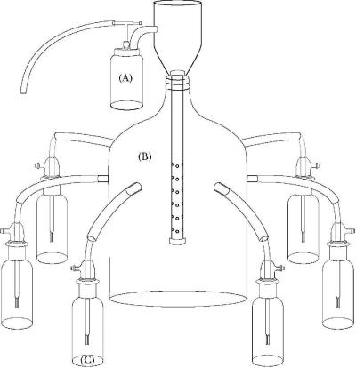
PRRSV diluted in PBS (1×) to a titer of 1 × 106.33 TCID50 was aerosolized using a 24-jet Collison nebulizer (model no. CN60; BGI, Inc., Waltham, MA) operated on compressed air (catalog no. 00916734000; Sears Roebuck, Hoffman Estates, IL) at 40 lb/in2 producing 80 liters of free air per minute. The aerosolized PRRSV flowed into a 5-gal glass reservoir (catalog no. B26; The Home Brewery, Inc., Ozark, MO), which was modified to allow simultaneous sample collection at six outlet ports (Fig. 1). Outlet ports were installed by drilling six equidistantly spaced holes at the circumference of the glass reservoir and permanently attaching a glass stem (inside diameter [ID], 0.5 in; length, 1.5 in) to each hole. Clear tubing (ID, 0.375 in; wall thickness, 0.125 in) (catalog no. 14-169-7H; Fisher Scientific, Hampton, NH) was used to connect the impingers to the outlet ports (Fig. 1). This arrangement made it possible to test up to six different treatments simultaneously on the same cloud of aerosolized PRRSV.
FIG. 1.
Illustration of the experimental design to optimize sampling time, collection media, and impingers for aerosolized PRRS virus. (A) Collison nebulizer. (B) Glass carboy. (C) AGI-30 impingers.
The concentration of PRRSV viral RNA in the suspension fluid was monitored during operation of the nebulizer. Samples were collected by inserting plastic tubing (ID, 0.050; length, 0.020 in) (catalog no. 14-170-15E; Fisher Scientific, Hampton, NH) through the nozzle of the nebulizer and into the virus solution. One-milliliter samples of the virus solution were collected at 5-min intervals using a syringe (catalog no. 14-823-69; Fisher Scientific, Hampton, NH) and hypodermic needle (catalog no. NC9062128; Fisher Scientific, Hampton, NH) inserted into the plastic tubing. Samples were assayed by quantitative RT-PCR.
Sampling of aerosolized PRRSV. (i) Collection medium.
Six collection medium treatments were compared on the basis of recovery of aerosolized PRRSV from the reservoir. PBS (1×) was used as the diluent in all treatments. Medium treatments were PBS; PBS and 1% activated carbon (Cal-Pacific, Fields Landing, CA) (wt/vol); PBS and 0.5% BSA (wt/vol); PBS and 20% ethylene glycol (vol/vol); PBS, 0.5% BSA, and 1% activated carbon; and PBS, 20% ethylene glycol, 0.5% BSA, and 1% activated carbon (Table 1).
TABLE 1.
Medium treatments examined for collection efficiency of aerosolized PRRS virus in experiment 2
| Medium treatment | Medium additivea
|
|||
|---|---|---|---|---|
| PBSb | Activated carbon, 1%c | BSA, 0.5%d | Ethylene glycol, 20%e | |
| 1 | + | |||
| 2 | + | + | ||
| 3 | + | + | ||
| 4 | + | + | ||
| 5 | + | + | + | |
| 6 | + | + | + | + |
+, presence of indicated additive.
PBS, catalog no. 10010-064 (Invitrogen, Carlsbad, CA).
Activated carbon (Cal-Pacific, Fields Landing, CA).
BSA, catalog no. A9418 (Sigma, St. Louis, MO).
Ethylene glycol, catalog no. 29,323-7 (Sigma, St. Louis, MO).
(ii) Impingers.
Three impinger models (AGI-30 [7540-10; Ace Glass, Vineland, NJ], AGI-4 [6 liter] [7541-10; Ace Glass, Vineland, NJ], and SKC BioSampler [225-9595; SCK, Inc., Eighty Four, PA]) were compared in terms of the recovery of aerosolized PRRSV. At a vacuum pressure of not more than −0.05 atm, the AGI-30 and SKC BioSampler operated at a flow rate of 12.5 liters per minute and the AGI-4 (6 liter) operated at 6.0 liters per minute. Vacuum pressure was maintained using oilless pumps (catalog no. S413801; Fisher, Hampton, NH) and was monitored using a vacuum pressure gauge (catalog no. G-S4LM20-VAC-100; Cato Western, Inc., Tucson, AZ). Flow rates of impingers in liters per minute were verified using a flow meter (catalog no. DW-806; Dwyer Instruments, Inc., Michigan City, IN).
(iii) Sampling time.
The effect of sampling time (0, 1, 2, 5, 10, 15, and 20 min) on the collection of aerosolized PRRSV was evaluated by medium treatment (n = 6) and impinger model (n = 3). For each replicate, each of six impingers of the identical model was filled with 20 ml of one of the six collection medium treatments to be tested. All six impingers sampled the same aerosol cloud for the designated sampling time, after which the collection fluid was harvested, aliquoted, and stored at −80°C. The experiment was repeated until each sampling time had been examined. The model was replicated three times for each of the three impinger models. A total of 63 experimental runs were completed with the six collection medium treatments (7 time points × 3 impinger models × 3 replications). Recovery of PRRSV was determined by quantitative RT-PCR. To reduce variability, RT-PCR was performed on the collection medium samples concurrently.
Statistical analysis.
Medium compound treatments evaluated in experiment 1 were compared by analysis of variance (ANOVA) (JMP; SAS Institute Inc., Cary, NC) using the data from the neutral red cell viability assays, microinfectivity assays, and quantitative RT-PCR assays. Results were reported as least square means. The null hypothesis stated that the means of the treatments and the means of the controls were equal. A significance level of <0.05 was used as the minimum acceptable P value. If the means were significantly different, individual pair-wise treatment comparisons were performed using Student's t test.
Quantitative RT-PCR data from collection medium treatments in experiment 2 were analyzed using repeated measures multivariate ANOVA (MANOVA) (JMP; SAS Institute Inc., Cary, NC) using sampling time as the repeating variable. The model included the main effects of impinger and collection media. Two-way and three-way interactions, i.e., impinger × time, collection media × time, impinger × collection media, and impinger × collection media × time, were included in the model. Results were reported as least square means. A significance level of <0.05 was required as the minimum acceptable P value. If the MANOVA was significant, one-way ANOVAs were performed at each time point. If the ANOVA was significant, individual pair-wise comparisons was performed at that time point using Student's t test.
RESULTS
Experiment 1: effects of medium compounds on cell viability, virus infectivity, and PCR performance. (i) Antifoams.
Two (A emulsion and C emulsion) of the six antifoams evaluated had no detrimental effect on the viability of either MARC-145 or MDCK cells (Table 2). That is, their NR assay absorbance values were not significantly different from the untreated control absorbance values (P > 0.05). The four remaining antifoams had absorbance values that were significantly lower than those of the untreated controls for MARC-145 and/or MDCK cells (P < 0.05), suggesting that these antifoams adversely affected one or both of the cell lines at the concentrations tested. Compared to controls, the exposure of PRRSV and SIV to antifoams did not significantly affect the titers of infectious virus (TCID50) or quantitative RT-PCR results (Table 3). On the basis of the overall results, antifoam A emulsion (0.01%) was selected for use in experiment 2.
TABLE 2.
Univariate effects of antifoams, protectants, and ethylene glycol on viability of MARC-145 and MDCK cell lines as measured by the neutral red assay
| Compound (n = 96) | Concn (%) | Neutral red assay value (absorbance at 540 nm) for:
|
|||
|---|---|---|---|---|---|
| MARC-145
|
MDCK
|
||||
| Meana | SEM | Mean | SEM | ||
| Antifoamsb | |||||
| Control | 0.00 | 0.782wx | 0.024 | 0.750wx | 0.027 |
| A emulsion | 0.01 | 0.816w | 0.024 | 0.861w | 0.027 |
| C emulsion | 0.01 | 0.738x | 0.024 | 0.832w | 0.027 |
| 204 | 0.01 | 0.641y | 0.024 | 0.729xy | 0.027 |
| O-30 | 0.01 | 0.606y | 0.024 | 0.653z | 0.027 |
| B emulsion | 0.01 | 0.588y | 0.024 | 0.598yz | 0.027 |
| SE-15 | 0.01 | 0.492z | 0.024 | 0.407v | 0.027 |
| Protectants and ethylene glycolc | |||||
| Control | 0.00 | 0.607w | 0.008 | 0.724w | 0.012 |
| BSA | 1.00 | 0.597w | 0.008 | 0.592y | 0.012 |
| Mucin | 1.00 | 0.547x | 0.008 | 0.665x | 0.012 |
| Gelatin | 1.00 | 0.502y | 0.008 | 0.646x | 0.012 |
| Ethylene glycol | 20.0 | 0.505y | 0.008 | 0.603y | 0.012 |
Values are least-square means of absorbance. Higher absorbance values indicate viable cells; lower absorbance values indicate cell damage or death. Values appearing with different letters differ (P < 0.01).
The catalog numbers for the antifoams used were as follows: for 204, A26426; A emulsion, A5758; B emulsion, A5757; C emulsion, A8011; O-30, A8082; and SE-15, A8582 (Sigma, St. Louis, MO).
The catalog numbers for the protectants used were as follows: for BSA, A9418; gelatin, G1890; and mucin, M1788 (Sigma, St. Louis, MO). The catalog number for the ethylene glycol used was 29,323-7 (Sigma, St. Louis, MO).
TABLE 3.
Univariate effects of antifoams, proteins, and ethylene glycol on virus infectivity and RT-PCR diagnostic performance
| Compound (n = 3) | Concn (%) | Value of effect fore:
|
|||||||
|---|---|---|---|---|---|---|---|---|---|
| PRRSV
|
SIV
|
||||||||
| TCID50f | SEM | PCRg | SEM | TCID50 | SEM | PCR | SEM | ||
| Antifoamsa | |||||||||
| Control | 0.00 | 2.63 | 0.14 | 5.68 | 0.15 | 4.75 | 0.20 | 4.15 | 0.49 |
| 204 | 0.01 | 2.70 | 0.14 | 5.60 | 0.15 | 4.40 | 0.20 | 4.16 | 0.49 |
| A emulsion | 0.01 | 2.80 | 0.14 | 5.70 | 0.15 | 4.10 | 0.20 | 2.83 | 0.49 |
| B emulsion | 0.01 | 2.80 | 0.14 | 5.76 | 0.15 | 4.40 | 0.20 | 4.33 | 0.49 |
| C emulsion | 0.01 | 2.46 | 0.14 | 5.73 | 0.15 | 4.00 | 0.20 | 4.06 | 0.49 |
| O-30 | 0.01 | 2.76 | 0.14 | 5.76 | 0.15 | 4.50 | 0.20 | 4.36 | 0.49 |
| SE-15 | 0.01 | 2.26 | 0.14 | 5.40 | 0.15 | 3.30 | 0.20 | 3.70 | 0.49 |
| Protectants and ethylene glycolb | |||||||||
| Control | 0.00 | 4.75y | 0.10 | 6.00x | 0.03 | 4.00 | 0.16 | 4.46x | 0.12 |
| BSA | 1.00 | 4.66y | 0.10 | 6.00x | 0.03 | 4.00 | 0.16 | 4.43x | 0.12 |
| Gelatin | 1.00 | 4.66y | 0.10 | 5.13y | 0.03 | 3.83 | 0.16 | 4.43x | 0.12 |
| Mucin | 1.00 | 2.28z | 0.10 | 6.00x | 0.03 | 4.00 | 0.16 | 2.83y | 0.12 |
| Ethylene glycol | 20.0 | 5.08x | 0.10 | 6.00x | 0.03 | 4.33 | 0.16 | 4.36x | 0.12 |
| Sorbents | |||||||||
| Control | 0.00 | 5.75x | 0.63 | 3.80x | 0.41 | ||||
| Carbon 1c | 2.00 | 5.68x | 0.45 | 3.10x | 0.29 | ||||
| Carbon 2d | 2.00 | 2.87y | 0.45 | 0.90y | 0.29 | ||||
The catalog numbers for the antifoams used were as follows: for 204, A26426; A emulsion, A5758; B emulsion, A5757; C emulsion, A8011; O-30, A8082; and SE-15, A8582 (Sigma, St. Louis, MO).
The catalog numbers for the protectants used were as follows: for BSA, A9418; gelatin, G1890; and mucin, M1788 (Sigma, St. Louis, MO). The catalog number for the ethylene glycol used was 29,323-7 (Sigma, St. Louis, MO).
Wood-based activated carbon, 2% (wt/vol) (Cal-Pacific, Fields Landing, CA).
Peat-based activated carbon, 2% (wt/vol) (catalog no. C9157; Sigma, St. Louis, MO).
Values appearing with different letters differ (P < 0.01).
Values are means of 50% tissue culture infective dose estimates calculated using the Spearman-Kärber method.
Values are means of quantitative RT-PCR based on TCID50 standards.
(ii) Protectants.
Solutions of BSA, gelatin, and mucin were tested for effects on the cell viability of MARC-145 and MDCK cells. Exposure to gelatin or mucin significantly (P < 0.05) reduced absorbance values for both MARC-145 and MDCK cells compared to controls (Table 2). BSA significantly reduced (P < 0.05) the NR assay absorbance values of MDCK, but not MARC-145, cells. Solutions of BSA, gelatin, and mucin were also evaluated for effects on virus infectivity (TCID50) and diagnostic performance (RT-PCR). Relative to controls, exposure to mucin significantly lowered the titer of infectious PRRSV (P = 0.001) but exposure to BSA or gelatin had no effect. The exposure of SIV to BSA, gelatin, or mucin had no significant effect on the titer of infectious virus. Compared to those of the controls, quantitative RT-PCR values were significantly reduced (P < 0.01) for PRRSV with the addition of gelatin and for SIV with the addition of mucin. On the basis of the overall results, BSA (1%) was selected for use in experiment 2.
(iii) Sorbents.
Activated carbon was cytotoxic to cell lines at all concentrations tested. Therefore, it was only possible to test the direct effect of activated carbon on diagnostic performance (RT-PCR). The addition of a wood-based activated carbon product (Cal-Pacific, Fields Landing, CA) to the collection medium had no effect on quantitative RT-PCR values for PRRSV or SIV compared to those for controls (P > 0.05). The addition of peat-based activated carbon product (catalog no. C9157; Sigma, St. Louis, MO) significantly (P < 0.001) reduced quantitative RT-PCR values for PRRSV and SIV compared to those for controls. On the basis of the overall results, wood-based activated carbon (1.0%) was selected for use in experiment 2.
(iv) Ethylene glycol.
Exposure of both MARC-145 and MDCK cells to a 20% ethylene glycol solution significantly reduced cell viability (P < 0.05) compared to that of controls. The addition of ethylene glycol to collection media did not reduce the infectivity or inhibit quantitative RT-PCR for PRRSV or SIV relative to controls. On the basis of the overall results, ethylene glycol (20%) was selected for use in experiment 2.
Experiment 2: effect of collection media, impinger, and sampling time on collection of aerosolized PRRSV. (i) Aerosolization of PRRSV.
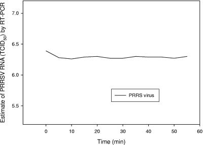
The concentration of PRRSV in the suspension fluid was measured during operation of the Collison nebulizer by sampling at intervals and then assaying the sample by quantitative RT-PCR (Fig. 2). The mean titer of PRRSV in the suspension fluid across sampling times was 1 × 106.33 TCID50. No difference was detected in the concentration of PRRSV when comparing the initial and subsequent samples across sampling times (P = 0.89). These results indicated that the concentration of aerosolized PRRSV was constant over the period of aerosolization.
FIG. 2.
Quantity of PRRSV in Collison nebulizer during operation.
(i) Sampling of aerosolized PRRSV.
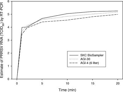
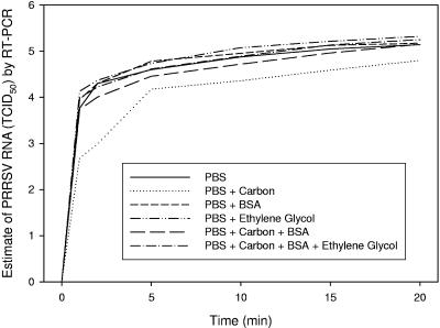
No interactions were detected between impinger, collection media, and time (P = 0.74) or between collection media and impinger (P = 0.20), but a statistically significant interaction existed both for collection media and time (P = 0.0002) and for impinger and time (P = 0.002). Therefore, the main effects of impinger and collection media were analyzed across time. The effect of impinger on PRRSV collection is presented in Fig. 3. The effect of collection media on PRRSV collection is presented in Fig. 4.
FIG. 3.
Collection of aerosolized PRRSV by impinger.
FIG. 4.
Collection of aerosolized PRRSV by collection medium treatment.
(ii) Collection media.
As estimated by quantitative RT-PCR, the mean titer (TCID50 equivalents) of PRRSV in collection medium treatments sampled seven times from 0 to 20 min was for PBS, 1 × 104.62; PBS and 1% activated carbon, 1 × 103.93; PBS and 0.5% BSA, 1 × 104.71; PBS and 20% ethylene glycol, 1 × 104.80; PBS, 0.5% BSA, and 1% activated carbon, 1 × 104.50; and PBS, 20% ethylene glycol, 0.5% BSA, and 1% activated carbon, 1 × 104.68. The analysis of variance of collection medium treatments indicated significant differences between collection medium treatments at all sampling points. Individual pair-wise comparisons of collection medium treatments at each sampling time indicated that a significantly lower quantity of PRRSV was detected in the PBS with activated carbon treatment at all sampling points. PBS with ethylene glycol had the greatest quantity of recovered PRRSV at all sampling points.
(iii) Impingers.
As estimated by quantitative RT-PCR, the mean titers of PRRSV collected across all time points were 1 × 104.67, 1 × 104.53, and 1 × 104.30 TCID50 for the SCK BioSampler, AGI-30, and AGI-4 (6 liter), respectively. The SCK BioSampler and AGI-30 impingers collected a significantly greater amount of PRRSV than did the AGI-4 (6 liter) impinger at sampling times of 10, 15, and 20 min. At sampling times of 15 and 20 min, the SKC BioSampler collected a significantly greater amount of PRRSV than did the AGI-30 or AGI-4 (6 liter) (Fig. 3).
(iv) Sampling time.
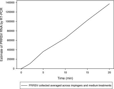
As measured by quantitative RT-PCR, the total quantity of PRRSV collected by impingers increased as sampling time increased. The mean titers by sampling time across impinger and media were 1 × 100, 1 × 103.66, 1 × 103.93, 1 × 104.57, 1 × 104.74, 1 × 104.96, and 1 × 105.17 TCID50 for 0, 1, 2, 5, 10, 15, and 20 min, respectively (Fig. 5).
FIG. 5.
Aerosolized PRRS virus collected over time.
DISCUSSION
The probability of detecting airborne viral pathogens is dependent on three primary factors: the concentration of airborne virus in the environment, the ability of the air-sampling system to recover airborne particles (collection efficiency), and the analytical sensitivity of the diagnostic assay(s) used to detect the target pathogen in the sample. In turn, each primary factor consists of component variables. For example, component variables recognized to affect the probability of recovery and detection of an airborne viral pathogen include collection medium composition, sampler type, sampling time, sampler flow rate (15, 25, 30, 42), particle size (30), rate of reentrainment (30), and the pathogen's affinity for the collection medium (31).
Collection media described for the recovery of airborne pathogens in air impingers are varied but include deionized water (30), buffered solutions (25), and mineral oil (31). Compounds added to collection medium to improve pathogen recovery include various proteins (38) and antifoaming agents (6, 23, 26, 37). However, the effect of collection medium composition on the recovery and detection of airborne viruses has not been quantified in direct comparisons.
Sampling times reported for the recovery of airborne pathogens using air impingers are variable, ranging from minutes to hours (12, 28, 29, 39). Likewise, flow rates described for the collection of airborne viruses are variable, ranging from 12.5 liters/min (14) to 450 liters/min (10). Counterintuitively, increased sampling time and/or flow rate does not necessarily result in an increase in the quantity of pathogen recovered. For example, foot-and-mouth disease virus was detected in 21 of 21 samples at a sampling time of 30 min but was detected in 0 of 4 samples at a sampling time of 4 h (33). Likewise, exotic Newcastle disease virus was detected in collection medium after 2 h of air sampling but not detected in collection medium after 8 h of air sampling (21). Possible explanations for a decrease in recovery and detection with longer sampling time include the destruction of infectious virus particles by shear forces during the process of impingement and/or reaerosolization (reentrainment) of captured particles. Lin et al. (31) hypothesized that the hydrophobic virus particles, i.e., enveloped viruses, may be reentrained more readily in liquid air impingers.
For the most part, the effects of component variables on the recovery of airborne viruses have not been systematically investigated. Our research examined specific component variables related to collection medium, sampling time, and impinger type in the context of the recovery and detection of PRRSV and SIV. Initially, various collection medium compounds were evaluated for their effects on the detection of infectious PRRSV and SIV. Subsequently, medium composition, impinger type, and sampling time were analyzed to optimize the recovery of aerosolized PRRSV.
In experiment 1, antifoams, activated carbons, protectants, and ethylene glycol were evaluated for direct effects on the infectivity of PRRSV and SIV, on the viability of continuous cell lines used for the isolation of these viruses, and on the performance of RT-PCR assays. Each compound tested was selected for a specific property related to the collection and/or detection of PRRSV and/or SIV: (i) antifoams are necessary to eliminate the excessive foaming that occurs during impingement when the collection medium contains proteins or carbohydrates (26, 37); (ii) activated carbon was evaluated for its potential to reduce reentrainment in impingers by adsorbing viral particles (32, 34, 36, 41); (iii) bovine serum albumin, mucin, and gelatin have been shown to reduce the rate of virus inactivation and preserve virus infectivity during the process of impingement or aerosolization (35, 38); and (iv) ethylene glycol was evaluated because it offered the possibility of collecting air samples at temperatures below 0°C.
The results of the first experiment showed that the addition of specific compounds to collection media can affect virus detection, i.e., the viability of continuous cell lines, virus infectivity, and/or performance of RT-PCR. These effects were not necessarily uniform within a group of compounds. For example, statistically significant differences were found among antifoams in their effect on continuous cell lines, with some antifoams exhibiting no effect on cells. Cytotoxic effects were not necessarily reflected in antiviral effects. Thus, although cytotoxic to cells, antifoams, protectants, and ethylene glycol had no effect on virus infectivity. The detection of PRRSV and SIV by RT-PCR was affected by the addition of protectants and activated carbon, but not antifoams or ethylene glycol. Overall, the variable effects of collection medium compounds on the detection of PRRSV and SIV demonstrated the need to examine each compound in the context of the compound's intended use in the air-sampling system and the pathogen of interest.
Questions related to the optimization of collection media remain. In particular, additional research on the use of activated carbon or other sorbents in collection medium is warranted. In this study, the addition of a peat-based activated carbon to collection medium significantly reduced the quantity of PRRSV and SIV detected by RT-PCR. Likewise, the addition of wood-based activated carbon reduced the quantity of PRRSV detected in impinger collection medium at all time points by RT-PCR. Activated carbon is known to adsorb viruses through nonspecific binding to viral surface proteins (7), but its effect on RT-PCR performance had not been described previously. The results in experiments 1 and 2 suggested two possibilities: activated carbons inhibited RT-PCR or virus was adsorbed to carbon but was rendered unavailable to RT-PCR assays under the conditions described in this experiment. If the latter is the case, then the possibility remains that activated carbons or other sorbents could increase collection efficiency by improving virus capture and/or reducing reentrainment by adsorption.
In experiment 2, the medium compounds selected in experiment 1 were evaluated in the context of recovery of aerosolized PRRSV in each of three impingers (AGI-30, AGI-4 [6 liter], and SKC BioSampler). The objective of experiment 2 was to optimize the quantity of virus recovered by manipulating the component variables of collection media, impinger model, and sampling time. To accomplish this, a method was introduced whereby direct comparisons among up to six independent treatments were possible. The validity of the comparisons was based upon the fact that all treatments sampled the same cloud of aerosolized virus. The results from the second experiment showed that the component variables of collection media, impinger model, and sampling time affected collection efficiency of aerosolized PRRSV. Specifically, the effects of impinger model and collection medium on recovery of PRRSV were consistent across sampling times. For example, the SKC BioSampler and AGI-30 impinger models recovered greater quantities of aerosolized PRRSV than the AGI-4 (6 liter) did at all sampling points. Likewise, PBS with ethylene glycol collection medium recovered the greatest quantity of PRRSV at each sampling time. As in experiment 1, the effects of collection media, impinger model, and sampling time on the recovery of PRRSV demonstrated the necessity to examine the effects of individual air-sampling variables on recovery of specific target pathogens.
Several points regarding these experiments are worth noting. First, the experiments focused on optimizing the recovery and detection of viral pathogens in aerosols. In general, this is an “underexplored” area of research, despite the commonly expressed fears of airborne spread of infectious agents. Second, this study described a method for direct comparisons of up to six treatments on total viral recovery. Although this simple method has not previously been described in the literature, it offers distinct advantages in terms of experimental efficiency and statistical validity. Third, it was demonstrated that a 20% solution of ethylene glycol collected the greatest quantity of aerosolized PRRSV, which suggests possible applications in sampling at temperatures below the freezing point. A review of the literature found no previous reports of the use of ethylene glycol in aerosol collection medium.
It should be noted that there are currently no standard methods for the recovery and detection of specific pathogens in aerosols. In the absence of standards, air-sampling protocols must be optimized and validated for each target pathogen. Results of the recovery and detection of the presence or absence of specific aerosolized pathogens made using unvalidated systems should be interpreted cautiously.
Acknowledgments
This project was funded in part by Check-Off dollars through the National Pork Board.
We are grateful to the virology staff and Karen Harmon of the Veterinary Diagnostic Laboratory, College of Veterinary Medicine, Iowa State University, for excellent technical assistance.
REFERENCES
- 1.Alexandersen, S., and A. I. Donaldson. 2002. Further studies to quantify the dose of natural aerosols of foot-and-mouth disease virus for pigs. Epidemiol. Infect. 128:313-323. [DOI] [PMC free article] [PubMed] [Google Scholar]
- 2.Babich, H., and E. Borenfreund. 1992. Neutral red assay for toxicology in vitro. Appl. Environ. Microbiol. 57:2101-2103. [DOI] [PMC free article] [PubMed] [Google Scholar]
- 3.Bayeva, L. Y., G. A. Shirman, V. A. Ginevskaya, Z. Kucharska, L. Dotlacilova, L. Danes, V. A. Kazantseva, and S. G. Drozdov. 1990. Using different sorbents for the concentration of enteroviruses. J. Hyg. Epidemiol. Microbiol. Immunol. 34:199-205. [PubMed] [Google Scholar]
- 4.Reference deleted.
- 5.Cage, B. R., K. Schreiber, C. Barnes, and J. Portnoy. 1996. Evaluation of four bioaerosol samplers in the outdoor environment. Ann. Allergy Asthma Immunol. 77:401-406. [DOI] [PubMed] [Google Scholar]
- 6.Chinivasagam, H. N., and P. J. Blackall. 2005. Investigation and application of methods for enumerating heterotrophs and Escherichia coli in the air within piggery sheds. J. Appl. Microbiol. 98:1137-1145. [DOI] [PubMed] [Google Scholar]
- 7.Clark, K. J., A. B. Sarr, P. G. Grant, T. D. Phillips, and G. N. Woode. 1998. In vitro studies on the use of clay, clay minerals and charcoal to adsorb bovine rotavirus and bovine coronavirus. Vet. Microbiol. 63:137-146. [DOI] [PMC free article] [PubMed] [Google Scholar]
- 8.Clavijo, A., D. B. Tresnan, R. Jolie, and E. M. Zhou. 2002. Comparison of embryonated chicken eggs with MDCK cell culture for the isolation of swine influenza virus. Can. J. Vet. Res. 66:117-121. [PMC free article] [PubMed] [Google Scholar]
- 9.Cookson, J. T., Jr. 1969. Mechanism of virus absorption on activated carbon. J. Am. Water Works Assoc. 61:52-56. [Google Scholar]
- 10.Dee, S. A., J. Deen, L. Jacobson, K. D. Rossow, C. Mahlum, and C. Pijoan. 2005. Laboratory model to evaluate the role of aerosols in the transport of porcine reproductive and respiratory syndrome virus. Vet. Rec. 16:501-504. [DOI] [PubMed] [Google Scholar]
- 11.De Jong, J. C., M. Harmsen, T. Trouwborst, and K. C. Winkler. 1974. Inactivation of encephalomyocarditis virus in aerosols: fate of virus protein and ribonucleic acid. Appl. Microbiol. 27:59-65. [DOI] [PMC free article] [PubMed] [Google Scholar]
- 12.Donaldson, A. I., C. F. Gibson, R. Oliver, C. Hamblin, and R. P. Kitching. 1987. Infection of cattle by airborne foot-and-mouth disease virus: minimal doses with O1 and SAT 2 strains. Res. Vet. Sci. 43:339-346. [PubMed] [Google Scholar]
- 13.Donaldson, A. I., N. P. Ferris, and J. Gloster. 1982. Air sampling of pigs infected with foot-and-mouth disease virus: comparison of Litton and cyclone samplers. Res. Vet. Sci. 33:384-385. [PubMed] [Google Scholar]
- 14.Elazhary, M. A., and J. B. Derbyshire. 1979. Aerosol stability of bovine parainfluenza type 3 virus. Can. J. Comp. Med. 43:295-304. [PMC free article] [PubMed] [Google Scholar]
- 15.Elazhary, M. A., and J. B. Derbyshire. 1979. Effect of temperature, relative humidity and medium on the aerosol stability of infectious bovine rhinotracheitis virus. Can. J. Comp. Med. 43:158-167. [PMC free article] [PubMed] [Google Scholar]
- 16.Gibson, C. F., and A. I. Donaldson. 1986. Exposure of sheep to natural aerosols of foot-and-mouth disease virus. Res. Vet. Sci. 41:45-49. [PubMed] [Google Scholar]
- 17.Gillespie, R. R., M. A. Hill, C. L. Kanitz, K. E. Knox, L. K. Clark, and J. P. Robinson. 2000. Infection of pigs by Aujeszky's disease virus via the breath of intranasally inoculated pigs. Res. Vet. Sci. 68:217-222. [DOI] [PubMed] [Google Scholar]
- 18.Grinshpun, S. A., K. Willeke, V. Ulevicius, J. Donnelly, X. Lin, and G. Mainelis. 1996. Collection of airborne microorganisms: advantages and disadvantages of different methods. J. Aerosol Sci. 27:S247-S248. [Google Scholar]
- 19.Harstad, J. B. 1965. Sampling submicron T1 bacteriophage aerosols. Appl. Microbiol. 13:899-908. [DOI] [PMC free article] [PubMed] [Google Scholar]
- 20.Hatch, M. T., and J. C. Warren. 1969. Enhanced recovery of airborne T3 coliphage and Pasteurella pestis bacteriophage by means of a presampling humidification technique. Appl. Microbiol. 17:685-689. [DOI] [PMC free article] [PubMed] [Google Scholar]
- 21.Hietala, S. K., P. J. Hullinger, A. A. Ardans, H. Kinde, M. McFarland, E. Skowronski, P. McCready, B. M. Schikora, L. M. Shih, and L. Lee. 2003. Air-sampling for the detection of exotic Newcastle disease virus in poultry houses, p. 72. Proceedings of the 46th Annual Conference of the American Association of Veterinary Laboratory Diagnosticians, San Diego, Calif.
- 22.Hubert, J. J. 1992. Bioassays, p. 73-76, 3rd ed. Kendall/Hunt Publishing Co., Dubuque, Iowa.
- 23.Ijaz, M. K., S. A. Sattar, C. M. Johnson-Lussenburg, V. S. Springthorpe, and R. C. Nair. 1985. Effect of relative humidity, atmospheric temperature, and suspending medium on the airborne survival of human rotavirus. Can. J. Microbiol. 31:681-685. [DOI] [PubMed] [Google Scholar]
- 24.Jensen, P. A., W. F. Todd, G. N. Davis, and P. V. Scarpino. 1992. Evaluation of eight bioaerosol samplers challenged with aerosols of free bacteria. Am. Ind. Hyg. Assoc. J. 53:660-667. [DOI] [PubMed] [Google Scholar]
- 25.Juozaitis, A., K. Willeke, S. A. Grinshpun, and J. Donnelly. 1994. Impaction onto a glass slide or agar versus impingement into a liquid for the collection and recovery of airborne microorganisms. Appl. Environ. Microbiol. 60:861-870. [DOI] [PMC free article] [PubMed] [Google Scholar]
- 26.Karim, Y. G., M. K. Ijaz, S. A. Sattar, and C. M. Johnson-Lussenburg. 1985. Effect of relative humidity on the airborne survival of rhinovirus-14. Can. J. Microbiol. 31:1058-1061. [DOI] [PubMed] [Google Scholar]
- 27.Kim, H. S., J. Kwang, I. J. Yoon, H. S. Joo, and M. L. Frey. 1993. Enhanced replication of porcine reproductive and respiratory syndrome (PRRS) virus in a homogenous subpopulation of MA-104 cells. Arch. Virol. 133:477-483. [DOI] [PubMed] [Google Scholar]
- 28.Lin, W. H., and C. S. Li. 1998. The effect of sampling time and flow rates on the bioefficiency of three fungal spore sampling methods. Aerosol Sci. Technol. 28:511-522. [Google Scholar]
- 29.Lin, W. H., and C. S. Li. 1999. Collection efficiency and culturability of impingement into a liquid for bioaerosols of fungal spores and yeast cells. Aerosol Sci. Technol. 30:109-118. [Google Scholar]
- 30.Lin, X., K. Willeke, V. Ulevicius, and S. A. Grinshpun. 1997. Effect of sampling time on the collection efficiency of the all-glass impingers. Am. Ind. Hyg. Assoc. J. 58:480-488. [Google Scholar]
- 31.Lin, X., T. Reponen, K. Willeke, Z. Wang, S. Grinshpun, and M. Trunov. 2000. Survival of airborne microorganisms during swirling aerosol collection. Aerosol Sci. Technol. 32:184-196. [Google Scholar]
- 32.Lipson, S. M., and G. Stotzky. 1985. Specificity of virus adsorption to clay minerals. Can. J. Microbiol. 31:50-53. [DOI] [PubMed] [Google Scholar]
- 33.Medaglia, S. P. 2001. SpinCon based air sampler foot and mouth disease virus test report, p. 14. Lares Corporation, Cambridge, Minn.
- 34.Moore, B. E., B. P. Sagik, and C. A. Sorber. 1979. Procedure for the recovery of airborne human enteric viruses during spray irrigation of treated wastewater. Appl. Environ. Microbiol. 38:688-693. [DOI] [PMC free article] [PubMed] [Google Scholar]
- 35.Sattar, S. A., Y. G. Karim, V. S. Springthorpe, and C. M. Johnson-Lussenburg. 1987. Survival of human rhinovirus type 14 dried onto nonporous inanimate surfaces: effect of relative humidity and suspending medium. Can. J. Microbiol. 33:802-806. [DOI] [PubMed] [Google Scholar]
- 36.Schaub, S. A., and B. P. Sagik. 1975. Association of enteroviruses with natural and artificially introduced colloidal solids in water and infectivity of solids-associated virions. Appl. Microbiol. 30:212-222. [DOI] [PMC free article] [PubMed] [Google Scholar]
- 37.Schoenbaum, M. A., J. J. Zimmerman, G. W. Beran, and D. P. Murphy. 1990. Survival of pseudorabies virus in aerosol. Am. J. Vet. Res. 51:331-333. [PubMed] [Google Scholar]
- 38.Stolze, B., and O. R. Kaaden. 1989. Efficient medium for impingement and storage of enveloped viruses. Zentbl. Vetmed. 36:161-167. [DOI] [PubMed] [Google Scholar]
- 39.Terzieva, S., J. Donnelly, V. Ulevicius, S. A. Grinshpun, K. Willeke, G. N. Stelma, and K. P. Brenner. 1996. Comparison of methods for detection and enumeration of airborne microorganisms collected by liquid impingement. Appl. Environ. Microbiol. 62:2264-2272. [DOI] [PMC free article] [PubMed] [Google Scholar]
- 40.Tseng, C. C., and C. S. Li. 2004. Collection efficiencies of aerosol samplers for virus-containing aerosols. J. Aerosol Sci. 36:593-607. [DOI] [PMC free article] [PubMed] [Google Scholar]
- 41.Turk, C. A., B. E. Moore, B. P. Sagik, and C. A. Sorber. 1980. Recovery of indigenous viruses from wastewater sludges, using a bentonite concentration procedure. Appl. Environ. Microbiol. 40:423-425. [DOI] [PMC free article] [PubMed] [Google Scholar]
- 42.Willeke, K., S. Grinshpun, C. W. Chang, A. Juozaitis, F. Liebhaber, A Nevalainen, and M. Thompson. 1992. Inlet sampling efficiency of bioaerosol samplers. J. Aerosol Sci. 23:S651-S654. [Google Scholar]


