Abstract
Bovine viral diarrhea virus (BVDV) membrane-anchored type I glycoprotein E2 is an ∼53-kDa immunodominant glycoprotein inducing the production of neutralizing antibodies in the animal host after natural infection or following immunization with live or killed vaccines. The E2 coding region lacking the transmembrane domain was constructed in a soluble secreted form (secE2) and expressed in the medium of a transiently transfected human cell line. The crude conditioned medium containing secE2 can be potentially employed to develop an enzyme-linked immunosorbent assay antigen for the diagnosis of BVDV infection or for vaccine purposes.
Pestiviruses are classified as a separate genus within the family Flaviviridae, which also includes flaviviruses and the hepatitis C virus group (12). Currently three pestivirus species are recognized, namely, bovine viral diarrhea virus (BVDV), classical swine fever virus, and border disease virus of sheep. The genomes of pestiviruses are positive-stranded RNAs, usually of about 12,300 nucleotides, which encode polyproteins of about 4,000 amino acids (3). Entire or partial genomic sequences of numerous BVDV, classical swine fever virus, and border disease virus isolates have been determined (1, 2), and their comparison demonstrated a high degree of sequence conservation among pestiviruses. The virion of pestiviruses consists, together with the RNA, of four structural proteins, the nucleocapsid C protein and the envelope glycoproteins Erns, E1, and E2 (11). Currently, 11 pestiviral proteins have been identified as products of polyprotein processing, which occurs co- and posttranslationally, due to the activity of viral and host cell proteases. In the hypothetical polyprotein, the proteins are arranged in the order Npro/C/Erns/E1/E2/NS2/NS3/NS4A/NS4B/NS5A/NS5B (10). The E2 protein of the BVDV NADL strain consists of about 370 amino acids and has a calculated molecular mass of 41 kDa. The N terminus of BVDV E2 is formed by Arg-690, and the C terminus is located around amino acid 1063. The C terminus of E2 includes approximately 30 amino acids, which could function as a transmembrane anchor for E2, and has a translocation signal for the downstream protein. Full-length E2 remains cell associated in virus-infected cells (9). In this work, we successfully expressed E2 in a secreted form in the medium of a mammalian cell line and the medium containing E2 could be directly employed for the development of a diagnostic enzyme-linked immunosorbent assay or immunization purposes.
Rational construct design.
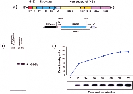
Starting from the assumption that the full length of E2 remains cell associated, we decided to construct a plasmid encoding a secreted form lacking the putative transmembrane domain of E2 (secE2), with pSecTag2HygroA (Invitrogen) as a backbone, and containing the cytomegalovirus (CMV) promoter and an immunoglobulin kappa light chain (Igk) leader sequence specifying secretion of heterologous proteins. Total RNA from BVDV strain NADL-infected MDBK (Madin-Darby bovine kidney) cells was reverse transcribed using the T-Primed first-strand kit (Amersham Biosciences). Three microliters of reverse transcribed RNA were amplified with a primer pair including an HindIII restriction site in the 5′ end (underlined) (sense primer, 5′-CCC GA AGC TTG CAC TTG GAT TGC AAA CCT GAA TTC-3′) in frame with the Igk signal peptide of the plasmid backbone and an XhoI restriction site in the 3′ end (underlined) (antisense, 5′-CCC CGC TC GAG AGT GGA CTC AGC GAA GTA ATC CCG-3′) in frame with a polar nonstructured 14-amino-acid peptide of the plasmid backbone. This would increase the solubility and secretion of the protein due to its polar characteristics (Fig. 1a).
FIG. 1.
Overall strategy employed for cloning and expression of BVDV glycoprotein E2 as a soluble secreted form in a mammalian cell line. (a) Diagram (not to scale) showing the structure of the BVDV genome with regions coding for structural and nonstructural (NS) proteins. The genomic RNA containing a single open reading frame is shown (the gene order is 5′-Npro-Crns-E1-E2-P7-NS2-3-NS4A-NS4B-NS5A-NS5B-3′); the glycoprotein E2 has been cloned with primers (black bars A and B) spanning the E2 coding region but missing the area coding for the transmembrane domain (TM). The E2ΔTM (blue box) was subcloned into an expression vector driven by the human immediate-early CMV promoter (CMVprom, black box) in frame with the Igκ (pink box) signal peptide and 14 extra amino acids (14aa, white box) on the 5′ and 3′ ends, respectively. (b) Western immunoblot showing the expression of secE2 in the medium of psecE2-transfected cells but not in mock-pEGFP-C1-transfected ones. (c) Time course of secE2 expression in the medium of psecE2-transfected cells at different times posttransfection (0, 12, 24, 30, 38, 48, 60, and 72 h) analyzed by Western immunoblotting and quantified through image analysis. The experiment was repeated three times with identical results.
secE2 is secreted in the culture medium of mammalian cells.
Next, we assessed the translation and secretion of secE2 in the culture medium of transiently transfected HEK 293T cells by means of Western blotting. Thirty micrograms of psecE2 was electroporated in HEK 293T cells from a confluent 75-cm2 flask, and a plasmid expressing green fluorescent protein under the control of the CMV promoter (pEGFP-C1) was identically transfected as a negative control and to monitor the efficiency of transfection through the number of green cells. Following electroporation, cells were recovered in a new 75-cm2 flask with 20 ml of Dulbecco's modified Eagle medium and 10% fetal bovine serum to allow cells to attach. At 8 h posttransfection, the medium was removed; cells were gently washed with 10 ml of serum-free F-12 (Sigma) medium to eliminate any trace of serum protein and incubated for 48 h with 10 ml of fresh serum-free F-12 medium. Thus, the only proteins present in the medium were cell-secreted proteins. Ten- and 5-μl aliquots of conditioned medium from psecE2 and 20 μl of mock-pEGFP-C1-transfected cells were loaded on a 12% sodium dodecyl sulfate-polyacrylamide gel and transferred to a nylon membrane by electroblotting (6). secE2 was probed with monoclonal anti-E2 antibody (VRMD, Pullman, WA), detected with horseradish peroxidase-labeled anti-mouse IgG antibody (Sigma), and visualized by chemiluminescence (ECL kit; Pierce, Rockford, IL). Correct protein expression of E2 was confirmed by the presence of a protein with a molecular mass of ∼53 kDa, which was absent in the pEGFP-C1-transfected negative control (Fig. 1b). These findings indicated that secE2 was translated and correctly processed by the cellular export apparatus, resulting in its secretion into the medium of cultured cells. To assess the maximum expression level of secE2, a time course was performed, sampling the medium at 12, 24, 30, 38, 48, 60, and 72 h posttransfection. The maximum expression level was observed between 60 and 72 h posttransfection (Fig. 1c and d).
secE2 retains native antigenic properties.
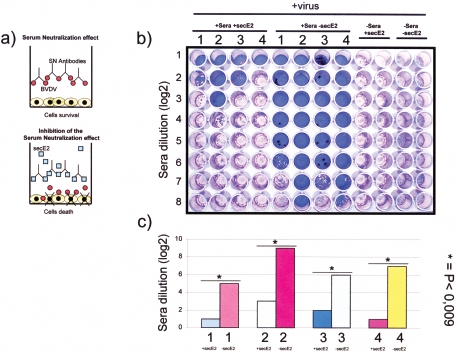
The E2 protein plays a major role in virus attachment and entry of BVDV (5). Furthermore, BVDV E2 is important for the induction of neutralizing antibodies (4) and protection against BVDV challenge in cattle (7, 8). Therefore, we explored secE2 antigenic properties by performing a serum neutralization inhibition test, based on the capability of secE2 to reduce or block the neutralizing activity of neutralizing antibodies against BVDV (Fig. 2a). Four heat-inactivated bovine sera that were positive for virus-neutralizing antibodies against BVDV were selected. Twenty-five microliters of each serum sample was added to the first row of 96-well plates. Twenty-five microliters of Dulbecco's modified Eagle medium was added to each well, and for each serum tested, serial twofold dilutions were made. An equal volume (25 μl) of conditioned medium containing secE2 was added to each well. Positive and negative controls, as well as virus controls, were included (Fig. 2b). After a 1-h incubation at room temperature, 25 μl of virus suspension containing 100 TCID50 (50% tissue cell infectious doses) of BVDV NADL was added to each well. After a 1-h incubation at 37°C, 50 μl of an MDBK cell suspension was added to each well and the plates were incubated for 5 days at 37°C. The expression of viral infectivity and serum-neutralizing activity through the cytopathic effect was detected by microscopy and crystal violet staining of the cell monolayer. The neutralization antibody titers were expressed as the reciprocal of the final dilution of serum that completely inhibited viral infectivity (Fig. 2c). A strong reduction of the neutralizing activity of the sera was obtained by the conditioned medium containing secE2 (Fig. 2b and c). No neutralizing inhibition was observed in the control assay performed with conditioned medium in the absence of secE2. These findings allowed us to show that secE2 maintains the native antigenic properties of BVDV E2. The aim of this work was to propose and describe a fast and easy method to produce a BVDV antigen in secreted and soluble form in the serum-free medium of transiently transfected mammalian cells, readily exploitable for diagnostic or vaccine purposes.
FIG. 2.
Inhibition of serum neutralization (SN) to assay the authentic antigenic characteristics of secE2. (a) Diagram showing the principle of the assay, where BVDV neutralizing antibodies preincubated with medium containing secE2 are blocked, allowing the virus to destroy the cell monolayer. (b) The quantitative assay performed in a 96-multiwell plate where 4 sera (1, 2, 3, and 4) containing neutralizing antibody against BVDV were tested at different dilutions (log2 of the serum dilution) in the presence of secE2 (+Sera +secE2) and in the absence of secE2 (+Sera −secE2). Control virus was established in the absence of sera and presence of secE2 (−Sera +secE2) and in the absence of sera and secE2 (−Sera −secE2). Crystal violet staining allows macroscopic evaluation of the integrity (blue wells) or the destruction (clear violet wells) of the cell monolayer. (c) Bar graph showing the quantification of serum neutralization made on the basis of three different experiments (P < 0.0009). Results are expressed as the reciprocal of the highest dilution of the serum that inhibited the development of virus-induced cytopathic effect in cell culture.
Acknowledgments
We thank Laura Kramer for correcting the English in the manuscript.
The cost of this work was supported by Italian Minister of University and Scientific Research (Italian National Grant MIUR, PRIN 2005, 2005078885).
REFERENCES
- 1.Becher, P., A. D. Shannon, N. Tautz, and H.-J. Thiel. 1994. Molecular characterization of border disease virus, a pestivirus from sheep. Virology 198:542-551. [DOI] [PubMed] [Google Scholar]
- 2.Becher, P., M. Konig, D. Paton, and H.-J. Thiel. 1995. Further characterization of border disease virus isolates: evidence for the presence of more than three species within the genus pestivirus. Virology 209:200-206. [DOI] [PubMed] [Google Scholar]
- 3.Bolin, S. R., and J. F. Ridpath. 1996. Glycoprotein E2 of bovine viral diarrhea virus expressed in insect cells provides calves limited protection from systemic infection and disease. Arch. Virol. 141:1463-1477. [DOI] [PubMed] [Google Scholar]
- 4.Bolin, S. R. 1993. Immunogens of bovine viral diarrhea virus. Vet. Microbiol. 37:263-271. [DOI] [PubMed] [Google Scholar]
- 5.Donis, R. O. 1995. Molecular biology of bovine viral diarrhea virus and its interactions with the host. Vet. Clin. North. Am. Food Anim. Pract. 11:393-423. [DOI] [PMC free article] [PubMed] [Google Scholar]
- 6.Donofrio, G., F. L. Heppner, M. Polymenidou, C. Musahl, and A. Aguzzi. 2005. Paracrine inhibition of prion propagation by anti-PrP single-chain Fv miniantibodies. J. Virol. 79:8330-8338. [DOI] [PMC free article] [PubMed] [Google Scholar]
- 7.Harpin, S., D. J. Hurley, M. Mbikay, B. Talbot, and Y. Elazhary. 1999. Vaccination of cattle with a DNA plasmid encoding the bovine viral diarrhoea virus major glycoprotein E2. J. Gen. Virol. 80:3137-3144. [DOI] [PubMed] [Google Scholar]
- 8.Meyers, G., T. Rumenapf, and H.-J. Thiel. 1989. Molecular cloning and nucleotide sequence of the genome of hog cholera virus. Virology 171:555-567. [DOI] [PubMed] [Google Scholar]
- 9.Rumenapf, T., G. Unger, J. H. Strauss, and H.-J. Thiel. 1993. Processing of the envelope glycoproteins of pestiviruses. J. Virol. 67:3288-3294. [DOI] [PMC free article] [PubMed] [Google Scholar]
- 10.Thiel, H.-J., P. G. W. Plagemann, and V. Moennig. 1996. Pestiviruses, p. 1059-1073. In B. N. Fields, D. M. Knipe, P. M. Howley, et al. (ed.), Virology, 3rd ed. Lippincott-Raven Publishers, Philadelphia, Pa.
- 11.Thiel, H.-J., R. Stark, E. Weiland, T. Rumenapf, and G. Meyers. 1991. Hog cholera virus: molecular composition of virions from a pestivirus. J. Virol. 65:4705-4712. [DOI] [PMC free article] [PubMed] [Google Scholar]
- 12.Wengler, G., D. W. Bradley, M. S. Collett, F. X. Heinz, R. W. Schlesinger, and J. H. Strauss. 1995. Flaviviridae, p. 415-427. In F. A. Murphy, C. M. Fauquet, D. H. L. Bishop, S. A. Ghabrial, A. W. Jarvis, G. P. Martelli, M. A. Mayo, and M. D. Summers (ed.), Virus taxonomy. Sixth report of the International Committee on Taxonomy of Viruses. Springer-Verlag, New York, N.Y.


