Abstract
The tear film is a complex mixture of secreted fluid, ions, proteins, glycoproteins, and lipids that lubricates and protects the ocular surface. Recently, several antimicrobial peptides have been described in the tear fluid. In this study, we describe the presence of the large secreted glycoprotein gp340 in the tear film. Western blot analysis showed that gp340 is abundant in secreted tears and in the lacrimal glands. Lesser amounts of gp340 were detected in the cornea and conjunctiva. Consistent with Western blot data, reverse transcription-PCR and real-time quantitative PCR showed that gp340 transcripts were abundant in lacrimal gland tissue and were also present in the cornea and conjunctiva. Immunohistochemistry localized gp340 to the acinar cells of the lacrimal gland and the deeper layers of the conjunctival epithelium. gp340 was not detected in conjunctival goblet cells. In the cornea, gp340 was present only in a peripheral band of basal epithelial cells, suggesting that gp340 may play a role in the cycle of corneal epithelial renewal. To determine if tear film gp340 may function as a bacterial agglutinin as it does in saliva, tears were incubated with streptococcal cells and the formation of bacterial aggregates was monitored. Addition of tears to late-exponential-phase Streptococcus mutans cells resulted in time- and dose-dependent aggregation of the bacteria. Furthermore, Western blot analysis confirmed the presence of cell-associated gp340 in isolated bacterial aggregates. The ocular pathogen Staphylococcus aureus, but not Pseudomonas aeruginosa, also aggregated when incubated with tears. These results suggest that gp340 is a normal component of the tear film and that the glycoprotein may function as a bacterial agglutinin.
The normal human tear film lubricates and protects the anterior surface of the eye. Its components include fluids and electrolytes, a striking variety of proteins, glycoproteins, and several lipid species. Each of these components functions to protect and preserve the smooth optical surface of the cornea. Conversely, a deficit of any specific component can arise in conjunction with ocular-surface disease. The ocular surface is constantly exposed to environmental pathogens that are prevented from colonizing the mucosal surface by the tear film antimicrobial defenses and the sweeping actions of the eyelids. Known antimicrobial components of the tear film include, defensins, MUC7, histatin, lysozyme, lactoperoxidase, transferrin, and surfactant protein D (SPD) (8, 13, 20, 22, 25).
gp340 is a large secreted glycoprotein of the scavenger receptor cysteine-rich family. gp340 is a multifunctional glycoprotein that has been implicated in pathogen clearance, regulation of epithelial growth, and linkage of the innate and adaptive immune systems (18). gp340 is heavily O and N glycosylated and is encoded by the “deleted in malignant brain tumor 1” (DMBT1) gene (18). In the rabbit, the orthologous protein, hensin, is involved in establishing polarity of cells of the kidney ducts (26). Murine orthologues include crp-ductin and eberin (18). gp340 strongly resembles secretory mucins, such as MUC5AC, in that it contains highly O-glycosylated tandem repeat regions, and it has been referred to as “muclin,” a mucin-like protein. Recent studies have shown that bovine pancreatic mucin is identical to muclin (4, 14). gp340 binds to a number of microbial pathogens, including streptococci, Haemophilus influenzae, and human immunodeficiency virus (5, 6). Saliva is known to agglutinate oral pathogens, and the salivary agglutinin (Sag) is an isoform of gp340 (5, 6). gp340 also binds to SPD at specific sites, and the two proteins apparently act synergistically to provide an innate defense for mucosal tissues (7). An earlier study by Schulz et al. found gp340/muclin in human tears following reduction and alkylation, separation by electrophoresis, and identification by matrix-assisted laser desorption ionization-time of flight analysis (24).
We now confirm and extend those results by demonstrating the presence and quantity of gp340 transcripts in ocular surface tissues, by comparing the abundancs of gp340 in tears and saliva, by examining the involvement of gp340 in aggregation of gram-positive and gram-negative bacteria, and by localizing the glycoprotein in the cornea conjunctiva and lacrimal gland. We demonstrate that gp340 is present in tears, is synthesized by ocular-surface epithelia, and promotes pathogen aggregation in a manner that resembles that of the salivary Sag.
MATERIALS AND METHODS
Collection of tears and conjunctival cells and tissues.
The Institutional Review Board, University of Louisville, approved human study protocols. Informed consent was obtained from healthy donors prior to collection of tears (three donors), saliva (one donor), and brush cytology samples (four donors). Tears (∼50 μl) were collected with a sterile pipette tip from a healthy individual for use in bacterial-aggregation assays. Saliva was collected from the sublingual space. Sterile swabs were used to gently remove conjunctival epithelial cells from four healthy adults (two male, two female) (27). Human cornea, conjunctiva, and lacrimal gland tissues (four donors each) were obtained from the Kentucky Lions Eye Bank (Louisville, KY) and the National Disease Research Interchange (Philadelphia, PA). For studies of protein and RNA, the peripheral cornea was trimmed away to prevent inclusion of scleral and/or conjunctival tissue.
RNA isolation and reverse transcription (RT)-PCR.
Total RNA was isolated (RNeasy kit; QIAGEN, Valencia, CA) according to the manufacturer's protocols. The quantity of RNA was measured by absorption at 260 nm, and RNA quality was assessed by electrophoresis on 1% agarose gels stained with ethidium bromide. To eliminate contamination by genomic DNA, total RNA was treated with DNase I (Sigma, St. Louis, MO). Pooled lung and salivary gland RNAs were obtained from BD Bioscience (San Diego, CA) and Clontech (Mountain View, CA), respectively. Reverse transcription of total RNA (SuperscriptII First Strand Synthesis System; InVitrogen, Chicago, IL) and the subsequent PCR (Amplitaq Gold; Roche, Branchburg, NJ) were completed according to the manufacturers' protocols. The gp340 primer pair, designed with Primer 3 software (Whitehead Institute for Biomedical Research, Cambridge, MA), consisted of hgp340L, TGC TCT GTC TGC CAA ATC AC, and hgp340R, GTC ATT GTC TGC CTG CTT GA (Integrated DNA Technologies, Coralville, IA) and amplified a 193-bp target near the 3′ end of the mRNA. PCR reagent concentrations included 2.5 mM MgCl2, 200 μM deoxynucleoside triphosphate, 0.625 μM primer mix, 10 ng of cDNA template, and 2.6 U/reaction Taq polymerase. Thermocycling conditions for gp340 included an initial 2-min denaturation at 94°C, followed by 35 cycles of denaturation at 94°C for 1 min, annealing at 51°C for 1 min, and extension at 72°C for 1 min. A final 5-min extension at 68°C completed the cycling. Total salivary gland and lung RNAs (Clontech, Palo Alto, CA) served as positive controls and β-2 microglobulin as the internal control (12, 13). Amplification products were separated on 1% agarose gels and visualized by ethidium bromide staining. The absence of contaminating genomic DNA was determined by using identical samples that had not been reverse transcribed. Purified PCR products were sequenced at the University of Louisville's Neurobiology DNA Core Facility.
Western blot analysis.
Antibody to gp340-specific peptide, Ac-CFGQGSGPIVLDDVR-amide coupled to keyhole limpet hemocyanin, was raised in a rabbit (New England Peptides, Gardner, MA) and used for detection of the glycoprotein in tears and tissues. Preliminary experiments showed that the antibody recognized only a high-molecular-weight protein and did not cross-react with the conjunctival mucin MUC5AC (11, 12). Preimmune serum did not react with gp340 or other tear film proteins.
Tissue to be used for Western blot analysis was homogenized in lysis buffer (10 mM Tris, pH 8.0, 1 mM EDTA, and a proteinase inhibitor cocktail) and stored at −80°C. Tissue, tear, and saliva samples were diluted in sodium dodecyl sulfate (SDS)-polyacrylamide gel electrophoresis sample buffer and used immediately in Western blot analysis or stored at −80°C. All samples and molecular weight standards were boiled for 5 min prior to being loaded onto 4 to 15% polyacrylamide gradient gels (Bio-Rad, Hercules, CA). The gels were run for 30 min at 50 V and 90 min at 100 V, washed in transfer buffer, and electroblotted onto nitrocellulose membranes for 1 h at 100 V. The blots were blocked overnight at 4°C with Tris-buffered saline (TBS) containing 3% milk and incubated for 2 h at room temperature with the anti-gp340 antibody at 1:1,000 or with the corresponding normal serum at the same dilution. Following three rinses, the blots were incubated with horseradish peroxidase (HRP)-conjugated rabbit anti-goat immunoglobulin G (1:2,000) for 1 h and developed using a chemiluminescent substrate (Dupont NEN, Boston, MA). Exposure times varied based on the strength of the gp340 signal.
Quantitative mRNA analysis by real-time RT-PCR.
The quality and quantity of conjunctival epithelial RNA collected from brush cytology samples were evaluated using the Agilent 2100 Bioanalyzer (Wilmington, DE). mRNA gene expression levels were determined after reverse transcription by real-time PCR using an Applied Biosystems PRISM 7300 Sequence Detection System (Foster City, CA). Total RNA was treated with DNase, and then cDNA was synthesized using random hexamers and the SuperScript first-strand synthesis kit (Invitrogen). The following primer-probe sets for real-time RT-PCR (Taqman) analysis were obtained as Assays on Demand reagents (Applied Biosystems): gp340 Hs00244827_m1 and 18S Hs99999901_s1.
Expression values were compared to those obtained for the endogenous control 18S rRNA, which we used rather than GAPDH (glyceraldehyde-3-phosphate dehydrogenase) or β-actin because of the variability of those two endogenous controls between tissues and among individuals (2, 17, 19). CT is defined as the threshold cycle at which the fluorescence produced by the cleavage of the Taqman fluorogenic probe rises above background level. Delta CT values (CT experimental minus CT 18S rRNA) were converted to a difference (n-fold) between experimental and 18S rRNA values and presented as [1/(n-fold difference)] × 10,000, so that stronger expression was represented by a higher value as previously described (19, 28). Each real-time RT-PCR assay was run in triplicate on four individual samples of conjunctival epithelial cDNA. Positive (salivary gland) and negative (muscle) mRNAs were purchased from BD Biosciences Clontech (Palo Alto, CA), reverse transcribed, and analyzed as described above.
Immunohistochemistry.
Normal human cornea and conjunctiva obtained from the Kentucky Lions Eye Bank and normal lacrimal gland from National Disease Research Interchange were fixed in freshly prepared 4% paraformaldehyde and delivered to the Ophthalmology Core Image Analysis Laboratory, where the tissues were embedded, cryosectioned (8 μm), and stored at −80°C prior to use. The sections were air dried, rehydrated in phosphate-buffered saline (PBS), and blocked with normal rabbit serum. Endogenous peroxidase was inactivated by incubation with 3% hydrogen peroxide for 5 min at room temperature. After being washed, the sections were incubated with rabbit anti-gp340 (1:100) and washed, and the bound antibody was detected using HRP-labeled anti-rabbit serum and 3,3′-diaminobenzidine according to the manufacturer's protocols (Vector, Burlingame, CA).
Aggregation assays and identification of gp340 in bacterial aggregates.
Aggregation assays were carried out essentially as previously described in the functional analysis of salivary gp340 (6). Overnight cultures of Streptococcus mutans KPSK2, Staphylococcus aureus ATCC 19095, or Pseudomonas aeruginosa PAO-1 were centrifuged at 5,000 × g for 10 min, and the cell pellets were washed twice and suspended in PBS to an optical density of approximately 6 at 675 nm. Tear samples were diluted up to 16-fold in PBS and incubated with the bacterial suspension (20 μl per reaction) at 37°C in a final reaction volume of 200 μl. Tear-mediated aggregation of bacteria was followed by measuring the optical density at 675 nm at 30-min intervals in a Wallace Victor3 microtiter plate reader. Control reactions that did not contain tear samples were also assayed to monitor the incidental settling of bacteria. For analysis of tear proteins adsorbed on aggregated bacteria, bacterial aggregates were carefully removed from the reaction mixtures, washed three times with PBS, and suspended in SDS gel buffer at 65°C. Insoluble material was collected by centrifugation at 14,000 × g, and the supernatant was analyzed for gp340 using the Western blot protocol described above. Staphylococcus aureus and Pseudomonas aeruginosa aggregation assays were performed in the same manner described above except that cultures were analyzed after a 90-min incubation in a 1:2 dilution of tears. Washed cells (2 μl) were also spotted directly onto nitrocellulose strips to visualize gp340 bound to intact bacteria. Filter strips were blocked with TBS-3% milk and reacted with anti-gp340 antibodies as described above. Duplicate blots were blocked with 2% milk and developed using the murine monoclonal antibody DF3 (Signet, Dedham, MA) to human MUC1 (20). Preliminary studies showed that neither the primary nor secondary antibodies used reacted with untreated bacteria.
RESULTS
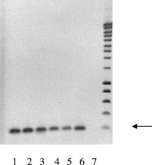
Ocular surface tissues, including cornea, conjunctiva, and lacrimal gland, were analyzed for the presence of mRNA encoding gp340 by RT-PCR. As shown in Fig. 1, positive reaction products, at 193 bp, were identified in all three mucosal tissues. An identical band was present in a sample of RNA derived from the salivary gland. The PCR product from the conjunctiva was cloned and sequenced and was found to be identical to gp340 (NCBI AF159456).
FIG. 1.
RT-PCR of cDNA from mucosal tissues. RNA samples from pooled lung and salivary gland and three ocular mucosal tissues derived from four individual donors were reverse transcribed, amplified, electrophoresed, and labeled with ethidium bromide. A 193-bp fragment was amplified from the cDNA of the lung (lane 1), two conjunctivae (lanes 2 and 3), the lacrimal gland (lane 4), the cornea (lane 5), and the salivary gland (lane 6). Lane 7 is a negative control lacking any target cDNA. The arrow indicates the 200-bp standard.
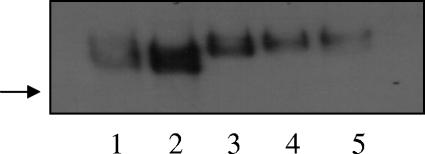
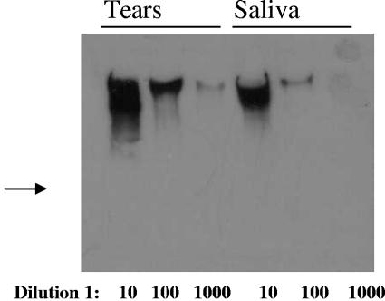
We examined normal tears for the presence of gp340 by Western blot analysis. As seen in Fig. 2, normal tears contain a high-molecular-mass protein that labels intensively with an antibody to gp340. gp340 is also abundant in the lacrimal gland and is detectable in the conjunctiva. The protein bands seen the tissue samples (lanes 1 and 2) are of lower molecular mass than those seen in tears. This may reflect the fact that the tissue samples were likely to include underglycosylated gp340 precursors, while the tear samples included only the mature glycoprotein. When tears are compared to saliva, identical bands are seen. Serial dilutions of human tears analyzed by Western blotting indicated that gp340 is more abundant in tears than in saliva (Fig. 3).
FIG. 2.
Samples were loaded onto a 4 to 12% SDS-polyacrylamide gel electrophoresis gel, electrophoresed, and transferred to nitrocellulose. Lane 1, 20 μg human conjunctival homogenate; lane 2, 20 ng human lacrimal gland homogenate; lanes 3 to 5, 20 nl human tears from three healthy adults. gp340 was detected using a specific antiserum. gp340 is present as a dispersed high-molecular-mass band in these samples. Tissue-derived gp340 is of lower molecular mass than the secreted form, probably due to incomplete glycosylation of the immature glycoprotein. The arrow indicates migration of a 250-kDa molecular mass marker.
FIG. 3.
gp340 in human tears and saliva. gp340 was detected in Western blots of serially diluted tears and saliva from a single donor; 20 μl of fluid, undiluted or diluted 1:10 or 1:100. gp340 is readily detectable at a 1:100 dilution in tears and therefore appears to be more abundant in tears than in saliva. The arrow shows the location of the 250-kDa molecular mass marker.
To determine the major source of ocular-surface gp340 mRNA, we used real-time quantitative PCR. The results, shown in Table 1, compare transcript abundances in cornea, conjunctiva, lacrimal gland, salivary gland, and muscle. Interestingly, gp340 transcripts were most abundant in the lacrimal gland and were expressed at a higher level than in the positive control tissue, salivary gland. The expression level in samples of whole conjunctiva was about sevenfold lower than that of the lacrimal gland. However, the level of gp340 in samples of conjunctival epithelium obtained by brush cytology was much lower than that of whole conjunctiva. Brush cytology samples primarily consist of the superficial layers of conjunctival epithelial cells. Therefore, these findings suggested that the major source of gp340 in whole conjunctiva must be cells in the deeper layers of the epithelium and/or stroma (see Immunohistochemistry above). Only a trace of gp340 mRNA expression was found in the central cornea. Muscle, a negative control tissue, expressed no detectable gp340 transcripts. Both gp340 mRNA and the glycoprotein itself are abundant in the human lacrimal gland, indicating that this gland is the major source of tear film gp340.
TABLE 1.
Expression of gp340 as determined by real-time RT-PCR
| Tissue | Valuea |
|---|---|
| Conjunctiva (whole) | 0.55 ± 0.31 |
| Conjunctival epithelium | 0.056 ± 0.032 |
| Cornea | 0.005 ± 0.0037 |
| Lacrimal gland | 3.55 ± 0.87 |
| Muscle | 0 |
| Salivary gland | 2.71 |
Taqman real-time RT-PCR results are expressed as [1/(n-fold difference)] × 10,000, as defined in Materials and Methods. Values for all ocular tissue samples are mean ± standard error for samples from four donors each. Muscle and salivary gland values were determined in triplicate on single pools from multiple donors; triplicate values varied by less than 4%.
We examined the cellular localization of ocular-surface gp340 by immunohistochemistry. Frozen sections were incubated with anti-gp340, and antibody-binding cells were visualized by indirect labeling with an HRP-labeled secondary antibody. Gp340 was localized throughout the conjunctival epithelium, with greater labeling in basal layers than in apical cells (Fig. 4a). In the lacrimal gland (Fig. 4b), only acinar cells were labeled with anti-gp340 with only background staining of vascular and connective tissues. Corneal gp340 was limited to a single layer of peripheral basal epithelial cells (Fig. 4c) and was absent in the central cornea (Fig. 4d). Incubation with preimmune serum resulted only in faint background labeling (not shown).
FIG. 4.
Normal human ocular mucosal tissues labeled with anti-gp340. (a) Normal human conjunctiva. Immunostaining with anti-gp340 shows localization in the basal layers of the conjunctiva and in isolated cells within the underlying loose connective tissue. The brace delineates the conjunctival epithelium, and the arrowheads indicate unlabeled goblet cells. (b) Normal human lacrimal gland. Immunostaining with anti-gp340 shows localization acini (circle), but not in connective tissue (*). (c) gp340-positive basal cells in the peripheral cornea (arrow). (d) Labeled cells are absent from the central cornea. Bar = 100 μM.
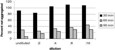
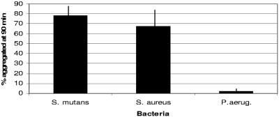
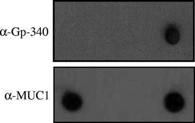
The abundance of tear fluid gp340 and the finding that the lacrimal gland, a recognized source of ocular-surface antimicrobials, appears to be a major source of this glycoprotein led us to examine the functional properties of the glycoprotein. The addition of tear fluid to S. mutans cells suspended in buffer resulted in time- and dose-dependent aggregation of the bacteria and concomitant reduction in nonaggregated cells (Fig. 5). Aggregated bacterial cells were collected by centrifugation, washed, and examined by Western blot analysis. Figure 6 shows that gp340 could be recovered from bacterial pellets and that the gp340 content of the pellets was directly proportional to the concentration of tears included in the incubation. These results suggest that gp340 in tears may function as a bacterial agglutinin, consistent with its reported activity in saliva. We also examined the participation of tear fluid gp340 in the agglutination of the ocular pathogens Pseudomonas aeruginosa and Staphylococcus aureus. Like S. mutans, S. aureus, but not P. aeruginosa, aggregated in tear fluid (Fig. 7). When intact bacterial cells were incubated with tear fluid and analyzed by dot blotting, S. aureus, but not P. aeruginosa, cells bound gp340 (Fig. 8). Interestingly, both bacterial species bound tear fluid MUC1 (Fig. 8).
FIG. 5.
Effect of tears on aggregation of S. mutans. Cells in log phase growth were incubated with the indicated dilutions of human tears for 30 to 90 min. The percentages of unaggregated cells were determined in triplicate wells. Tears caused dose- and time-dependent aggregation of S. mutans. By 90 min, undiluted tears resulted in aggregation of >90% of these bacteria.
FIG. 6.
Western blot of gp340 from bacterial aggregates. Cells were incubated with serially diluted tears for 90 min. The aggregated cells were centrifuged, washed, lysed, and analyzed by Western blotting. The amounts of gp340 in the aggregates (lanes 1 to 5) were compared to that in a 1:10 dilution of tears (lane 6).
FIG. 7.
Aggregation of ocular pathogens by tear fluid. Log phase bacterial cultures were incubated with tear fluid (1:2 dilution) for 90 min. The percentages of aggregated cells were determined in triplicate wells. The addition of tear fluid resulted in aggregation of S. mutans and S. aureus but negligible aggregation of P. aeruginosa. Means plus SEM are shown (n = 3).
FIG. 8.
Dot blot analysis of ocular pathogens following incubation with tear fluid. Cells were incubated with tear fluid for 90 min, centrifuged, washed, and resuspended in TBS. An aliquot was applied to nitrocellulose, and the blot was reacted with anti-gp340 (top) or the DF3 antibody to MUC1 (bottom). gp340 was associated with S. aureus (right lane), but not P. aeruginosa (left lane). The middle lane is blank. MUC1 associated with both bacterial species.
We conclude that gp340 is a normal glycoprotein component of the normal human tear fluid and that this mucin-like glycoprotein may participate in ocular-surface protection by agglutinating bacteria. The primary source of tear film gp340 appears to be the lacrimal gland, with lesser contributions from the conjunctiva and cornea.
DISCUSSION
Infection of the ocular surface can result in corneal ulceration and loss of sight. The blinking of the eyelids and the secretions of ocular cells and glands protect the visual surface of the eye from both environmental insults and pathogens. Protection of the ocular surface from pathogenic colonization clearly depends on the complex and coordinated functions of a number of tear film proteins. The current studies indicate that gp340 is one such protein and that it may function to aggregate opportunistic pathogens, thereby facilitating their removal to the nasal-lacrimal drainage system. The range of antimicrobial activity of gp340 is unknown. However, it is well known that gp340 interacts with oral streptococci, inducing the formation of aggregates that are subsequently cleared from the oral cavity (5, 6). Given the apparent similarity between salivary and lacrimal gp340 glycoproteins, we tested samples of tears and found that they, like saliva, induced aggregation of oral streptococci.
The major source of tear film gp340 is the lacrimal gland. Lacrimal gp340 apparently originates in acinar cells and is secreted into the tears. gp340 transcripts were also present in conjunctival mRNA, and the corresponding protein was detected in conjunctival samples. The levels, however, were low compared to those in the tears and lacrimal gland. One possible explanation is that conjunctival gp340 is largely soluble and leaches out of donor tissue stored in liquid medium. The histological appearance of conjunctival tissue labeled for gp340 to some extent supports this supposition, as gp340 is more abundant in basal than in apical epithelium, from which it would first leach. gp340 transcripts were detected in two of four donor corneas, and the mature glycoprotein was not detected in Western blots of this tissue. This is consistent with the results of immunohistochemistry, which show that gp340-positive cells are limited to the peripheral corneal epithelium. To avoid false-positive results from accidental inclusion of scleral and conjunctival tissues, only the central cornea was used for mRNA and protein preparations. It is likely that peripheral cells were variably included in these preparations.
The serous tear and salivary glands share common developmental patterns, innervation, and histology. In many ways, the protective challenge of the ocular-surface mucosa resembles that of the oral cavity. Both sites are repeatedly exposed to a variety of foreign substances and organisms and possess efficient mechanisms that limit infection. The eyes and the oral cavity are bathed in fluid that is secreted by the tear or salivary glands, which secrete mucins and antimicrobial peptides and proteins, including MUC7, histatin, lysozyme, and, as is now known, gp340 (13, 22).
Furthermore, our studies suggest that the antimicrobial properties of salivary gp340 (salivary agglutinin) are conserved in the tear isoform of gp340. Salivary agglutinin interacts with various oral streptococci via both sialic acid-containing carbohydrate (6, 10) and specific peptide determinants (1), leading to aggregation of the organisms. Exposure of oral streptococci to tears also induces bacterial aggregation, and high levels of gp340 are recovered from the aggregates. This suggests that the antimicrobial carbohydrate and peptide motifs are present in tear gp340.
gp340 is a large multifunctional protein and is capable of interacting directly with pathogens, including streptococci, H. influenzae, and human immunodeficiency virus (5, 6, 7). Furthermore, SPD, a lipophilic molecule that opsonizes invading bacteria, is present in the tears and has been shown to inhibit cornea invasion by Pseudomonas (20). SPD is also known to interact with lung gp340, and the complex possesses antiviral activity (7). Thus, gp340, either alone or in concert with SPD, may play a major role in preventing or controlling bacterial and viral infection of the ocular surface. The participation of gp340 in the agglutination of oral streptococci has been well characterized (5, 6). We have now shown that gp340 participates in aggregation of the ocular pathogen S. aureus but neither binds to nor aggregates P. aeruginosa. An earlier study showed that CRP-ductin, the murine homologue of gp340, bound to three of three gram-positive bacteria and two of three gram-negative species tested (18). The current report appears to be the first examination of the interaction of P. aeruginosa PAO-1 with gp340. While this gram-negative bacterial strain did not bind gp340 or agglutinate in tears, it did bind to tear fluid MUC1, presumably through the bacterial receptor protein flagellin (15). The function of binding of MUC1 by S. aureus is unknown, but Muc1 null mice have been shown to have an increased incidence of ocular staphylococcal infection (16).
gp340 also appears to play a role in epithelial differentiation. In the kidney, the gp340 orthologue hensin is involved in establishing the cell polarity necessary for renal filtration (26). gp340 also binds epidermal growth factor and may therefore buffer its concentration in mucosal fluids (14). In the cornea, gp340 expression is limited to a ring of peripheral basal corneal epithelium. As the hensin orthologue of gp340 regulates kidney tubule polarity, we hypothesize that gp340/hensin is involved in the cycle of corneal epithelial proliferation, migration, and differentiation (23). Hensin/gp340 polymerization is regulated by galectin-3, a known protein component of the cornea that participates in the wound-healing response (3, 21). Galectin-3 is found in the cornea and conjunctiva, but not in the lacrimal gland, and is up-regulated in ocular-surface disease (3, 9). gp340/hensin, a molecule that both inhibits microbial invasion and promotes epithelial differentiation, could be a key player in ocular mucosal protection.
Acknowledgments
This study was supported by NIH/EY10736 (M.M.J.), NIH/EY015134 (W.W.Y.), NIH/DE014605 (D.R.D.), NIH/EY015636, an unrestricted grant from Research to Prevent Blindness, and the Kentucky Lions Eye Foundation.
Editor: J. F. Urban, Jr.
REFERENCES
- 1.Bikker, F. J., A. J. Ligtenberg, C. End, M. Renner, S. Blaich, S. Lyer, R. Wittig, W. van't Hof, E. C. Veerman, K. Nazmi, J. M. de Blieck-Hogervorst, P. Kioschis, A. V. Nieuw Amerongen, A. Poustka, and J. Mollenhauer. 2004. Bacteria binding by DMBT1/SAG/gp340 is confined to the VEVLXXXXW motif in its scavenger receptor cysteine-rich domains. J. Biol. Chem. 279:47699-47703. [DOI] [PubMed] [Google Scholar]
- 2.Bustin, S. A. 2001. Absolute quantification of mRNA using real-time reverse transcription polymerase chain reaction assays. J. Mol. Endocrinol. 25:169-193. [DOI] [PubMed] [Google Scholar]
- 3.Cao, Z., N. Said, S. Amin, H. K. Wu, A. Bruce, M. Garate, D. K. Hsu, I. Kuwabara, F. T. Liu, and N. Panjwani. 2002. Galectins-3 and -7, but not galectin-1, play a role in re-epithelialization of wounds. J. Biol. Chem. 27:42299-422305. [DOI] [PubMed] [Google Scholar]
- 4.De Lisle, R. C., M. Petitt, J. Huff, K. S. Isom, and A. Agbas. 1997. MUCLIN expression in the cystic fibrosis transmembrane conductance regulator knockout mouse. Gastroenterology 113:521-532. [DOI] [PubMed] [Google Scholar]
- 5.Demuth, D. R., C. A. Davis, A. M. Corner, R. Lamont, P. S. Leboy, and D. Malamud. 1988. Cloning and expression of surface antigen from Streptococcus sanguis M5 that interacts with a human salivary agglutinin. Infect. Immun. 56:2484-2490. [DOI] [PMC free article] [PubMed] [Google Scholar]
- 6.Demuth, D. R., M. S. Lammey, M. Huck, E. T. Lally, and D. Malamud. 1990. Comparison of Streptococcus mutans and Streptococcus sanguis receptors for human salivary agglutinin. Microb. Pathog. 9:99-211. [DOI] [PubMed] [Google Scholar]
- 7.Hartshorn, K. L., M. R. White, T. Mogues, T. Ligtenberg, E. Crouch, and U. Holmskov. 2003. Lung and salivary scavenger receptor glycoprotein-340 contribute to the host defense against influenza A viruses. Am. J. Physiol. Lung Cell. Mol. Physiol. 285:L1066-L1076. [DOI] [PubMed] [Google Scholar]
- 8.Haynes, R. J., P. J. Tighe, and H. S. Dua. 1999. Antimicrobial defensin peptides of the human ocular surface. Br. J. Ophthalmol. 83:737-741. [DOI] [PMC free article] [PubMed] [Google Scholar]
- 9.Hrdlickova-Cela, E., J. Plzak, K. Smetana, Jr., Z. Melkova, H. Kaltner, M. Filipec, F. T. Liu, and H. J. Gabius. 2001. Detection of galectin-3 in tear fluid at disease states and immunohistochemical and lectin histochemical analysis in human corneal and conjunctival epithelium. Br. J. Ophthalmol. 85:1336-1340. [DOI] [PMC free article] [PubMed] [Google Scholar]
- 10.Jakubovics, N. S., S. W. Kerrigan, A. H. Nobbs, N. Stromberg, C. J. van Dolleweerd, D. M. Cox, C. G. Kelly, and H. F. Jenkinson. 2005. Functions of cell surface-anchored antigen I/II family and Hsa polypeptides in interactions of Streptococcus gordonii with host receptors. Infect. Immun. 73:6629-6638. [DOI] [PMC free article] [PubMed] [Google Scholar]
- 11.Jumblatt, J. E., L. T. Cunningham, Y. Li, and M. M. Jumblatt. 2002. Characterization of human ocular mucin secretion mediated by 15(S)-HETE. Cornea 21:818-824. [DOI] [PubMed] [Google Scholar]
- 12.Jumblatt, M. M., R. W. McKenzie, and J. E. Jumblatt. 1999. MUC5AC mucin is a component of the human precorneal tear film. Investig. Ophthalmol. Vis. Sci. 40:43-49. [PubMed] [Google Scholar]
- 13.Jumblatt, M. M., R. W. McKenzie, P. S. Steele, C. G. Emberts, and J. E. Jumblatt. 2003. MUC7 expression in the human lacrimal gland and conjunctiva. Cornea 22:41-45. [DOI] [PubMed] [Google Scholar]
- 14.Kang, W., and K. B. Reid. 2003. DMBT1, a regulator of mucosal homeostasis through the linking of mucosal defense and regeneration? FEBS Lett. 540:21-25. [DOI] [PubMed] [Google Scholar]
- 15.Kardon, R., R. E. Price, J. Julian, E. Lagow, S. C. Tseng, S. J. Gendler, and D. D. Carson. 1999. Bacterial conjunctivitis in Muc1 null mice. Investig. Ophthalmol. Vis. Sci. 40:1328-1335. [PubMed] [Google Scholar]
- 16.Lillehoj, E. P., B. T. Kim, and K. C. Kim. 2002. Identification of Pseudomonas aeruginosa flagellin as an adhesin for Muc1 mucin. Am. J. Physiol. Lung Cell. Mol. Physiol. 28:L751-L756. [DOI] [PubMed] [Google Scholar]
- 17.Livak, K. J., and T. D. Schmittgen. 2001. Analysis of relative gene expression data using real-time quantitative PCR and the 2(-delta delta C(T)) method. Methods 25:402-408. [DOI] [PubMed] [Google Scholar]
- 18.Madsen, J., I. Tornoe, O. Nielsen, M. Lausen, I. Krebs, J. Mollenhauer, G. Kollender, A. Poustka, K. Skjodt, and U. Holmskov. 2003. CRP-ductin, the mouse homologue of gp340/deleted in malignant brain tumors (DMBT1), binds gram-positive and gram-negative bacteria and interacts with lung surfactant protein D. Eur. J. Immunol. 33:2327-2336. [DOI] [PubMed] [Google Scholar]
- 19.Medhurst, A. D., D. C. Harrison, S. J. Read, C. A. Campbel, M. J. Robbins, and M. N. Pangalos. 2001. The use of TaqMan RT-PCR assays for semiquantitative analysis of gene expression in CNS tissues and disease models. J. Neurosci. Res. 98:9-20. [DOI] [PubMed] [Google Scholar]
- 20.Ni, M., D. J. Evans, S. Hawgood, E. M. Anders, R. A. Sack, and S. M. Fleiszig. 2005. Surfactant protein D is present in human tear fluid and the cornea and inhibits epithelial cell invasion by Pseudomonas aeruginosa. Infect. Immun. 73:2147-2156. [DOI] [PMC free article] [PubMed] [Google Scholar]
- 21.Plzak, J., K. Smetana, Jr., E. Hrdlickova, R. Kodet, Z. Holikova, F. T. Liu, B. Dvorankova, H. Kaltner, J. Betka, and H. J. Gabius. 2001. Expression of galectin-3-reactive ligands in squamous cancer and normal epithelial cells as a marker of differentiation. Int. J. Oncol. 19:59-64. [PubMed] [Google Scholar]
- 22.Sack, R. A., A. Beaton, S. Sathe, C. Morris, M. Willcox, and B. Bogart. 2003. Towards a closed eye model of the pre-ocular tear layer. Prog. Retin. Eye Res. 19:649-668. [DOI] [PubMed] [Google Scholar]
- 23.Schermer, A., S. Galvin, and T. T. Sun. 1986. Differentiation-related expression of a major 64K corneal keratin in vivo and in culture suggests limbal location of corneal epithelial stem cells. J. Cell Biol. 103:49-62. [DOI] [PMC free article] [PubMed] [Google Scholar]
- 24.Schulz, B. L., D. Oxley, N. H. Packer, and N. G. Karlsson. 2002. Identification of two highly sialylated human tear-fluid DMBT1 isoforms: the major high-molecular-mass glycoproteins in human tears. Biochem. J. 366:511-520. [DOI] [PMC free article] [PubMed] [Google Scholar]
- 25.Steele, P. S., and M. M. Jumblatt. 2004. Defense proteins of the ocular surface, abstr. 3792. Annu. Meet. Assoc. Res. Vis. Ophthalmol. Association for Research in Vision and Ophthalmology, Rockville, Md.
- 26.Takito, J., and Q. Al-Awqati. 2004. Conversion of ES cells to columnar epithelia by hensin and to squamous epithelia by laminin. J. Cell Biol. 166:1093-1102. [DOI] [PMC free article] [PubMed] [Google Scholar]
- 27.Tsubota, K., K. Kajiwara, S. Ugajin, and T. Hasegawa. 1990. Conjunctival brush cytology. Acta Cytol. 34:233-235. [PubMed] [Google Scholar]
- 28.Young, W. W., Jr., D. R. Holcomb, K. G. Ten Hagen, and L. A. Tabak. 2003. Expression of UDP-GalNAc:polypeptide N-acetylgalactosaminyltransferase isoforms in murine tissues determined by real-time PCR: a new view of a large family. Glycobiology 13:549-557. [DOI] [PubMed] [Google Scholar]


