Abstract
Background
Study of the normal development of the intestinal epithelium has been hampered by a lack of suitable model systems, in particular ones that enable the introduction of exogenous genes. Production of such a system would advance our understanding of normal epithelial development and help to shed light on the pathogenesis of intestinal neoplasia. The criteria for a reliable culture system include the ability to perform real time observations and manipulations in vitro, the preparation of wholemounts for immunostaining and the potential for introducing genes.
Results
The new culture system involves growing mouse embryo intestinal explants on fibronectin-coated coverslips in basal Eagle's medium+20% fetal bovine serum. Initially the cultures maintain expression of the intestinal transcription factor Cdx2 together with columnar epithelial (cytokeratin 8) and mesenchymal (smooth muscle actin) markers. Over a few days of culture, differentiation markers appear characteristic of absorptive epithelium (sucrase-isomaltase), goblet cells (Periodic Acid Schiff positive), enteroendocrine cells (chromogranin A) and Paneth cells (lysozyme).
Three different approaches were tested to express genes in the developing cultures: transfection, electroporation and adenoviral infection. All could introduce genes into the mesenchyme, but only to a small extent into the epithelium.
However the efficiency of adenovirus infection can be greatly improved by a limited enzyme digestion, which makes accessible the lateral faces of cells bearing the Coxsackie and Adenovirus Receptor. This enables reliable delivery of genes into epithelial cells.
Conclusion
We describe a new in vitro culture system for the small intestine of the mouse embryo that recapitulates its normal development. The system both provides a model for studying normal development of the intestinal epithelium and also allows for the manipulation of gene expression. The explants can be cultured for up to two weeks, they form the full repertoire of intestinal epithelial cell types (enterocytes, goblet cells, Paneth cells and enteroendocrine cells) and the method for gene introduction into the epithelium is efficient and reliable.
Background
The gastrointestinal tract of vertebrates is lined by different epithelia that develop from the embryonic endoderm under the influences of developmental signals from the associated splanchnic mesoderm [1]. In the developing rodent intestine, the epithelium is initially composed of a pseudostratified layer which converts into a single-layered epithelium at around embryonic day 13.5–14.5 (E13.5–14.5d) [2]. The other cell layers of the gut are derived from the mesoderm. The epithelium, together with the underlying connective tissue (the lamina propria) and a thin muscle layer (the muscularis mucosa), is referred to as the mucosa.
The adult intestinal epithelium is organized into villi, which project into the gut lumen. At the base of the villi are the crypts of Lieberkuhn which contain the stem cell compartment [3-5]. There are three differentiated cell types located along the villus. These are the enterocytes, goblet cells and enteroendocrine cells. The enterocytes are absorptive and secrete enzymes such as sucrase-isomaltase [6]. Goblet cells secrete mucus thereby providing a protective lining for the intestinal cells against the proteolytic action of the digestive enzymes. The enteroendocrine cells secrete hormones including secretin and glucagon-like peptide-1 [7,8]. In addition, Paneth cells are found at the bottom of the crypts. These secrete antimicrobial agents such as lysozyme, cryptidins and defensins [9]. All four epithelial cell types are believed to differentiate from common pluripotent stem cells located in the crypt compartment of the intestine [10,11].
Study of the normal development of the intestinal epithelium has been hampered by lack of suitable model systems [12], in particular ones that enable the introduction of exogenous genes. Production of such a system would advance our understanding of normal epithelial development and help to shed light on the pathogenesis of intestinal neoplasia. Several attempts have been made to produce culture systems that mimic normal intestinal development. The main problem is that once intestinal cells are removed from the basement membrane and underlying stroma, apoptosis is initiated within a few hours [13,14]. Explants of embryonic gut will develop successfully when transplanted under the kidney capsule of syngeneic or immunocompromised hosts [15] or the coelomic cavity [16]. However, with such in vivo cultures the tissue is then inaccessible to study. A recently developed in vitro system involves culture of embryonic intestinal explants by attachment to filters (catenary cultures) [17-19]. Some morphogenesis of the tissue was observed but the authors did not look at the full complement of epithelial cell types. Abud and colleagues [18] reported some success introducing genes by electroporation but we have not been able to replicate these results (see below).
We sought to develop an in vitro organ culture system in which cultures were easily established and the culture conditions supported the differentiation of the various intestinal cell lineages. The criteria for a reliable culture system include: the ability to perform real time observations and manipulations in vitro, the preparation of wholemounts for immunostaining and the potential for introducing genes. We have previously described the development of in vitro systems for mouse embryonic pancreas and oesophagus based on culture of tissue explants on fibronectin-coated glass coverslips [20-23]. We have now extended this culture system to intestine and furthermore developed a new procedure for gene introduction involving enzyme treatment and adenovirus infection. This culture system meets all of the criteria. The explants can be cultured for up to two weeks, they form the full repertoire of intestinal epithelial cell types (enterocytes, goblet cells, Paneth cells and enteroendocrine cells) and the method for gene introduction into the epithelium is efficient and reliable.
Results
Development of intestinal embryonic tissue in vitro
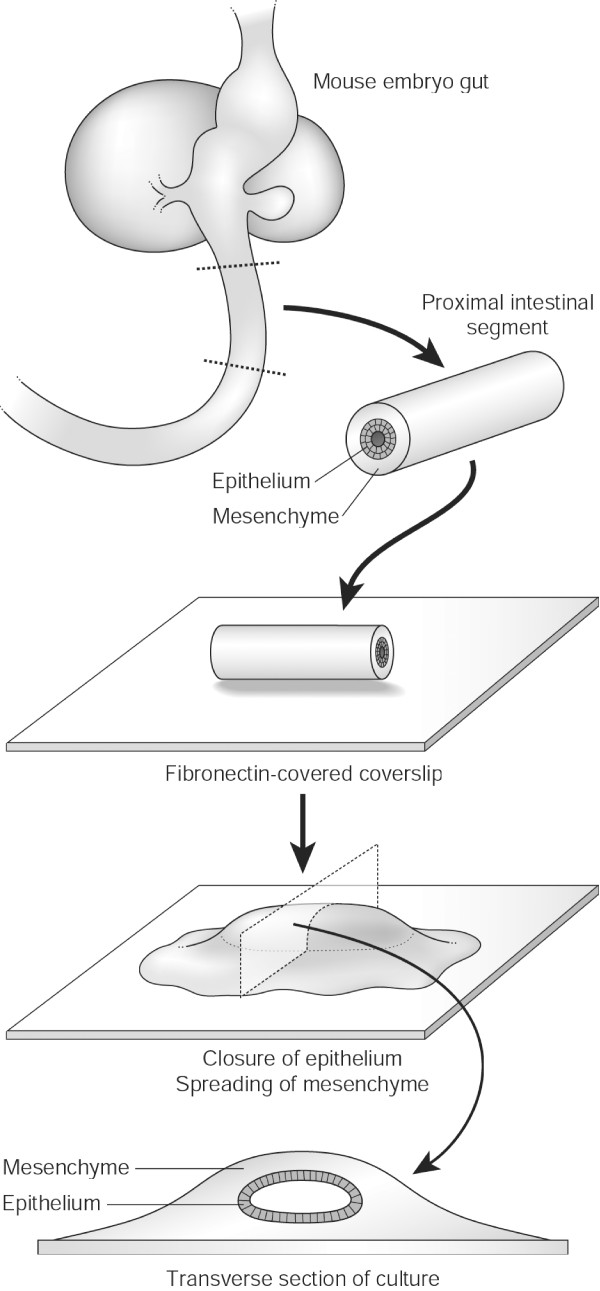
E13.5d intestinal explants were isolated and grown in culture for up to 11 days (Fig 1). Intestinal tissue from younger embryos (E11.5d) was also used successfully (results not shown). We investigated the development in vitro, initially focusing on morphology and differentiation. The explants adhere to the fibronectin substrate within a few hours and gradually flatten out over the first 1–2 days. The cut ends of the epithelium close and the mesenchymal cells spread rapidly out of the explant to form a monolayer of cells on the substrate, with several cell layers surrounding the epithelial tube in the centre. Over the next few days, the epithelium extends and the mesenchyme continues to expand, increasing the area of the culture (Fig 2A–C). The flattening of the whole culture with time is accompanied by collapse of the lumen although this remains as a virtual space, i.e. with opposing epithelial layers apposed. Initially the explants did not exhibit any contractile activity but after 2–3 days in culture, random peristaltic movements were apparent. These are slow contractions lasting a couple of seconds and occurring once or twice a minute.
Figure 1.
The establishment of an intestinal explant culture and its internal structure.
Figure 2.
Development of the murine embryonic intestine in vitro. A-C) Phase contrast images of small intestine after 1 (A), 7 (B) and 11 (C) days of culture. An epithelial tube persists in the centre, with the surrounding mesenchyme spreading out to form a monolayer. D-E) Cdx2 expression in 1 day cultures, wholemount (D) and confocal section through epithelium (E). F-H) Epithelium and mesenchyme form concentric layers. The columnar epithelium is visualised by immunostaining for cytokeratin 8 (F) and the mesenchyme by staining for smooth muscle actin (G). Scale bars = 100 μm A-D, F-H; 20 μm E.
Expression of intestinal markers in the epithelium
We initially anlaysed the expression of the intestinal transcription factor Cdx2. The cdx2 gene encodes a homeodomain transcription factor whose expression normally distinguishes between the upper and lower regions of the alimentary canal epithelia [24,25]. Cdx2 is expressed in vivo by E9.5d [24]. Twenty-four hours after isolation, the epithelium of the E13.5d intestinal explants still expressed Cdx2 (Fig 2D,E) and this expression persisted for the duration of the culture period (Fig 3A–D).
Figure 3.
Expression of intestinal markers in culture. A-D) Continued Cdx2 expression after 7 (A, B) and 11 (C, D) days of culture. E-G) Concentric expression of the columnar epithelial marker cytokeratin 8 (red) and mesenchymal smooth muscle actin (green) persists on day 7 (E, F) and 11 (G) of culture. H-DD) Expression of differentiated epithelial markers. H-M) alkaline phosphatase, visualised by histochemistry, is absent after 1 day of culture (H, I) and is expressed in enterocytes after 7 (J, K) and 11 days (L, M). N-Q) Sucrase-isomaltase, visualised by immunostaining, is absent at 1 (N) and 7 days (O), thereafter demonstrated in enterocytes at day 11 of culture (P, Q). R-T) Goblet cells visualised by PAS staining are seen at 7 days (R, S) and persist for the duration of culture (T). U-X) Chromogranin A, visualised by immunostaining, is expressed in enteroendocrine cells at 7 (U, V) and 11 (W, X) days. Y-AA) The antimicrobial agent lysozyme is demonstrated by immunostaining in Paneth cells at 7 (Y) and 11 (Z, AA) days. BB-DD) HNF-4 α expression, visualised by immunostaining, shows a similar pattern (BB, CC 7 days; DD 11 days of culture). Scale bars = 100 μm A, C, E, H, J, L, N-P, R, T, U, W, Z, BB; 20 μm B, D, F, G, I, K, M, Q, S, V, X, Y, AA, CC, DD.
We proceeded to examine the expression of epithelial and mesenchymal markers in the cultures. Simple columnar epithelia express cytokeratin 8 (K8), a type II acidic protein that normally heterodimerises with another intermediate filament, the type I cytokeratin 18 (K18) to form the keratin intermediate filament structure [26]. Fig 2F–H shows a specimen cultured for one day then stained for K8 to show the columnar epithelium and smooth muscle actin to show the mesenchyme. This concentric arrangement of epithelium and mesenchyme persisted for the duration of the culture period (Fig 3E–G).
As previous studies have only described limited characterisation of differentiation, we studied the formation of all four differentiated epithelial cell types in the culture system using various markers (Fig 3H–DD). The enterocyte marker alkaline phosphatase was detected by day 7 in culture by histochemical staining and sucrase-isomaltase by day 11. PAS-positive goblet cells were demonstrated at day 7. Chromogranin A, produced by enteroendocrine cells, and lysozyme-positive Paneth cells were both detected by day 7. The transcription factor hepatocyte nuclear factor 4α (HNF-4α), a member of the steroid hormone receptor superfamily important in regulating intestinal gene expression, was also seen by day 7 in culture. The appearance of these markers is consistent with development in vivo [27-29] and, once present, all the markers persisted for the duration of the culture period.
Ectopic gene expression in intestinal explants
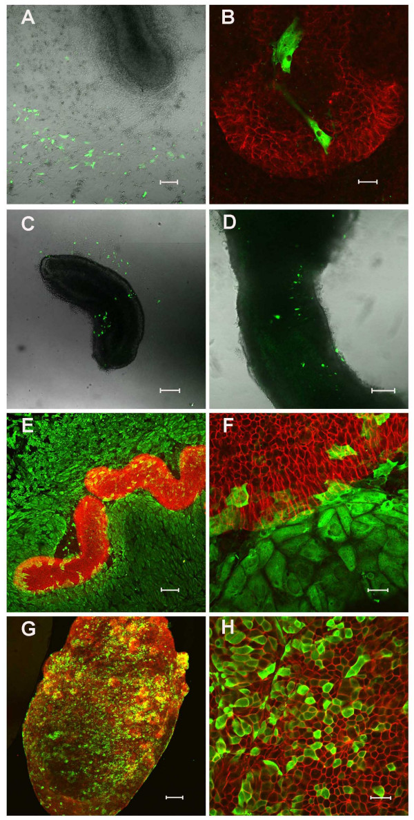
We were especially interested in the potential of the intestinal culture system for ectopic gene expression. We initially tried a commercially available transfection reagent, GeneJuice, to introduce DsRed into the intestinal epithelium. Mesenchymal but not epithelial expression of DsRed could be detected under epifluorescence following 24 hours of culture (Fig 4A,B). Since there was no epithelial expression we decided to examine alternative methods. Electroporation has been successfully developed for manipulating gene expression in chick [30,31] and there have been reports of electroporation of mouse tissues [18,32]. We tested a wide variety of electroporation conditions using the pcDNA3nucGFP2 plasmid. The optimum condition was 40 V, 3 pulses of 50 ms, pulse interval 500 ms, but even with these settings only 0.9 ± 0.3% epithelial cells in the centre of the cultures took up the plasmid and expressed GFP (Fig 4C,D). A number of GFP-positive cells were also found in the mesenchyme.
Figure 4.
Gene delivery to embryonic intestine. (A) and (B) DsRed expression 24 hours after GeneJuice transfection (DsRed is here coloured green to permit direct comparison with the images in the rest of the figure). There is no expression of DsRed in the epithelium (E-cadherin, red). (C) and (D) Electroporation of pcDNA3nucGFP2 into the intestine. GFP is visualised predominantly in the mesenchyme after 24 hours culture. (E) and (F) Infection of intestine explants with Ad-CMV-LacZ adenoviral vector. E-cadherin is shown in red and β-galactosidase in green 48 hours after infection. (G) and (H) Pretreatment of intestinal explants with dispase substantially enhances adenoviral-mediated gene delivery to the epithelium. E-cadherin is shown in red and β-galactosidase in green. Representative cultures are shown for each regime. Scale bars = 100 μm A, C-E, G; 20 μm B, F, H.
Since epithelial expression was poor both with transfection and electroporation, we set out to determine whether active genes could be introduced into the epithelium of the cultures using adenoviral vectors. We infected intestinal epithelium with a first generation adenovirus expressing LacZ as described in the Methods. The intestinal explants were opened to ensure good access to the epithelium then exposed to high titre virus for 2 hours. We observed abundant expression of β-galactosidase in the mesenchyme (Fig 4E,F) but only in 2.3 ± 1.3% cells of the epithelium (Table 2). Adenovirus infection requires the interaction of a viral coat protein with the Coxsackie and Adenovirus Receptor (CAR) in the target cell. In polarised epithelial cells such as those of the intestine, CAR is found on the lateral membranes associated with the tight junctions [37]. Because of this we reasoned that a light enzyme digestion might succeed in opening up the tight junctions, so allowing access of the virus to CAR, while not degrading the tissue so much that the epithelium broke down. A number of trials were made with enzymes including trypsin and collagenase, but the best results were obtained using dispase. As before, the intestinal segments were opened, and then treated at various times and concentrations, followed by application of the virus. The optimum regime was a 1–2 minute treatment with dispase at 50 caseinolytic units/ml. This enhanced β-galactosidase expression in the epithelium, verified by the co-expression with E-cadherin (Fig 4G,H).
Table 2.
Percentage of labeled epithelium following transfection, electroporation or adenoviral infection. The percentage of epithelial cells labeled represents the average counts from 100 cells over five fields from three different buds.
| % explants with any expression | % explants with epithelial expression | % epithelial cells | |
| GeneJuice | 75 (3/4) | 0 (0/4) | 0 |
| Electroporation | 73.8 (31/42) | 11.9 (5/42) | 0.9 ± 0.3 |
| AdLacZ - Dispase | 100 (24/24) | 25 (6/24) | 2.3 ± 1.3 |
| AdLacZ + Dispase | 100 (25/25) | 68 (17/25) | 29.1 ± 1.1*** |
*** p < 0.001. One-way ANOVA (non-parametric). SEM given for % epithelial cells labeled.
Table 2 shows the percentage of labeled epithelium following transfection, electroporation or adenoviral infection, showing quantitatively that infection with adenovirus following dispase treatment is the most efficient method (p < 0.001).
Discussion
Many previous in vitro culture systems have been utilized to investigate intestinal development. Such systems include embryonic gut transplanted under the kidney capsule of syngeneic or immunocompromised hosts [15], intestinal explants attached to filters [17-19] or small slices or biopsies of fetal or postnatal intestinal tissue cultured in vitro for 3 days [38-40]. However, these models suffer from a number of different problems. Tissue cultured using these techniques are not accessible to experimental manipulation and the medium surrounding the explants cannot be manipulated. In some of these systems [38-40], differentiation of the cultures is poor due to reduced viability and only limited differentiation has been documented in other organ systems. In the present system, we provide evidence for the differentiation of the embryonic intestine based on the expression of markers of epithelial cell types. We therefore conclude that the differentiation program is normal.
Although we tried a number of techniques for overexpression including transfection, electroporation and adenoviral infection, none of them produced significant epithelial expression. This was somewhat surprising given the previous published protocols [18]. We therefore sought to improve on the adenoviral system. We believe that the dispase digestion method works because it slightly separates the cells and makes accessible the lateral faces which carry the adenovirus receptor. This dispase/adenovirus method provides the first reliable technique for introducing exogenous genes into the intestinal epithelium. Because the action of many genes can be specifically antagonised by dominant negative constructs or siRNA, gene introduction can also enable inhibition of the action of specific endogenous genes. This new method will provide an additional resource to study changes in intestinal gene expression and differentiation, and will enable the study of intestinal development by both gene overexpression and specific ablation.
Conclusion
The current system offers a number of advantages to those previously described. It (i) is simple and depends largely on the presence of a suitable substrate and a simple medium (ii) maintains the gut tube with intact mesenchymal and epithelial layers (iii) permits differentiation of all of the normal intestinal cell types on a normal time scale (iv) remains accessible to observation and manipulation during the culture period (v) is suitable for wholemount immunostaining, providing three-dimensional visualisation of anatomical features and (vi) is suitable for ectopic gene expression.
Methods
Mice and isolation of embryos
Animal husbandry and embryo isolation were carried out in accordance with UK Home Office regulations. Stage-specific embryos were isolated from timed matings with CD1 mice, based on the observation of a copulatory plug representing E0.5d. Pregnant animals were killed by cervical dislocation and the uteri dissected free into ice-cold sterile phosphate buffered saline A (PBSA). E13.5d embryos were removed from the decidua, transferred to ice-cold Minimum Essential Medium (MEM) with Hanks' salts (Sigma), 10% fetal bovine serum (FBS, Invitrogen), 2 mM L-glutamine (Sigma) and 20 μg/ml gentamicin (Invitrogen) and the gut (from the pharynx to the intestine) was dissected free. An intestinal segment was then removed as shown in Fig 1.
Isolation and culture of embryonic intestine
E13.5d intestines were cultured on coverslips subbed with 3-aminopropyltriethoxysilane (APTES, Sigma) and coated with bovine plasma fibronectin (Invitrogen) at 50 μg/ml in sterile water [23]. Initially, a cloning ring was placed over the fibronectin-coated area in order to ensure that the explant stayed on the substrate. Basal Medium Eagle (BME) with Earle's salts (Sigma), 20% FBS, 2 mM L-glutamine and 20 μg/ml gentamicin was pipetted dropwise into the cloning ring. Twenty four hours later the cloning ring was removed, the medium decanted and fresh medium added. The cultures were grown at 37°C, 95% air/5% CO2 in a humidified incubator for up to 11 days. The medium was changed every 2 days.
Histology and immunohistochemistry
We performed immunohistochemistry on wholemounts as described previously[33,34]. Briefly, intestinal cultures (1–11 days) were fixed for 5 minutes in acetone/methanol (1:1 ratio) at -20°C for immunostaining of cytoskeleton proteins or 30 minutes in MEMFA pH7.4 (3.8% formaldehyde, 0.15 M MOPS, 2 mM EGTA, 1 mM MgSO4) at room temperature for immunostaining of membrane, cytosolic and nuclear proteins. After fixation, cultures were washed three times in PBSA and stored in PBSA at 4°C. Cultures were permeabilised by adding 1% Triton X-100 (Sigma) in PBSA for 30 minutes prior to immunostaining. Antigen retrieval was performed using citrate buffer pH6 (Lab Vision Corporation) for 1 hour at 37°C. The cultures were washed three times in PBSA. Non-specific binding sites were blocked for at least 1 hour in 2% Blocking Reagent (Roche). Primary antibodies were applied overnight at 4°C and the following day the samples were washed three times in PBSA. The fluorescent secondary antibody was applied for 3 hours in the dark at room temperature, and the coverslips were rewashed and then mounted in Gel/Mount (Biomeda corp).
Microscopy and photography
During the culture period, the live cultures were observed using a Leica DMIRB inverted microscope and the immunostained or histochemically stained wholemounts were visualised under a Leica DMRB compound microscope. The images were taken using a color SPOT RT camera (Diagnostic Instruments) operated with Advanced Spot RT 3.0 software. A Zeiss LSM510 confocal microscope was used to take high resolution images of optical sections. The images were cropped and arranged using Adobe Photoshop 7.0.
Antisera
The antibodies were obtained and diluted as described in Table 1.
Table 1.
Antibodies
| Experiment | Primary | Supplier | Secondary | Supplier |
| AdLacZ | mouse monoclonal anti-E-cadherin 1/50 | BD Transduction Laboratories | horse anti-mouse Texas Red conjugated IgG 1/100 | Vector |
| rabbit polyclonal anti-β-galactosidase 1/300 | ICN | goat anti-rabbit fluorescein isothiocyanate (FITC) conjugated IgG 1/100 | Vector | |
| GeneJuice | mouse monoclonal anti-E-cadherin 1/50 | BD Transduction Laboratories | horse anti-mouse FITC conjugated IgG 1/100 | Vector |
| rabbit polyclonal anti-DsRed 1/3000 | Clontech | Swine anti-rabbit tetramethylrhodamine isomer R (TRITC) conjugated IgG 1/100 | DAKO | |
| Small Intestine characterisation | Rat monoclonal anti-E-cadherin 1/100 | Zymed | goat anti-rat Texas Red conjugated IgG 1/100 | Vector |
| rat monoclonal anti-Troma-1 (cytokeratin 8) 1/50 | DSHB, University of Iowa | goat anti-rat Texas Red conjugated IgG 1/100 | Vector | |
| mouse monoclonal anti-α smooth muscle actin 1/100 | Sigma | horse anti-mouse FITC conjugated IgG 1/100 | Vector | |
| mouse monoclonal anti-sucrase-isomaltase 1/200 | Gift (Dr Hans-Peter Hauri) | horse anti-mouse FITC conjugated IgG 1/100 | Vector | |
| mouse monoclonal anti-cdx2 1/100 | BioGenex | horse anti-mouse FITC conjugated IgG 1/100 | Vector | |
| rabbit polyclonal anti-lysozyme 1/100 | DAKO | goat anti-rabbit FITC conjugated IgG 1/100 | Vector | |
| rabbit polyclonal anti-chromogranin A 1/100 | DAKO | goat anti-rabbit FITC conjugated IgG 1/100 | Vector | |
| goat polyclonal anti-HNF-4α 1/100 | Santa Cruz | rabbit anti-goat FITC conjugated IgG 1/100 | Vector |
Histochemical methods
Enterocytes
Vector Blue alkaline phosphatase substrate kit was used according to the manufacturer's instructions. Briefly, 2 drops of reagent 1 were added to 5 ml 100 mM Tris-HCl pH8.2, followed by 2 drops of reagent 2 and 2 drops of reagent 3. Embryonic buds were incubated for 30 minutes at room temperature in the dark, washed in PBSA for 5 minutes, rinsed in tap water before dehydration and mounting in DePex (BDH).
Goblet cells
Goblet cells were stained by the Periodic Acid Schiff (PAS) method. MEMFA-fixed embryonic buds were washed in running tap water for 3 minutes, then treated with 1% periodic acid (BDH) for 10 minutes at room temperature, washed for 3 minutes in water and then immersed in Schiff's reagent (Sigma) in the dark for 2 minutes. Samples were washed in running tap water for 20 minutes and then 3 minutes in distilled water. The buds were then counterstained with haematoxylin before dehydration and mounting in DePeX.
Overexpression of reporter genes
Transfection protocol
E13.5d small intestine explants were isolated and cultured for 48 hours as described above. For each explant, 3 μl of GeneJuice Transfection Reagent (Novagen) was added to 100 μl of Optimem I medium (Gibco) and mixed by vortexing. The mixture was then incubated for 5 minutes at room temperature in semi-darkness. 1 μg pcDNA3DsRed was added to the tube and incubated for a further 15 minutes at room temperature. The culture medium was changed and, keeping the cloning ring in position, the transfection complex was added dropwise into the cloning ring. Cultures were then incubated in 95% air/5% CO2 at 37°C for 24 hours before MEMFA fixation and immunostaining.
Electroporation conditions
pcDNA3nucGFP2 was constructed by subcloning nucGFP2 as a BamHI/XbaI fragment into pcDNA3 (Invitrogen). 0.01% Fast Green was added to 1 μg/μl pcDNA3nucGFP2 plasmid DNA for easy visualization. Borosilicate standard wall (1.0 mm O.D./0.58 mm I.D.) glass capillaries (Harvard Apparatus) were pulled using a P-97 micropipette puller (Sutter Instrument Co.) with the following conditions: heat = ramp+25, pull = 20, velocity = 255, time = 150. The micropipette was back-filled with DNA solution and Fast Green, DNA injected into the lumen of the isolated intestine and electroporation performed with an Electro Square Porator (BTX, ECM830) using an electrode gap width of 4 mm. The voltage was varied from 25–100 V in a unipolar direction and the number of pulses from 3x–6x. The tissues were then examined for GFP expression by confocal microscopy after 24 hours of culture. The following settings proved optimal in terms of epithelial cells expressing GFP: 40 V, 3 pulses of 50 ms, pulse interval 500 ms.
Recombinant adenoviral vectors
A first-generation, replication-defective, recombinant adenoviral vector was used in these experiments. Ad-CMV-LacZ was constructed according to a standard protocol using two plasmids, pJM17 and pXCX2, as described previously [35]. The Ad-CMV-LacZ adenovirus was propagated in the E1-containing human embryonic kidney (HEK) cell line 293 [36]. HEK 293 cells were maintained in Dulbecco's Modified Eagle Medium (Invitrogen) supplemented with 10% heat-inactivated fetal calf serum (Sigma). Ad-CMV-LacZ contains the constitutive cytomegalovirus (CMV) promoter driving the expression of the Escherichia coli-derived β-galactosidase gene (LacZ). To make it, the CMV promoter was cloned into the Klenow-blunted XbaI site of pXCX2. The resulting plasmid was cut with HindIII/BamHI and a 3.7 kb HindIII/BamHI fragment from pCH110 (Pharmacia) containing LacZ and an SV40 polyadenylation signal was ligated in. The resulting plasmid was cotransfected with pJM17 into HEK 293 cells by calcium phosphate precipitation and the cells were overlaid with agarose. Individual viral plaques were picked and plaque-purified three times. The DNA structure of the new viral vector was verified by restriction diagnostics. The virus was grown to high titre, released from the cells by rapidly freeze/thawing three times and then purified twice by caesium chloride density ultra-centrifugation. The buffer was exchanged with 10 mM Tris.HCl pH 7.5, 1 mM MgCl2 on a PD-10 Sephadex column (Amersham Biosciences), before the virus was passed through a sterile 0.22 μm Millex filter (Millipore) and frozen in 10 μl aliquots using dry-ice. The viral stocks were stored at -80°C. The titer of the vector was 7 × 10⁹ infectious units/ml, which was calculated using the Adeno-X™ Rapid Titer Kit (Stratagene).
Ad-CMV-LacZ infection
E13.5d small intestine was isolated as previously described and opened with a tungsten needle to expose the luminal epithelium. The segments were then incubated in dispase (activity 50 caseinolytic units/ml (BD Biosciences)) at 37°C for 1 to 2 minutes to improve viral access to the epithelium. The reaction was terminated by washing the intestine in supplemented BME and the organ buds transferred to the centre of a cloning ring. 20 μl Ad-CMV-LacZ was added to the cloning ring and incubated in 95% air/5% CO2 at 37°C for 2 hours. A further 40 μl supplemented BME was then added to the cloning ring. The culture medium was changed at 24 hours and the cloning ring removed. Cultures were maintained for up to 7 days with medium changes (2 ml) every 2 days and fixed in MEMFA.
Immunostaining of infected explants
MEMFA-fixed intestinal cultures were treated with acetone/methanol 1:1 at -20°C for 5 minutes and immunostained as described above.
Authors' contributions
WYY and JMQ carried out the trials of culture conditions, studies of differentiation and gene introduction. MAH prepared the recombinant adenovirus. DT and JMWS conceived of the study, and supervised its execution. The manuscript was mostly written by JMQ and JMWS, with input from all authors. All authors read and approved the final manuscript.
Acknowledgments
Acknowledgements
We wish to thank Cancer Research UK (JMQ), Overseas Research Studentship (ORS) fund (WYY), the Biotechnology and Biological Sciences Research Council, Sulis Seedcorn, Medical Research Council and the Wellcome Trust (JMWS and DT) for financial support. We gratefully acknowledge the gift of sucrase-isomaltase antibody from Dr Hans-Peter Hauri (University of Basel), and the suggestion of Mr Wan Chun Li to try dispase to facilitate virus infection.
Contributor Information
Jonathan M Quinlan, Email: bspjmq@bath.ac.uk.
Wei-Yuan Yu, Email: wei-yuan.yu@kcl.ac.uk.
Mark A Hornsey, Email: bssmaah@bath.ac.uk.
David Tosh, Email: d.tosh@bath.ac.uk.
Jonathan MW Slack, Email: j.m.w.slack@bath.ac.uk.
References
- Slack JMW. Essential Developmental Biology. 2. Blackwell Scientific; 2006. Development of endodermal organs; pp. 249–263. [Google Scholar]
- Mathan M, Moxey PC, Trier JS. Morphogenesis of fetal rat duodenal villi. Am J Anat. 1976;146:73–92. doi: 10.1002/aja.1001460104. [DOI] [PubMed] [Google Scholar]
- Potten CS, Loeffler M. Stem cells: attributes, cycles, spirals, pitfalls and uncertainties. Lessons for and from the crypt. Development. 1990;110:1001–1020. doi: 10.1242/dev.110.4.1001. [DOI] [PubMed] [Google Scholar]
- Potten CS. Stem cells in gastrointestinal epithelium: numbers, characteristics and death. Phil Trans R Soc London Ser B. 1998;353:821–830. doi: 10.1098/rstb.1998.0246. [DOI] [PMC free article] [PubMed] [Google Scholar]
- Sancho E, Batlle E, Clevers H. Signaling pathways in intestinal development and cancer. Ann Rev Cell Dev Biol. 2004;20:695–723. doi: 10.1146/annurev.cellbio.20.010403.092805. [DOI] [PubMed] [Google Scholar]
- Real FX, Xu M, Vila MR, de Bolos C. Intestinal brush-border associated enzymes: co-ordinated expression in colorectal cancer. Int J Cancer. 1992;51:173–181. doi: 10.1002/ijc.2910510203. [DOI] [PubMed] [Google Scholar]
- Hocker M, Wiedenmann B. Molecular mechanisms of enteroendocrine differentiation. Ann NY Acad Sci. 1998;859:160–174. doi: 10.1111/j.1749-6632.1998.tb11120.x. [DOI] [PubMed] [Google Scholar]
- Roth KA, Gordon JL. Spatial differentiation of the intestinal epithelium: analysis of enteroendocrine cells containing immunoreactivity serotonin, secretin, and substance P in normal and transgenic mice. Proc Natl Acad Sci USA. 1990;87:6408–6412. doi: 10.1073/pnas.87.16.6408. [DOI] [PMC free article] [PubMed] [Google Scholar]
- Porter EM, Bevins CL, Ghosh D, Ganz T. The multifaceted Paneth cell. Cell Mol Life Sci. 2002;59:156–170. doi: 10.1007/s00018-002-8412-z. [DOI] [PMC free article] [PubMed] [Google Scholar]
- Cheng H, Leblond CP. Origin, differentiation, and renewal of the four main epithelial cell types in the mouse small intestine. V Unitarian Theory of the origin of the four epithelial cell types. Am J Anat. 1974;141:537–562. doi: 10.1002/aja.1001410407. [DOI] [PubMed] [Google Scholar]
- Wright NA. Epithelial stem cell repertoire in the gut: clues to the origin of cell lineages, proliferative units and cancer. Int J Exp Pathol. 2000;81:117–143. doi: 10.1046/j.1365-2613.2000.00146.x. [DOI] [PMC free article] [PubMed] [Google Scholar]
- Whitehead RH, Demmler K, Rockmann SP, Watson NK. Clonogenic growth of epithelial cells from normal colonic mucosa from both mice and humans. Gastroenterology. 1999;117:858–865. doi: 10.1016/S0016-5085(99)70344-6. [DOI] [PubMed] [Google Scholar]
- Hall PA, Coates PJ, Ansari B, Hopwood D. Regulation of cell number in the mammalian gastrointestinal tract: the importance of apoptosis. J Cell Sci. 1994;107:3569–3577. doi: 10.1242/jcs.107.12.3569. [DOI] [PubMed] [Google Scholar]
- Strater J, Koretz K, Gunthert AR, Moller P. In situ detection of enterocytic apoptosis in normal colonic mucosa and in familial adenomatous polyposis. Gut. 1995;37:819–825. doi: 10.1136/gut.37.6.819. [DOI] [PMC free article] [PubMed] [Google Scholar]
- Tait IS, Evans GS, Kedinger M, Flint N, Potten CS, Campbell FC. Progressive morphogenesis in vivo after transplantation of cultured small bowel epithelium. Cell Transplant. 1994;3:33–40. doi: 10.1177/096368979400300106. [DOI] [PubMed] [Google Scholar]
- Simon-Assmann P, Kedinger M. Heterotypic cellular cooperation in gut morphogenesis and differentiation. Semin Cell Biol. 1993;4:221–230. doi: 10.1006/scel.1993.1026. [DOI] [PubMed] [Google Scholar]
- Hearn CJ, Young HM, Ciampoli D, Lomax AE, Newgreen D. Catenary cultures of embryonic gastrointestinal tract support organ morphogenesis, motility, neural crest cell migration, and cell differentiation. Dev Dyn. 1999;214:239–247. doi: 10.1002/(SICI)1097-0177(199903)214:3<239::AID-AJA7>3.0.CO;2-O. [DOI] [PubMed] [Google Scholar]
- Abud HE, Lock P, Heath JK. Efficient gene transfer into the epithelial cell layer of embryonic mouse intestine using low-voltage electroporation. Gastroenterology. 2004;126:1779–1787. doi: 10.1053/j.gastro.2004.03.006. [DOI] [PubMed] [Google Scholar]
- Abud HE, Watson N, Heath JK. Growth of intestinal epithelium in organ culture is dependent on EGF signaling. Exp Cell Res. 2005;303:252–262. doi: 10.1016/j.yexcr.2004.10.006. [DOI] [PubMed] [Google Scholar]
- Percival AC, Slack JMW. Analysis of pancreatic development using a cell lineage label. Exp Cell Res. 1999;247:123–132. doi: 10.1006/excr.1998.4322. [DOI] [PubMed] [Google Scholar]
- Horb LD, Slack JMW. Role of cell division in branching morphogenesis and differentiation of the embryonic pancreas. Int J Dev Biol. 2000;44:791–796. [PubMed] [Google Scholar]
- Shen C-N, Seckl JR, Slack JMW, Tosh D. Glucocorticoids suppress beta cell development and induce hepatic metaplasia in embryonic pancreas. Biochem J. 2003;375:41–50. doi: 10.1042/BJ20030140. [DOI] [PMC free article] [PubMed] [Google Scholar]
- Yu W-Y, Slack JMW, Tosh D. Conversion of columnar to stratified squamous epithelium in the developing mouse oesophagus. Dev Biol. 2005;284:157–170. doi: 10.1016/j.ydbio.2005.04.042. [DOI] [PubMed] [Google Scholar]
- Silberg DG, Swain GP, Suh ER, Traber PG. Cdx1 and Cdx2 expression during intestinal development. Gastroenterology. 2000;119:961–971. doi: 10.1053/gast.2000.18142. [DOI] [PubMed] [Google Scholar]
- Yuasa Y. Control of gut differentiation and intestinal-type gastric carcinogenesis. Nature Rev Cancer. 2003;3:592–600. doi: 10.1038/nrc1141. [DOI] [PubMed] [Google Scholar]
- Owens DW, Lane EB. The quest for the function of simple epithelial keratins. Bioessays. 2003;25:748–758. doi: 10.1002/bies.10316. [DOI] [PubMed] [Google Scholar]
- Stappenbeck TS, Gordon JI. Rac1 mutations produce aberrant epithelial differentiation in the developing and adult mouse small intestine. Development. 2000;127:2629–2642. doi: 10.1242/dev.127.12.2629. [DOI] [PubMed] [Google Scholar]
- Bry L, Falk P, Huttner K, Ouellette A, Midvedt T, Gordon JI. Paneth cell differentiation in the developing intestine of normal and transgenic mice. Proc Natl Acad Sci USA. 1994;91:10335–10339. doi: 10.1073/pnas.91.22.10335. [DOI] [PMC free article] [PubMed] [Google Scholar]
- Zhong W, Mirkovitch J, Darnell JE., Jr Tissue-specific regulation of mouse hepatocyte nuclear factor 4 expression. Mol Cell Biol. 1994;14:7276–7284. doi: 10.1128/mcb.14.11.7276. [DOI] [PMC free article] [PubMed] [Google Scholar]
- Grapin-Botton A, Majithia AR, Melton DA. Key events of pancreas formation are triggered in gut endoderm by ectopic expression of pancreatic regulatory genes. Genes Dev. 2001;15:444–454. doi: 10.1101/gad.846001. [DOI] [PMC free article] [PubMed] [Google Scholar]
- Itasaki N, Bel-Vislar S, Krumlauf R. "Shocking" developments in chick embryology:electroporation and in ovo gene expression. Nature Cell Biology. 1999;1:E203–E207. doi: 10.1038/70231. [DOI] [PubMed] [Google Scholar]
- Saito T, Nakatsuji N. Efficient gene transfer into embryonic mouse brain using in vivo electroporation. Dev Biol. 2001;240:237–246. doi: 10.1006/dbio.2001.0439. [DOI] [PubMed] [Google Scholar]
- Shen C-N, Slack JMW, Tosh D. Molecular basis of transdifferentiation of pancreas to liver. Nature Cell Biology. 2000;2:879–887. doi: 10.1038/35046522. [DOI] [PubMed] [Google Scholar]
- Tosh D, Shen C-N, Slack JMW. Differentiated properties of hepatocytes induced from pancreatic cells. Hepatology. 2002;36:534–543. doi: 10.1053/jhep.2002.35060. [DOI] [PubMed] [Google Scholar]
- Graham FL, Smiley J, Russell WC, Nairn R. Characteristics of a human cell line transformed by DNA from human adenovirus type 5. J Gen Virol. 1977;36:59–74. doi: 10.1099/0022-1317-36-1-59. [DOI] [PubMed] [Google Scholar]
- Graham FL, Prevec L. Adenovirus-based expression vectors and recombinant vaccines. Biotechnology. 1992;20:363–390. doi: 10.1016/b978-0-7506-9265-6.50022-1. [DOI] [PubMed] [Google Scholar]
- Cohen CJ, Shieh JTC, Pickles RJ, Okegawa T, Hsieh JT, Bergelson JM. The coxsackievirus and adenovirus receptor is a transmembrane component of the tight junction. Proc Natl Acad Sci USA. 2001;98:T15191–15196. doi: 10.1073/pnas.261452898. [DOI] [PMC free article] [PubMed] [Google Scholar]
- Black BL, Moog F. Alkaline phosphatase and maltase activity in the embryonic chick intestine in culture. Dev Biol. 1978;66:232–249. doi: 10.1016/0012-1606(78)90287-7. [DOI] [PubMed] [Google Scholar]
- Bleich HL, Boro ES, Trier JS. Organ-culture methods in the study of gastrointestinal-mucosal function and development. N Engl J Med. 1976;295:150–155. doi: 10.1056/NEJM197607152950308. [DOI] [PubMed] [Google Scholar]
- DeRitis G, Falchuk ZM, Trier JS. Differentiation and maturation of cultured fetal rat jejunum. Dev Biol. 1975;45:304–317. doi: 10.1016/0012-1606(75)90068-8. [DOI] [PubMed] [Google Scholar]


