Abstract
flg22, a peptide corresponding to the most conserved domain of bacterial flagellin, acts as a potent elicitor in plants. Here, we have used an iodinated derivative of flg22 (125I-labeled Tyr-flg22) as a molecular probe for the flagellin receptor in tomato cells. This radioligand showed rapid binding to a single class of specific, saturable, high-affinity receptor sites in intact cells and membrane preparations. Binding, although essentially nonreversible under physiological conditions, was not covalent, and chemical cross-linking was required to specifically label a single polypeptide of 115 kD. Intact flagellin and elicitor-active flagellin peptides but not biologically inactive analogs efficiently competed for binding of radioligand. Peptides lacking the C terminus of the conserved domain, previously found to act as competitive antagonists of elicitor action in tomato cells, also competed for binding of radioligand. Thus, this novel, high-affinity binding site exhibited all the characteristics expected of a functional receptor of bacterial flagellin. For a model of receptor activation, we propose a two-step mechanism according to the address–message concept, in which binding of the N terminus (address) is the first step and activation of responses with the C terminus (message) is the second step.
INTRODUCTION
Recognition of potential pathogens plays a central role in the induction of active defense responses in plants (Yang et al., 1997). In plant–pathogen interactions that follow the classic gene-for-gene interaction, resistance depends on specific resistance (R) genes that function as recognition systems for the products of the corresponding avirulence (avr) genes expressed by the pathogens (Hammond-Kosack and Jones, 1997; Parker and Coleman, 1997). In addition to recognition systems for specific pathogens, plants have evolved perception systems for microbial components that are characteristic of entire classes of microorganisms in general. Various fungal structures act as elicitors of responses associated with plant defense (Boller, 1995). For several of these general elicitors, specific binding sites residing in the plasma membranes of plant cells have been demonstrated (Hahn, 1996; Nürnberger, 1999). Assays for specific binding sites provide the basis for biochemical purification and cloning of the corresponding genes involved, as exemplified by the binding site for the heptaglucan elicitor (Umemoto et al., 1997). Identification and characterization of specific, high-affinity binding sites also provide essential information for understanding the processes underlying the perception of these microbial stimuli by plants.
Recently, we identified flagellin, the protein subunit building up the filament of the bacterial flagellum, as an elicitor of defense-related responses in cells from various plant species (Felix et al., 1999). Flagellins from different bacterial species vary in their central part but show conservation of their N-terminal and C-terminal regions (Schuster and Khan, 1994). Elicitor activity could be attributed to the most conserved domain within the N-terminal part (Felix et al., 1999). Synthetic peptides spanning a core of 15 amino acid residues within this domain exhibited elicitor activity when applied at subnanomolar concentrations. Flagellins from the plant-associated bacteria Agrobacterium tumefaciens and Rhizobium meliloti are exceptionally divergent in this domain, and peptides synthesized according to those sequences were inactive as elicitors. When applied to intact seedlings of Arabidopsis, the elicitor-active flagellin peptides induced defense-related genes, an oxidative burst, the deposition of callose, and a marked inhibition in growth (Gómez-Gómez et al., 1999).
In suspension-cultured tomato cells, flagellin-derived elicitors induced a set of rapid responses, including medium alkalinization, oxidative burst, and increased biosynthesis of ethylene. These responses, previously observed also after treatment with fungal elicitors such as chitin fragments, xylanase, ergosterol, and high-mannose–type glycopeptides (Basse et al., 1993), served as rapid, quantifiable bioassays for studying the structure–activity relationship of flagellin-derived peptides (Felix et al., 1999). Peptides spanning the 15 amino acids in the core of the conserved domain induced responses when applied in subnanomolar concentrations, whereas peptides comprising only 12 to 14 amino acid residues exhibited much less activity. Interestingly, peptides lacking the C-terminal part of this domain were inactive as agonists but acted as specific, competitive antagonists for flagellin-related elicitors. The sensitivity of the cells suggested perception by means of a receptor with high affinity for the flagellin elicitor. In this article, we demonstrate the presence of such a binding site on the surface of tomato cells. Using a radioiodinated derivative of the highly active flagellin peptide flg22, we observed rapid, high-affinity binding to intact cells as well as to microsomal membrane preparations. Binding by the most active flagellin peptides was essentially irreversible under physiological conditions. Moreover, the binding was specific for flagellin and flagellin-derived peptides that exhibited biological activity as agonists or antagonists, indicating that this binding site acts as the functional receptor of flagellin.
RESULTS
Activation and Suppression of the Alkalinization Response by Flagellin-Derived Peptides
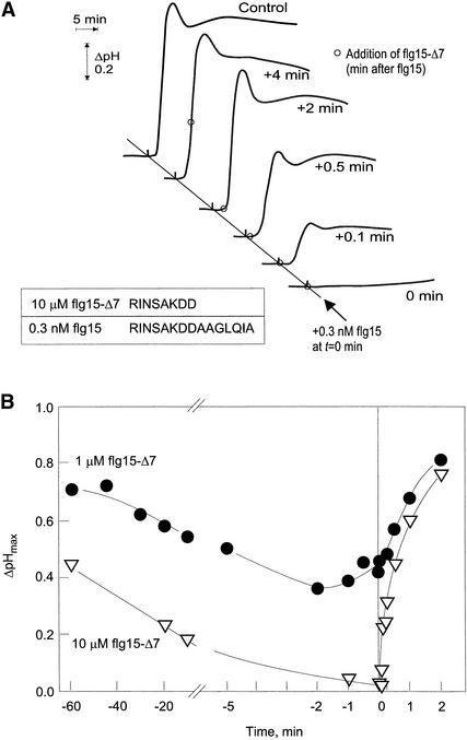
Alkalinization of the culture medium is a rapid, easily measurable response of cultured plant cells treated with the bacterial surface protein flagellin or with synthetic peptides that span the most conserved N-terminal domain of flagellin (Felix et al., 1999). Peptides comprising the 15 amino acid residues RINSAKDDAAGLQIA (flg15) at the core of this domain exhibited potent elicitor activity and induced alkalinization of the medium at EC50 (concentration for half-maximal biological activity) ⩽0.1 nM. Peptides lacking 4 to 7 amino acid residues from the C terminus, termed flg15-Δ4 to flg15-Δ7, showed no activity as elicitors but acted as competitive antagonists for elicitor-active flagellin peptides and intact flagellin protein (Felix et al., 1999). In the experiments shown in Figure 1, the antagonistic effect of flg15-Δ7 (RINSAKDD) was tested with respect to its addition either before or after the addition of the agonist flg15. In cells treated with 0.3 nM flg15 alone (Figure 1A, control), the extracellular pH started to increase after a short lag of ∼2 min and rapidly exceeded the initial pH of 5.1 by >0.8 pH units. When added concomitantly with flg15, a 10-μM excess of flg15-Δ7 inhibited this alkalinization response (Figure 1A, 0 min). However, when applied only 6 sec after the agonist flg15, the same dose of flg15-Δ7 suppressed alkalinization only partially, and application toward the end of the lag phase had little effect on the further progress of the response (Figure 1A).
Figure 1.
Antagonism of Peptide flg15-Δ7 to Alkalinization Response of Suspension-Cultured Tomato Cells Treated with Agonist flg15.
(A) Extracellular alkalinization in suspension-cultured tomato cells treated with 0.3 nM flg15 (RINSAKDDAAGLQIA) alone (control) or in combination with 10 μM flg15-Δ7 (RINSAKDD) added at the times indicated (open circles). Extracellular pH of untreated cells was 5.1.
(B) Alkalinization response of cells treated with combinations of 0.3 nM flg15 and either 1 μM flg15-Δ7 (closed circles) or 10 μM flg15-Δ7 (open inverted triangles). The x axis indicates the time of flg15-Δ7 addition with respect to the addition of flg15. Thus, negative values indicate a pretreatment with flg15-Δ7, and positive values indicate addition of flg15-Δ7 after flg15. Control cells treated with 0.3 nM flg15 alone showed an alkalinization response with a mean ΔpHmax of 0.86 (sd = 0.03, n = 4 replicates). The time dependence of inhibition by flg15-Δ7 was reproduced in three independent series of experiments with different batches of cells.
These results indicate that flg15 exerts its effect very rapidly and in a way that is not readily reversed by subsequent application of the antagonist. We tested whether the antagonist had a similar, nonreversible effect when used to pretreat the cells (Figure 1B). Addition of 1 μM flg15-Δ7 reduced alkalinization of the medium by ∼50% when given concomitantly with 0.3 nM flg15, an antagonistic effect that was only slightly stronger in cells pretreated for 1 or 2 min with the antagonist (Figure 1B). Preincubation for ⩾5 min led to less pronounced inhibition by the antagonist. A progressive decrease of suppressor activity was also observed after prolonged pretreatment with 10 μM flg15-Δ7 (Figure 1B). Similar results were obtained with antagonists flg15-Δ4, flg15-Δ5, and flg15-Δ6 in various combinations with flg15 and flg22 (data not shown). In summary, the antagonistic effect of the truncated peptides flg15-Δ4 through flg15-Δ7 was reversible and required a >1000-fold molar excess over the agonist, indicating a lower affinity of the antagonists for the putative receptor site. The alkalinization response initiated by the agonists flg15 and flg22, in contrast, appeared to involve a process that, once initiated, could not be reversed by subsequent addition of antagonist. This irreversible step might take place at the level of elicitor perception, for example, by persistent binding of the stimulus to its receptor site, or could involve a continued switch-like activation of a subsequent step in signal transduction.
Binding of 125I-Labeled Tyr-flg22 to Tomato Cells and Microsomal Membranes
The sensitivity of the plant cells for specific peptides spanning the conserved domain of bacterial flagellin suggested a receptor-mediated activation process involving a high-affinity binding site. As a strategy to search for such a binding site, we chose 125I as the radioactive isotope to obtain a radioligand with a high specific radioactivity. flg22 was extended by adding an extra tyrosine residue at the N terminus to obtain Tyr-flg22. This peptide, as well as its nonradioactively iodinated derivative, exhibited the same specific biological activity as flg22 and induced the alkalinization response with an EC50 of 0.03 nM. Monoiodination of the peptide with 125I resulted in 125I-labeled Tyr-flg22 ([3-125I]iodotyrosine-flg22, or 125I-Tyr-flg22), a radioligand with a specific radioactivity of >2000 Ci/mmol. We investigated the binding of this ligand under in vivo conditions by incubating tomato cells (60 mg fresh weight) at room temperature with ∼60 fmol of radioligand (0.12 nM). Association was very rapid, reaching maximum after 15 min, and the label associated with the cells remained stable throughout the 120-min experiment. Nonspecific binding, assayed in the presence of an excess of 1 μM unlabeled Tyr-flg22, was initially very low but steadily increased during prolonged incubation (Figure 2A), indicating that specific binding reached a peak after ∼10 min and then slowly decreased. Most interestingly, binding was not readily reversible, and no clear release of label bound to the cells was observed when an excess of 1 μM unlabeled Tyr-flg22 was added after 20 min of incubation. Binding kinetics of cells incubated on ice exhibited characteristics similar to those of cells observed at room temperature (Figure 2B). In particular, an excess of unlabeled Tyr-flg22 nearly completely inhibited binding when added concomitantly with the radioligand but did not displace bound radioligand when added after 20 min. Because the processes of internalization and active uptake can be expected to be greatly reduced in cells incubated at 4°C, the nonreversibility of binding appeared to be due to very slow dissociation of the radioligand from its binding site.
Figure 2.
Binding of 125I-Tyr-flg22 to Intact Tomato Cells and Membranes.
Kinetics of binding was reproducible in three independent series of experiments with different batches of membranes and two independent series of experiments with cells.
(A) and (B) Binding kinetics of 125I-Tyr-flg22 to intact tomato cells at room temperature (A) and at 4°C (B). Aliquots of the cell suspension (0.06 g, fresh weight) were supplied with 0.12 nM 125I-Tyr-flg22 alone (solid circles) or in combination with 1 μM Tyr-flg22 added at t = 0 min (open circles) or at 20 min (open diamonds). Radioactivity retained on the cells was measured by γ-counting after washing of the cells.
(C) Binding kinetics of 125I-Tyr-flg22 to microsomal membranes prepared from tomato cells. Aliquots containing 100 μg of microsomal protein were supplied with 0.4 nM 125I-Tyr-flg22 (solid circles) alone or in combination with 1 μM Tyr-flg22 added at t = 0 min (open circles) or at 20 min (open diamonds). The inset shows results from a separate experiment with dissociation kinetics over an extended time span; note that the scale of the x axis in the inset is in days rather than minutes.
On the basis of size and charge, flagellin peptides are unlikely to be membrane permeable, and perception of them is expected to occur at the cell surface, most likely by way of a binding site residing in the plasma membrane. Therefore, we looked for the flagellin binding site in membrane preparations obtained from tomato cell cultures. Binding to microsomal membranes at 4°C was very rapid, reached a maximum within 20 min, and remained nearly constant for at least 120 min (Figure 2C). As observed with intact cells, binding was not readily reversible, and addition of 1 μM unlabeled Tyr-flg22 after 20 min did not lead to clear release of label from the membranes within the 120 min of the experiment (Figure 2C). In a separate experiment, dissociation of ligand from its binding site on membranes was assayed over an extended span of 12 days (Figure 2C, inset). Under these conditions, we observed a slow, partial decrease of bound label. However, this decrease was not exponential and did not allow determination of a rate constant for dissociation. In summary, the ligand–binding site complex appears to be extremely stable over several days.
125I-Tyr-flg22 Bound to Cells Is Protected from Degradation
Flagellin peptides were not stable in suspension-cultured cells, and elicitor activity rapidly disappeared when the supernatant of cells treated with flg22 peptide was assayed for its capacity to induce alkalinization of the medium in fresh aliquots of cells (Figure 3). Activity also disappeared with a half-life of ∼6 min during incubation in culture medium from which the cells had been removed. In contrast, no inactivation occurred after heat treatment of the medium, indicating an enzymatic, most likely proteolytic process. Similarly, radioligand 125I-Tyr-flg22 added to cell suspensions in binding assays at room temperature was rapidly degraded. Analysis by reversed-phase HPLC showed rapid disappearance of 125I-Tyr-flg22 and no intact radioligand remaining in the cell supernatant after 20 min of incubation (Figure 4A). In contrast, the radioligand that bound to the cells appeared intact over a much more prolonged period (Figure 4B). In moderately acidic conditions (pH 2.0 for 0.5 min), label bound to cells was released. At all times tested up to 120 min, most of the label released from cells coeluted from the C18 column with authentic 125I-Tyr-flg22 (Figure 4B), demonstrating that the binding to living cells occurred in a tight but noncovalent manner that protects the peptide ligand from degradation.
Figure 3.
Inactivation of the Alkalinization-Inducing Activity of flg22.
flg22 was incubated in cell suspension cultures of tomato cells in culture medium from which the cells had been removed by filtration or in cell-free medium treated for 5 min at 95°C. Alkalinization-inducing activity remaining in the samples was assayed by quantifying the alkalinization response in fresh aliquots of cells. Rapid inactivation of flg22 activity was observed in five independent experiments, and the rate of inactivation was determined twice (yielding a half-life of elicitor activity of ∼5 and ∼8 min).
Figure 4.
Stability of Radioligand 125I-Tyr-flg22 in Cell Suspension at Room Temperature.
HPLC analysis of free radioligand and radioligand bound to cells was reproduced for four time points (between 1 and 120 min of incubation) in two independent series of experiments with different batches of cells.
(A) Analysis of the amount of radioligand 125I-Tyr-flg22 remaining free in cell suspension. Supernatant was freed of cells by filtration after 1, 5, and 20 min of incubation as indicated and analyzed by reversed-phase HPLC on a C18 column. Radioactivity in the eluate was detected with an online radioactivity detector (Raytest Ramona-90; Raytest Isotopenmessgeräte, Straubenhardt, Germany). Shaded area indicates the time of elution for untreated radioligand 125I-Tyr-flg22.
(B) Analysis of radioligand bound to cells. After incubation with radioligand for 5 or 120 min as indicated, cells were washed as described for the binding assays. Radioactivity bound to cells was released by treatment under acidic conditions (0.5 min at pH 2.0) and analyzed by HPLC as described in (A).
Flagellin peptides and radiolabel were rapidly degraded at room temperature (Figures 3 and 4A) but proved more stable in assays with cells and microsomal preparations at lower temperatures. After 20 min of incubation on ice, the radioligand in supernatants of cells and membrane preparations was still active when assayed for binding, and flagellin peptides still retained activity as elicitors of the alkalinization response (data not shown).
Saturation of 125I-Tyr-flg22 Binding to Intact Cells and Microsomal Membranes
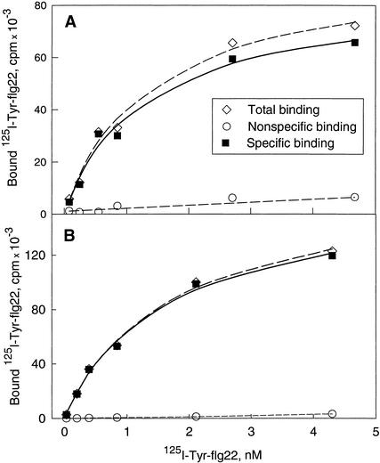
Incubation of intact cells or microsomal membranes with increasing concentrations of 125I-Tyr-flg22 showed saturable numbers of binding sites (Figure 5). Maximal binding, calculated by fitting data of specific binding to hyperbolic curves, was 3 pmol g−1 cells and 2 pmol mg−1 microsomal membrane protein, respectively. Half-maximal binding was observed at concentrations of 1.3 nM for intact cells and 1.6 nM for microsomal membranes, respectively. Further experiments confirmed that >90% saturation was reached with ligand concentrations >10 nM (data not shown). Although at saturating conditions an equilibrium of binding can be assumed, equilibrium of binding was not necessarily reached for lower concentrations. Thus, the concentrations required to obtain half-maximal binding represent only estimates, probably upper limits, of the dissociation constant Kd.
Figure 5.
Saturation of Binding to Intact Cells and Microsomal Membranes.
Saturation of binding (>90%) was reached at ligand concentrations >10 nM in two repetitions of these saturation assays. For different batches of cells and membrane preparations (n > 3), the number of binding sites varied by two- to threefold.
(A) Various amounts of 125I-Tyr-flg22 diluted with nonradioactive Tyr-flg22 to the specific radioactivity of 200 Ci/mmol were incubated with tomato cells (0.5-mL aliquots containing 0.06 g, fresh weight, of cells) at 4°C for 20 min in the absence (total binding) or in the presence of 10 μM Tyr-flg22 (nonspecific binding). To determine specific binding, nonspecific binding was subtracted from total binding.
(B) Various amounts of 125I-Tyr-flg22 diluted with nonradioactive Tyr-flg22 to the specific radioactivity of 200 Ci/mmol were incubated with microsomal membranes (100 μg of protein) at 4°C for 20 min in the absence (total binding) or in the presence of 10 μM Tyr-flg22 (nonspecific binding). To determine specific binding, nonspecific binding was subtracted from total binding.
Specificity of 125I-Tyr-flg22 Binding
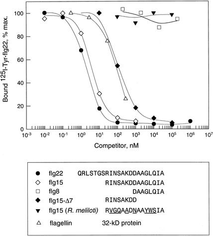
Specificity of binding was tested in competition assays with 125I-Tyr-flg22 and increasing concentrations of unlabeled intact flagellin protein or various peptides exhibiting a range of different activities as agonists or antagonists of flagellin. Figure 6 shows examples of competition curves in binding assays with microsomal membranes incubated for 20 min at 4°C. The concentrations required to inhibit binding of the radioligand by 50% (IC50) were approximated from the curves. Table 1 summarizes these IC50 values and the IC50 values obtained with a variety of other peptides in analogous competition assays and compares them with the EC50 values in the alkalinization response described earlier (Felix et al., 1999). Intact flagellin protein efficiently competed with radioligand binding (IC50 = 120 nM). The flagellin-derived peptides Tyr-flg22, flg22, and flg15, which act as the most potent inducers of the alkalinization response, were also most active as competitors of binding, having IC50 values of 1, 2, and 7 nM, respectively. Thus, the peptide flg15 spans the core domain necessary for binding and biological activity in tomato cells. In comparison with flg15, further shortening of the peptide from both ends led to rapid loss of both elicitor activity and the capacity to compete in the binding assays. However, shortening or modifying the N terminus, as in peptides flg13 or flg15-swap8-9, affected biological activity less drastically than it did the binding competition. In contrast, truncation at the C terminus, as in peptides flg15-Δ3, flg15-Δ4, and flg15-Δ7, reduced agonist activity more drastically than it did the activity in competitive binding assays. As described earlier, flg15-Δ4 through flg15-Δ7 exhibit no measurable agonist activity but act as competitive antagonists for flagellin elicitors. Peptides that showed neither agonist nor antagonist activity in bioassays, such as flg8 or the structurally related peptides flg15R.mel. and flg15A.tum., had no activity as competitors (IC50 >>105 nM). Also, no competition was observed with structurally unrelated peptides such as systemin.
Figure 6.
Competition of 125I-Tyr-flg22 Binding by Flagellin and Different Peptides.
Binding assays with 125I-Tyr-flg22 (0.4 nM) and various concentrations of unlabeled flagellin protein or flagellin-derived peptides flg22, flg15, flg8, flg15-Δ7, and flg15R.mel. (the underlined amino acids in flg15 from R. meliloti indicate differences from flg15 of Pseudomonas). Results were obtained with different batches of microsomal membrane preparations and are presented as the percentage of specific binding. Total binding for these different batches was between 16,000 and 18,000 cpm, and nonspecific binding was between 800 and 1200 cpm. Competition of binding was tested in at least two independent assays for all competitors. IC50 values for flg22, used as a reference in the different experiments, varied between 0.9 and 3.7 nM (mean ±sd = 1.9 ± 1.2 nM in four different experiments).
Table 1.
Alkalinization-Inducing Activity and Binding Activity of Flagellin and Flagellin-Derived Peptides
| Protein or Peptide | Amino Acid Sequencea | Biological Activity EC50 (nM)b |
Binding Affinity IC50 (nM)c |
|---|---|---|---|
| Flagellin | Intact protein, 33 kD | 0.30 | 120 |
| Tyr-flg22 | YQRLSTGSRINSAKDDAAGLQIA | 0.03 | 1 |
| flg22 | QRLSTGSRINSAKDDAAGLQIA | 0.03 | 2 |
| flg15 | RINSAKDDAAGLQIA | 0.06 | 7 |
| flg15-Δ3 | RINSAKDDAAGL | 15.00 | 80 |
| flg15-Δ4 | RINSAKDDAAG | Antagonist | 400 |
| flg15-Δ7 | RINSAKDD | Antagonist | 400 |
| flg13 | NSAKDDAAGLQIA | 75.00 | 600,000 |
| flg8 | DAAGLQIA | Inactive | Inactive |
| flg15-swap8-9 | RINSAKDADAGLQIA | 14.00 | 12,000 |
| flg15A.tum. | RVGDASDNAAYWSIA | Inactive | Inactive |
| flg15R.mel. | RVGQAADNAAYWSIA | Inactive | Inactive |
| flg15E.coli | RINSAKDDAAGQAIA | 0.20 | 6 |
| Systemin | AVQSKPPSKRDPPKMQTD | Inactive | Inactive |
Amino acid sequence of synthetic peptides used. Amino acids underlined indicate differences from the sequence in Pseudomonas aeruginosa.
Concentration that induces half-maximal alkalinization response in tomato cells. Some of the values are reproduced from Felix et al. (1999). Inactive, no significant alkalinization response when tested in concentrations up to 10 μM (EC50 >> 10,000 nM).
Concentration required to reduce binding of 125I-Tyr-flg22 to microsomal membranes by 50% when measured after 20 min of incubation at 4°C. Inactive, no significant competition when tested in concentrations up to 100 μM (IC50 >> 100,000 nM).
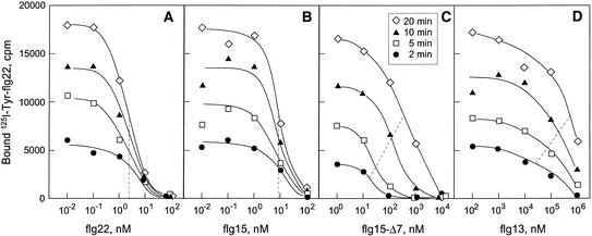
Higher IC50 values in competition assays indicated lower affinities for the binding site—whether because of lower rates of association or higher rates of dissociation (or both) than for 125I-Tyr-flg22. Regarding the nonreversibility of radioligand binding, IC50 values might also depend on the time of incubation. We compared competition of binding by flg22, flg15, flg13, and flg15-Δ7 after different periods of incubation (Figure 7). The IC50 values for flg22 and flg15 were low and stable for assays of 2, 5, 10, and 20 min (Figures 7A and 7B), thereby indicating rates of association and dissociation similar to those of 125I-Tyr-flg22 and, consequently, demonstrating essentially nonreversibility of binding for these peptides. The IC50 values for flg13 and flg15-Δ7 were higher and increased with the duration of the competition assays (Figures 7C and 7D), thus demonstrating weaker and reversible association of these peptides with the binding site.
Figure 7.
Time-Dependence of Binding Competition by Flagellin-Derived Peptides.
Time dependence of binding competition was confirmed in at least two independent sets of experiments for all peptides.To assess the competitive binding of 125I-Tyr-flg22 (0.4 nM) and various concentrations of peptides, radioligand, competitors, and microsomal membranes were incubated at 4°C for 2 (closed circles), 5 (open squares), 10 (closed triangles), and 20 min (open diamonds) as indicated. Dashed lines connect the IC50 values observed at the different times.
(A) flg22.
(B) flg15.
(C) flg15-Δ7.
(D) flg13.
Rather than representing equilibrium or inhibition “constants,” IC50 values indicate the relative efficiency of the competitors to inhibit tracer binding for a given time of incubation. Nevertheless, for all peptides tested, the relative efficiency of competing in binding assays correlated with their biological activity as agonists and antagonists. This provides strong evidence that the binding assays are dealing with a site relevant for biological activity.
Affinity Cross-Linking of 125I-Tyr-flg22
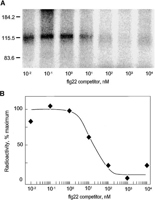
Covalent affinity cross-linking of labeled ligands and their binding sites has been used previously to estimate the molecular mass of binding sites for a heptaglucan elicitor (Frey et al., 1993), an N-acetylchitooligosaccharide elicitor (Ito et al., 1997), and the Pep13 elicitor derived from Phytophthora sojae (Nürnberger et al., 1995). In experiments with microsomal membrane preparation derived from tomato cells, we observed specific cross-linking of 125I-Tyr-flg22 to a single polypeptide migrating with an apparent molecular mass of ∼115 kD on SDS-PAGE. Figure 8 shows an example of cross-linking with the homo-bifunctional chemical cross-linker dithiobis(succinimidyl propionate). Increasing concentrations of unlabeled flg22 competitively inhibited the labeling of this protein. In good agreement with the competitive binding studies described above, a 50% reduction of labeling was reached at ∼10 nM flg22. Specific labeling of a single 115-kD polypeptide was also visible with other cross-linkers, notably disuccinimidyl suberate, and SDS-PAGE under reducing conditions (data not shown). On the basis of the total specific binding of ligand by microsomal membranes under the assay conditions used, the estimated efficiency of cross-linking the radioligand to its binding site was only ∼1%. Nevertheless, this efficiency was much higher than nonspecific cross-linking to other proteins, as inferred from the fact that separating the receptor–ligand complexes from free 125I-Tyr-flg22 before cross-linking was not necessary, in contrast to many other binding studies with plant membranes.
Figure 8.
Chemical Cross-Linking of 125I-Tyr-flg22 to Microsomal Membranes.
Specific labeling of a single 115-kD polypeptide was reproduced in two independent experiments with dithiobis(succinimidyl propionate) and in one experiment with disuccinimidyl suberate as the cross-linkers.
(A) Aliquots of microsomal membranes were incubated with 125I-Tyr-flg22 and different concentrations of unlabeled Tyr-flg22 for 30 min on ice. Cross-linking was initiated by addition of dithiobis(succinimidyl propionate), and radiolabeled proteins were analyzed (with a PhosphorImager) after separation by SDS-PAGE. Numbers and dashes at left denote positions and molecular masses in kilodaltons of standard proteins.
(B) Quantitative analysis of radioactivity in the band migrating at 115 kD in (A).
DISCUSSION
In the present work, we used a radioiodinated derivative of flg22 as a molecular probe to identify and characterize a high-affinity binding site on the surface of tomato cells. This binding site, present in intact cells and membrane preparations derived from these cells, showed rapid, specific, and saturable binding of radioligand. Most strikingly, the binding was nonreversible under physiological conditions, and bound ligand could not be displaced even by prolonged incubation in the presence of excess nonlabeled ligands. Nonreversibility with intact cells could reflect an uptake process, but the binding of 125I-Tyr-flg22 to membrane preparations exhibited the same characteristics. Preliminary data show that the binding site also retains these characteristics after solubilization with detergents (T. Meindl, unpublished results). In prolonged dissociation experiments, >50% of the label still bound to the microsomal membrane proteins 5 days after addition of competitor, indicating a very slow rate of dissociation and an extremely stable receptor–ligand complex. However, the binding was not covalent and ligand was released from the binding site by heating, treating with 1% SDS, or using moderately acidic conditions.
Irreversibility of binding or very slow rates of dissociation are common phenomena for many human and animal effector–receptor interactions. Such agonist “locking” has been reported in particular for G-protein–coupled receptors, for example, the somatostatin receptor (Schönbrunn et al., 1993). The locking of ligands to these receptors is often reversed by treatment with low concentrations of guanine nucleotides (GTP), which lead to greatly accelerated rates of agonist release. Preliminary experiments with 125I-Tyr-flg22 bound to solubilized tomato membrane proteins, however, showed no accelerated dissociation after addition of GTPγS (T. Meindl, unpublished results). A slow rate of dissociation has been reported also for the fungal elicitor AVR9, which dissociated from its binding site in tomato membranes with a half-life of ∼4 hr (Kooman-Gersmann et al., 1996). However, association of AVR9 with its binding protein also proceeded slowly, requiring >60 min to approach a maximum, even at 37°C. In contrast, association of 125I-Tyr-flg22 with its binding site occurred rapidly, even at 4°C (Figure 2). This rapid binding is in good agreement with the onset of physiological responses such as medium alkalinization, which start after a lag phase of <2 min.
Ligand saturation experiments showed a binding capacity of ∼3 pmol g−1 intact cells or, assuming a mass of 5 ng per cell, a number in the order of ∼104 binding sites per cell. Half-saturation of binding sites was reached at a ligand concentration of ∼1 nM. Although this value does not represent a dissociation constant derived from a binding equilibrium, it can be compared with the dose dependence for stimulation of the alkalinization response. For Tyr-flg22, the half-maximal response is observed at only 0.03 nM, and a concentration of 1 nM induces a full response. Accordingly, occupancy of binding site and strength of the alkalinization response do not show a linear, one-to-one, relationship. Rather, the full alkalinization response is triggered under conditions in which only a fraction of the binding sites are occupied. A surplus of receptor sites, also termed “spare” receptors, is characteristic of many receptor-mediated signaling processes in animals and has also been observed with elicitor-active chitin fragments in tomato cells (Baureithel et al., 1994). The presence of a relatively high number of functional receptor sites might explain the extreme sensitivity of the tomato cells for the most active flagellin elicitors. In turn, a high or complete occupancy of the binding sites by antagonists appears necessary for efficient inhibition of agonist action, thus explaining the requirement of a high molar excess of antagonists over agonists for full suppression of the agonist.
Competitive antagonists such as flg15-Δ7 suppressed the induction of biological responses most efficiently when added concomitantly with the agonists. A much weaker suppression was observed when the antagonists were applied only a few seconds after the agonists (Figure 1). The binding kinetics provide an explanation for these aspects of agonist and antagonist actions: binding of flg15-Δ7 to the binding site is readily reversible, whereas agonists such as Tyr-flg22, flg22, and flg15 bind in a manner that is essentially nonreversible. The duration of response activation for many receptor-mediated processes in animals and plants depends on continuous activation of the receptor and continuous presence of the respective ligands. In the tomato cells used in this study, continuous presence of elicitor stimuli has been found to be required for continuous stimulation of responses with fungal elicitors (Felix et al., 1991; Basse et al., 1993). For the flagellin-derived peptide elicitors studied in this report, we observed rapid disappearance of free elicitor from the cell suspension, whereas ligand bound to the binding site was protected from degradation over several hours. Thus, a “hit and stick” rather than a “hit and go” mechanism could cause the prolonged activation of elicitor responses such as alkalinization of medium observed for the most active flagellin peptides.
Specificity of the binding site was tested in binding competition assays (Table 1). Given the essentially nonreversible binding of the radioligand, these IC50 values are not equilibrium constants and do not represent inhibition constants or binding constants for the competitors. In particular, for ligands with relatively weak affinities, these values represent underestimations with regard to equilibrium conditions. However, they probably represent overestimations with regard to the binding conditions during the first minutes—the time relevant for receptor activation and induction of biological responses. Despite these limitations, the relative capacity to compete with radioligand binding strongly correlated with the relative activity of the agonists or antagonists in bioassays for all compounds tested. Strongest competition was observed for the strongest agonists: Tyr-flg22, flg22, flg15, and intact flagellin. Strong competition was found also for antagonists, such as flg15-Δ7. Flg8, comprising the 8 amino acids at the C terminus, showed no affinity for the binding site and no apparent activity in bioassays. Nevertheless, the C terminus is essential for activation of the biological responses. Apparently, binding and activation can be attributed to distinct N- and C-terminal domains. However, applying peptides flg15-Δ7 and flg8 in combination, even at very high doses, did not result in induction of biological responses (data not shown), indicating that binding and activation most likely occur as linked processes. Therefore, we hypothesize that perception of the flagellin elicitor involves two consecutive, linked steps, as tentatively schematized in Figure 9: the N terminus of the elicitor peptide binds to a high-affinity binding site in a first step and activates the receptor with its C terminus in a second step. The second step could be envisaged as an intramolecular process of isomerization, as shown in the model, or it could involve interaction with a second protein that functions as a coreceptor. Locking and essentially nonreversible binding of the most active agonists might result from the product of the two steps: strong bimolecular interaction with the binding site, and the isomerization process. Accordingly, the antagonistic peptides lacking the active C-terminal domain apparently interact with the high-affinity binding site but do not induce the isomerization step; consequently, no activation of responses occurs, the ligand is not locked, and binding remains reversible. In contrast, Flg13, a peptide truncated at the N terminus, shows low affinity for the binding site in competition assays; nonetheless, it efficiently induces the second step of isomerization and activation of the receptor by its functional C-terminal domain. Cautious interpretation of IC50 values from binding competition and the concept of spare receptors discussed above might provide an explanation for the apparent discrepancy between binding affinity and biological activity observed for this peptide (cf. IC50 and EC50 values in Table 1).
Figure 9.
Model for the Interaction of Flagellin with Its Receptor.
Model depicting the interaction of the flagellin elicitor with its putative receptor as a two-step process. Binding of the N terminus is presented as the first step, and activation by the C terminus is the second step.
Perception of flagellin-derived peptides exhibits remarkable similarities to the perception of the plant wound hormone systemin by tomato (Meindl et al., 1998). This 18–amino acid peptide also binds with the N terminus to its receptor but needs the C-terminal domain to activate it. On the basis of the biological activity of different systemin analogs and their apparent affinity for the receptor site, we proposed a model of systemin perception that involved “induced fit” on the receptor site. That is, binding of the N terminus of the peptide would lead to a conformational change that in a subsequent step would enable its C terminus to bind and activate the receptor. Bipartite function has also been described previously for fungal-derived glycopeptide elicitors (Basse and Boller, 1992). The glucan parts of these elicitors are inactive as agonists but act as competitive antagonists and also compete with the complete glycopeptides for binding to the high-affinity binding site present on tomato membranes. Bipartite function is well known in animal hormones, and this characteristic has led to the concept of “message–address” (Schwyzer, 1977): part of the signaling molecule is required for binding, conferring selectivity for the receptor (address), and part for the biological action, mediating signal transduction (message). Examples of such signaling molecules in animals include several hormones that are active in the nervous system, such as ACTH (Schwyzer, 1977), opioids (Schwyzer, 1986; Portoghese, 1989), and neurokinins (Schwyzer, 1987; Seelig et al., 1996).
Chemical cross-linking of radiolabeled elicitor ligands to their respective high-affinity binding sites has previously been used to identify specific binding proteins in plants. Examples include polypeptides specifically cross-linked to the P. sojae–derived elicitors hepta-β-glucoside (Cosio et al., 1992) and Pep-13 (Nürnberger et al., 1995). Chemical cross-linking of the radioiodinated flagellin peptide used in this study labeled a single membrane-derived protein that migrated with a molecular mass of ∼115 kD on SDS-PAGE. The coupling of labeled ligand to this polypeptide was inhibited by unlabeled flg22 with a concentration dependence corresponding to that found in binding assays. Together, these data provide evidence that the covalently labeled 115-kD polypeptide represents at least part of the high-affinity receptor for bacterial flagellin.
In intact seedlings of Arabidopsis, flagellin-derived peptides cause pronounced growth inhibition (Gómez-Gómez et al., 1999). Recently, this effect was successfully used to isolate several mutants with affected responses to flagellin that map to more than one locus (Gómez-Gómez and Boller, 2000). One of the genes, termed fls2, involved in flagellin perception was found by mapping, sequencing, and complementation to code for a protein that has the structure of a transmembrane receptor kinase with leucine-rich repeats (Gómez-Gómez and Boller, 2000). Current work aims at studying the role of the fls2 protein in binding of the flagellin elicitor. Preliminary results show reduced binding in flagellin mutants affected in fls2 but also in mutants affected in different genes (Z. Bauer, unpublished results). Thus, at present, we cannot attribute binding of flagellin to the gene product of fls2 alone.
In summary, our data provide strong evidence that the novel high-affinity binding site described in this report represents the functional receptor for the bacterial elicitor flagellin. Binding characteristics of agonists and antagonists can explain molecular aspects of receptor activation through an address–message mechanism and locking of the agonist. The biochemical approach described in this article, together with the genetic approach used with Arabidopsis, should provide the basis for further efforts concentrating on the isolation and characterization of elements involved in flagellin perception and in the early steps of intracellular signal transduction.
METHODS
Flagellin-Derived Peptides and Radioiodination
The flagellin-derived peptides were synthesized according to the consensus sequence for the most highly conserved region in the N terminus of eubacterial flagellin (Felix et al., 1999). flg22 (QRLSTGSRINSAKDDAAGLQIA), flg15 (RINSAKDDAAGLQIA), flg15-Δ7 (DAAGLQIA), flg8 (RINSAKDD), flg15R.mel. (RVGQAADNAAYWSIA), and flg15A.tum. (RVGDASDNAAYWSIA) were synthesized by F. Fischer (Friedrich Miescher-Institute). Peptides were dissolved in water (stock solutions of 1 to 10 mM) and diluted in a solution containing 0.1% BSA and 0.1 M NaCl. For iodination, flg22 was N-terminally extended by adding a Tyr residue (Tyr-flg22). Tyr was iodinated nonradioactively with Chloramine-T as described previously (Meindl et al., 1998) or labeled with 125I, also using Chloramine-T, to yield [3-125I]iodoty-rosine-flg22 (125I-Tyr-flg22) with a specific radioactivity of >2000 Ci/mmol by Anawa Trading SA (Wangen, Switzerland).
Cell Cultures and Measurement of Alkalinization Response
Cell cultures of tomato were subcultured as described previously (Felix et al., 1991). For assaying the alkalinization response, aliquots of the cell suspension, 5 to 8 days after subculture, were incubated at room temperature in open flasks on a rotary shaker at 120 cycles per min. Extracellular pH was measured with a small, combined, glass pH electrode (Metrohm, Herisau, Switzerland); continuous measurements were registered with a pen recorder.
Depending on the age of the culture, the density of cells, and the extracellular pH, different batches of cells showed maximal pH increases (ΔpHmax) to saturating doses of flg22 that ranged from 0.7 to 1.4 pH units (Felix et al., 1999). Within one batch of cells, as used in bioassays for the experiments shown, alkalinization to replicate treatments with the same dose of flg22 varied little (mean sd <10%).
Binding Assays with Intact Cells and Microsomal Membrane Preparations
For binding assays with intact cells, cultured tomato cells were used 5 to 8 days after subculture. Aliquots (0.5 mL) of suspension containing ∼60 mg cells (fresh weight) were incubated with 125I-Tyr-flg22 (60 fmol in standard assays) for the times indicated. Radioactivity bound or retained by the cells was determined by γ-counting after washing with 15 mL of ice-cold wash buffer (25 mM Tris-Mes, pH 7.4, 100 mM MgCl2, 100 mM KCl, 10 mM KI, 0.1% tryptone, and 0.1% BSA).
Microsomal membranes were prepared from suspension-cultured cells of tomato as described previously (Grosskopf et al., 1990), except that the cells were broken in a cell disruption bomb (Parr Instrument Co., Moline, IL) rather than a French press. For binding assays, aliquots of microsomal membranes containing 100 μg of protein were incubated with 60 fmol of 125I-Tyr-flg22 (>2000 Ci /mmol) in a total volume of 100 μL of binding buffer. Binding buffer, designed to minimize nonspecific binding, contained 25 mM Tris-Mes, pH 7.4, 10 mM MgCl2, 10 mM KI, 5 mM 3-iodo-l-tyrosine, 5 mM l-lysine, 0.1% tryptone, and 0.1% BSA. In long-term binding assays extending over several days, the binding buffer was supplemented with 3 mM NaN3 to prevent growth of microbial contaminants. After incubation on ice for the times indicated, cells and microsomal membranes were collected by vacuum filtration on paper filters (MN 713; Machery-Nagel, Düren, Germany) and glass fiber filters (GF/B; Whatman), respectively, and washed for 10 sec with 15 mL of ice-cold wash buffer (see above). Radioactivity retained on filters was measured by γ-counting.
In assays studying binding saturation with intact cells and membrane preparations, the specific radioactivity of 125I-Tyr-flg22 was decreased to 200 Ci/mmol by dilution with nonradioactive Tyr-flg22.
Replicate measurements in standard binding assays showed a mean sd <10% with membrane preparations and <20% with intact cells.
Chemical Cross-Linking
Microsomal membranes were prepared in PBS buffer, pH 8.0. Aliquots (100 μL) containing 100 μg of protein and 60 fmol of 125I-Tyr-flg22 were incubated in the presence or absence of unlabeled Tyr-flg22 for 30 min on ice. Cross-linking was initiated by addition of dithiobis(succinimidylpropionate) or disuccinimidyl suberate (both from Pierce Chemical, Rockford, IL) to a final concentration of 8 mM. After incubation for 1 hr on ice and an additional 1 hr at room temperature, the samples were solubilized in SDS sample buffer (50 mM Tris-HCl, pH 6.8, 5% [w/v] SDS, and 10% [v/v] glycerol) under nonreducing conditions (15 min at room temperature, 5 min at 95°C). Proteins were separated by SDS-PAGE on gels containing 7% (w/v) acrylamide. Gels were dried, analyzed, and quantified using a PhosphorImager (Molecular Dynamics, Sunnyvale, CA).
Acknowledgments
We thank Franz Fischer (Friedrich Miescher-Institute) for the synthesis of various peptides, Martin Regenass for maintaining the cell cultures, and Dr. Arnd Sturm for critically reading the manuscript.
References
- Basse, C.W., and Boller, T. (1992). Glycopeptide elicitors of stress responses in tomato cells. N-linked glycans are essential for activity but act as suppressors of the same activity when released from the glycopeptides. Plant Physiol. 98, 1239–1247. [DOI] [PMC free article] [PubMed] [Google Scholar]
- Basse, C.W., Fath, A., and Boller, T. (1993). High affinity binding of glycopeptide elicitor to tomato cells and microsomal membranes and displacement by specific glycan suppressors. J. Biol. Chem. 268, 14724–14731. [PubMed] [Google Scholar]
- Baureithel, K., Felix, G., and Boller, T. (1994). Specific, high affinity binding of chitin fragments to tomato cells and membranes. Competitive inhibition of binding by derivatives of chitooligosaccharides and a Nod factor of Rhizobium. J. Biol. Chem. 269, 17931–17938. [PubMed] [Google Scholar]
- Boller, T. (1995). Chemoperception of microbial signals in plant cells. Annu. Rev. Plant Physiol. Plant Mol. Biol. 46, 189–214. [Google Scholar]
- Cosio, E.G., Frey, T., and Ebel, J. (1992). Identification of a high-affinity binding protein for a hepta-β-glucoside phytoalexin elicitor in soybean. Eur. J. Biochem. 204, 1115–1123. [DOI] [PubMed] [Google Scholar]
- Felix, G., Grosskopf, D.G., Regenass, M., Basse, C.W., and Boller, T. (1991). Elicitor-induced ethylene biosynthesis in tomato cells: Characterization and use as a bioassay for elicitor action. Plant Physiol. 97, 19–25. [DOI] [PMC free article] [PubMed] [Google Scholar]
- Felix, G., Duran, J.D., Volko, S., and Boller, T. (1999). Plants have a sensitive perception system for the most conserved domain of bacterial flagellin. Plant J. 18, 265–276. [DOI] [PubMed] [Google Scholar]
- Frey, T., Cosio, E.G., and Ebel, J. (1993). Affinity purification and characterization of a binding protein for a hepta-β-glucoside phytoalexin elicitor in soybean. Phytochemistry 32, 543–549. [Google Scholar]
- Gómez-Gómez, L., and Boller, T. (2000). A LRR receptor-like kinase is involved in the perception of the bacterial elicitor, flagellin, in Arabidopsis. Mol. Cell 5, 1003–1011. [DOI] [PubMed] [Google Scholar]
- Gómez-Gómez, L., Felix, G., and Boller, T. (1999). A single locus determines sensitivity to bacterial flagellin in Arabidopsis thaliana. Plant J. 18, 227–284. [DOI] [PubMed] [Google Scholar]
- Grosskopf, D.G., Felix, G., and Boller, T. (1990). K-252a inhibits the response of tomato cells to fungal elicitors in vivo and their microsomal protein kinase in vitro. FEBS Lett. 275, 177–180. [DOI] [PubMed] [Google Scholar]
- Hahn, M.G. (1996). Microbial elicitors and their receptors in plants. Annu. Rev. Phytopathol. 34, 387–412. [DOI] [PubMed] [Google Scholar]
- Hammond-Kosack, K.E., and Jones, J.D.G. (1997). Plant disease resistance genes. Annu. Rev. Plant Physiol. Plant Mol. Biol. 48, 575–607. [DOI] [PubMed] [Google Scholar]
- Ito, Y., Kaku, H., and Shibuya, N. (1997). Identification of a high-affinity binding protein for N-acetylchitooligosaccharide elicitor in the plasma membrane of suspension-cultured rice cells by affinity labeling. Plant J. 12, 347–356. [DOI] [PubMed] [Google Scholar]
- Kooman-Gersmann, M., Honée, G., Bonnema, G., and De Wit, P.J.G.M. (1996). A high-affinity binding site for the avr9 peptide elicitor of Cladosporium fulvum is present on plasma membranes of tomato and other solanaceous plants. Plant Cell 8, 929–938. [DOI] [PMC free article] [PubMed] [Google Scholar]
- Meindl, T., Boller, T., and Felix, G. (1998). The plant wound hormone systemin binds with the N-terminal part to its receptor but needs the C-terminal part to activate it. Plant Cell 10, 1–11. [DOI] [PMC free article] [PubMed] [Google Scholar]
- Nürnberger, T. (1999). Signal perception in plant pathogen defense. Cell. Mol. Life Sci. 55, 167–182. [DOI] [PMC free article] [PubMed] [Google Scholar]
- Nürnberger, T., Nennstiel, D., Hahlbrock, K., and Scheel, D. (1995). Covalent cross-linking of the Phytophthora megasperma oligopeptide elicitor to its receptor in parsley membranes. Proc. Natl. Acad. Sci. USA 92, 2338–2342. [DOI] [PMC free article] [PubMed] [Google Scholar]
- Parker, J.E., and Coleman, M.J. (1997). Molecular intimacy between proteins specifying plant–pathogen recognition. Trends Biochem. Sci. 22, 291–296. [DOI] [PubMed] [Google Scholar]
- Portoghese, P.S. (1989). Bivalent ligands and the message-address concept in the design of selective opioid receptor antagonists. Trends Pharmacol. Sci. 10, 230–235. [DOI] [PubMed] [Google Scholar]
- Schönbrunn, A., Lee, A.B., and Brown, P.J. (1993). Characterization of a biotinylated somatostatin analog as a receptor probe. Endocrinology 132, 146–152. [DOI] [PubMed] [Google Scholar]
- Schuster, S.C., and Khan, S. (1994). The bacterial flagellar motor. Annu. Rev. Biophys. Biomol. Struct. 23, 509–539. [DOI] [PubMed] [Google Scholar]
- Schwyzer, R. (1977). ACTH: A short introductory review. Ann. N.Y. Acad. Sci. 297, 3–26. [DOI] [PubMed] [Google Scholar]
- Schwyzer, R. (1986). Molecular mechanism of opioid receptor selection. Biochemistry 25, 6335–6342. [DOI] [PubMed] [Google Scholar]
- Schwyzer, R. (1987). Membrane-assisted molecular mechanism of neurokinin receptor subtype selection. EMBO J. 6, 2255–2259. [DOI] [PMC free article] [PubMed] [Google Scholar]
- Seelig, A., Alt, T., Lotz, S., and Hoelzemann, G. (1996). Binding of substance P agonists to lipid membranes and to the neurokinin-1 receptor. Biochemistry 35, 4365–4374. [DOI] [PubMed] [Google Scholar]
- Umemoto, N., Kakitani, M., Iwamatsu, A., Yoshikawa, M., Yamaoka, N., and Ishida, I. (1997). The structure and function of a soybean beta-glucan-elicitor–binding protein. Proc. Natl. Acad. Sci. USA 94, 1029–1034. [DOI] [PMC free article] [PubMed] [Google Scholar]
- Yang, Y., Shah, J., and Klessig, D.F. (1997). Signal perception and transduction in plant defense responses. Genes Dev. 11, 1621–1639. [DOI] [PubMed] [Google Scholar]


