Abstract
Background
The cryptochrome 1 and 2 genes (cry1 and cry2) are necessary for the generation of circadian rhythms, as mice lacking both of these genes (cry1,2-/-) lack circadian rhythms. We studied sleep in cry1,2-/- mice under baseline conditions as well as under conditions of constant darkness and enforced wakefulness to determine whether cryptochromes influence sleep regulatory processes.
Results
Under all three conditions, cry1,2-/- mice exhibit the hallmarks of high non-REM sleep (NREMS) drive (i.e., increases in NREMS time, NREMS consolidation, and EEG delta power during NREMS). This unexpected phenotype was associated with elevated brain mRNA levels of period 1 and 2 (per1,2), and albumin d-binding protein (dbp), which are known to be transcriptionally inhibited by CRY1,2. To further examine the relationship between circadian genes and sleep homeostasis, we examined wild type mice and rats following sleep deprivation and found increased levels of per1,2 mRNA and decreased levels of dbp mRNA specifically in the cerebral cortex; these changes subsided with recovery sleep. The expression of per3, cry1,2, clock, npas2, bmal1, and casein-kinase-1ε did not change with sleep deprivation.
Conclusions
These results indicate that mice lacking cryptochromes are not simply a genetic model of circadian arrhythmicity in rodents and functionally implicate cryptochromes in the homeostatic regulation of sleep.
Keywords: circadian genes, oscillatory network of transcriptional factors, EEG slow-wave activity
Background
Sleep is regulated by both circadian and homeostatic mechanisms. As a consequence of a signal from the circadian clock, located in the suprachiasmatic nuclei (SCN) of the anterior hypothalamus in mammals [1], sleep is more likely to occur at certain times of the 24-h day than others, thereby determining the daily sleep-wake distribution [2]. Sleep is homeostatically regulated in the sense that sleep drive accumulates in the absence of sleep and decreases during sleep. Changes in sleep drive are thus driven by the sleep-wake history. Homeostatic and circadian mechanisms interact to determine the duration and quality of sleep and wakefulness [3-5]. Homeostatic regulation of sleep can still be observed in animals that lack circadian rhythms after lesioning of the SCN [6-9], suggesting that circadian rhythms and sleep homeostasis are independent processes.
Homeostatic regulation of sleep can be quantified objectively after a period of enforced wakefulness (i.e., sleep deprivation), although a similar relationship between sleep parameters and spontaneous wakefulness can be quantified under baseline conditions as well [10,11]. The compensatory responses in time spent asleep, sleep consolidation (i.e., sleep bout duration; [12]), and/or sleep intensity observed after an extended period of wakefulness are all taken as evidence that sleep drive is increased during wakefulness and thus that sleep is homeostatically regulated. Non-REM sleep (NREMS) intensity, quantified as EEG power in the delta frequency range (1–4 Hz), and NREMS consolidation are in a quantitative and predictive relationship with sleep history: both variables increase with the duration of prior wakefulness and subsequently decline during NREMS [3,4,10,12,13]. These variables are thus quantitative markers of NREMS homeostasis and are presumed to reflect an underlying physiological drive for sleep [4].
The time constants describing the dynamics of the increasing NREMS drive during wakefulness and decreasing NREMS drive during NREMS [10,14] are compatible with a role for changes in gene expression in their regulation. Although past studies have shown that extended periods of wakefulness cause changes in gene expression (for review see [15]), no causal relationship between changes in gene expression and sleep homeostasis has been identified. A recent study in Drosophila melanogaster implicates transcriptional regulation by the circadian gene cycle, a homolog of the mammalian bmal1 gene, in the homeostatic regulation of rest [16]. While rest in flies shares several features with sleep in mammals [17,18], it remains to be determined whether a similar role for BMAL1 or related transcriptional regulators is necessary for the homeostatic regulation of sleep.
In both flies and mammals, circadian rhythms are thought to be generated by transcriptional/translational feedback loops comprising a network of transcriptional regulators [19-21]. The core of this self-sustained molecular oscillation consists of positive and negative elements. In mammals, the positive elements are two basic helix-loop-helix (bHLH) PAS-domain-containing transcription factors, CLOCK and BMAL1, that form heterodimers that can drive the transcription of three period (per) genes per1–3 and two cryptochromes (cry1,2). PER1,2 and CRY1,2 proteins suppress CLOCK:BMAL1-mediated transcription thereby forming the negative elements in the feedback loop. Consistent with their central role in circadian rhythm generation, genetic inactivation of both cryptochromes (cry1,2-/-) results in circadian arrhythmicity in mice [22,23]. Given the widespread expression of the elements of the molecular clock in the brain (and elsewhere) and the large number of genes regulated by bHLH-PAS transcription factors (e.g. [24]), the role of cryptochromes may extend beyond circadian clock function. To determine whether transcriptional regulation by CRY1,2 influences the homeostatic regulation of sleep, we studied sleep in cry1,2-/- mice under baseline conditions, under conditions of constant darkness, and after sleep deprivation (SD). The expression of circadian genes that are regulated by cryptochromes were evaluated in the brain of cry1,2-/- mice and in sleep deprived wild type mice and rats.
Results
Sleep regulation in cry1,2-/- mice
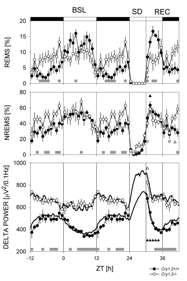
In baseline conditions, wild type mice showed the sleep-wake distribution typical for this nocturnal species with high values of sleep in the light period and low values in the dark period (Figure 1, Table 1). The sleep-wake distribution in cry1,2-/- mice was distinct from that of wild type controls (Figure 1) in that sleep variables did not differ between the 12-h light (L) and dark (D) periods (Table 1). Nevertheless, as in wild type controls, the light-to-dark transition was accompanied by a pronounced decrease in sleep time in cry1,2-/- mice (Figure 1).
Figure 1.
Time course of sleep (upper panels) and NREMS EEG delta power (lower panel). Data from baseline (BSL), sleep deprivation (SD), and recovery (REC) are shown. Open (Cry1,2-/-) and closed (Cry1,2+/+) symbols designate mean hourly values ± 1 SEM. In the lower panel, thicker lines connect mean predicted delta power values based on the sleep-wake distribution in individual mice; (see Methods). Triangles mark intervals in which recovery values differed from corresponding baseline values within each genotype (triangle orientation designates direction of deviation; P < 0.05, post-hoc paired t-tests). Gray bars at the bottom of each panel mark intervals with significant genotype differences (P < 0.05, post-hoc t-tests). The baseline dark period was depicted twice to illustrate the changes at the dark-to-light transition.
Table 1.
Sleep in 12-h light and dark periods under baseline conditions in cry1,2+/+ and cry1,2-/- mice.
| NREMS amount [%] | |||
| light | dark | Difference | |
| cry1,2+/+ | 48.8 ± 1.9# | 31.3 ± 2.3 | 17.5 ± 3.9 |
| cry1,2-/- | 49.5 ± 1.9 | 45.4 ± 1.9* | 4.2 ± 1.6* |
| NREMS bout duration [min] | |||
| light | dark | difference | |
| cry1,2+/+ | 2.9 ± 0.2 | 2.9 ± 0.2 | 0.1 + 0.2 |
| cry1,2-/- | 3.9 ± 0.2* | 4.1 ± 0.4* | -0.2 + 0.2 |
| REMS amount [%] | |||
| light | dark | Difference | |
| cry1,2+/+ | 11.9 ± 1.1# | 3.8 ± 1.0 | 8.2 ± 1.5 |
| cry1,2-/- | 8.5 ± 0.5* | 7.8 ± 0.7* | 0.8 ± 0.8* |
Post-hoc comparisons: * P < 0.05 vs. wild type, unpaired t-tests; # P < 0.05 vs. dark period, within same genotype, paired t-tests. Differences represent the mean of individual light-dark differences.
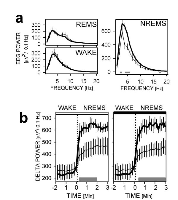
During baseline, total daily NREMS time was significantly greater in cry1,2-/- mice than in wild type mice (683 ± 25 vs. 576 ± 12 min, P < 0.002, unpaired t-test), whereas the daily time spent in REM sleep did not differ (118 ± 7 vs. 113 ± 10 min). The difference between genotypes in NREMS time was present only in the dark period and not in the light (Table 1; Figure 1). Average NREMS bout duration, a measure of NREMS consolidation that is positively correlated with a high homeostatic sleep pressure [13], was greater by 34% in cry1,2-/- mice relative to wild type mice during the light period, and by 41% during the dark period (Table 1). Consistent with this increase in sleep consolidation, NREMS delta power was significantly higher in cry1,2-/- than in cry1,2+/+ mice during most of baseline (Figure 1). This difference in EEG delta power was specific for NREMS and did not extend to other EEG frequencies (Figure 2A), indicating that the increase in delta power is not due to a difference in EEG amplification. This state-specific EEG difference in delta power was especially evident at the wake-to-NREMS transition. cry1,2-/- mice exhibited higher delta power values than cry1,2+/+ immediately after the onset of NREMS (Figure 2B).
Figure 2.
EEG power in the 1–20 Hz range for NREMS, REM sleep (REMS) and wake during baseline. (A) EEG spectral power in cry1,2-/- (thick lines) and wild type mice (thin lines). Differences between the genotypes are limited to NREMS delta power. (B) Delta power (1–4 Hz) during wake-to-NREMS transitions in the baseline light (left) and dark (right panel) period. Gray horizontal bars underneath the curves indicate significant genotype differences (P < 0.05; post-hoc t-tests). Error bars span ± 1 SEM.
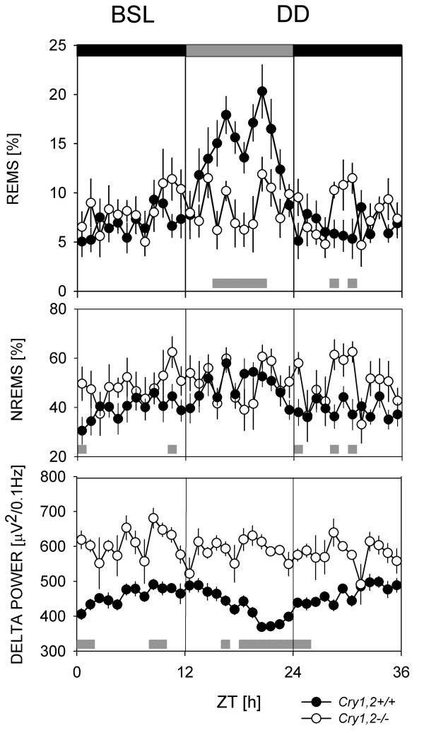
The same genotypic differences in sleep were observed in constant dark (DD) conditions (Figure 3). Mice lacking cry1,2 spent more time in NREMS (719 ± 29 vs. 629 ± 29 min, P = 0.053, unpaired t-test), NREMS bouts were longer (3.8 ± 0.3 vs. 2.6 ± 0.1 min, P < 0.001, unpaired t-test), and EEG delta power in NREMS was higher in cry1,2-/- compared to cry1,2+/+ controls (594 ± 21 vs. 448 ± 52 μV2/0.1 Hz, P < 0.04, unpaired t-test; Figure 3). The decrease in sleep time prior to the onset of the dark period (Figure 1) was still present in DD in cry1,2+/+ mice; sleep time decreased in the latter half of the subjective day and minimum average sleep time was reached immediately after the onset of the subjective night (Figure 3). The anticipatory decrease in sleep time that occurred prior to the onset of dark in cry1,2-/- mice in LD did not occur under DD conditions (Figure 3). Under DD conditions, less REMS was present in cry1,2-/- mice than in cry1,2+/+ mice (119 ± 7 vs. 149 ± 9 min, P < 0.03, unpaired t-test) due to an increase in REMS in cry1,2+/+ mice in DD (149 ± 9 min) relative to LD (113 ± 10 min, P < 0.03, paired t-test).
Figure 3.
Time course of sleep and NREMS EEG delta power during constant dark conditions (DD). Layout and symbols are same as in Figure 1. Gray bars at the bottom of each panel mark intervals with significant genotype differences (P < 0.05, post-hoc t-tests). The subjective day is marked with a gray horizontal bar at the top of the upper panel. The first 12-h represents the last dark-period under baseline (BSL) light-dark conditions.
To further investigate the homeostatic regulation of sleep in cry1,2-/- mice, we assayed the compensatory response to a 6 h SD (see Figure 1). After SD, wild type mice displayed the typical increase in REMS and NREMS time, NREMS bout duration, and EEG delta power above baseline levels ([10,13] Table 2, Figure 1). In contrast, cry1,2-/- mice did not exhibit significant increases in REMS, NREMS time or NREMS bout duration after SD; only a brief increase in NREMS delta power was observed (lasting 1 h in cry1,2-/- vs. 5 h in wild type mice; Figure 1). The initial increase in delta power (relative to baseline), measured over the first recovery hour, was significantly smaller in cry1,2-/- than the increase observed in wild type mice (Table 2). Delta power (Figure 1, Table 2) and NREMS bout duration (Tables 1, 2), in cry1,2-/- mice is maintained at a level only attained by the wild type mice when their sleep pressure was highest, i.e., at the end of the active or dark period and after SD.
Table 2.
Effect of 6 h SD on sleep time and delta power in cry1,2+/+ and cry1,2-/- mice.
| NREMS amount [min] | |||
| baseline | post-SD | difference | |
| cry1,2+/+ | 264 ± 9 | 297 ± 15# | 33 ± 8 |
| cry1,2-/- | 312 ± 9* | 318 ± 9 | 5 ± 4* |
| NREMS bout duration [min] | |||
| baseline | post-SD | difference | |
| cry1,2+/+ | 2.6 ± 0.1 | 3.3 ± 0.3# | 0.7 ± 0.3 |
| cry1,2-/- | 4.2 ± 0.4* | 3.7 ± 0.2 | -0.4 ± 0.3* |
| NREMS Delta power [μV2/0.1 Hz] | |||
| baseline | post-SD | difference [%] | |
| cry1,2+/+ | 407 ± 59 | 704 ± 125# | 170 ± 14 |
| cry1,2-/- | 619 ± 67* | 805 ± 40# | 134 ± 8* |
| REMS amount [min] | |||
| baseline | post-SD | difference | |
| cry1,2+/+ | 52 ± 6 | 63 ± 3# | 11 ± 7 |
| cry1,2-/- | 53 ± 4 | 58 ± 1 | 5 ± 3 |
Post-hoc comparisons: * P < 0.05 vs. wild type, unpaired t-test; # P < 0.05, vs. baseline day, within same genotype, paired t-test. Delta power was calculated over the first hour after the SD (ZT6-ZT7). REMS and NREMS amount and bout duration were calculated over 12 hours (ZT6-ZT18). Differences indicate mean individual post-SD – baseline differences except for delta power which is calculated as percentage of baseline.
In wild type mice, EEG delta power still varied as a function of the sleep-wake history with high values at the end of the baseline dark period and after the SD and low values at the end of the light or major rest period (Figure 1). Presumably due to the altered sleep-wake distribution, the daily range of EEG delta power values was smaller in cry1,2-/- mice than in wild type mice (Table 1, Figure 1). We tested the assumption of a relationship between delta power and the sleep-wake history by using a mathematical method that predicts the level of EEG delta power occurring in individual NREMS bouts based on the 42 h sequence of 10-sec behavioral state scores for individual animals [10]. With this analytical tool, the time constants of the increasing delta power during wakefulness and its decrease during NREMS are estimated. For both genotypes, delta power in both baseline and recovery from SD could be reliably predicted on the basis of sleep-wake history (Figure 1), which is underscored by the highly significant correlations between empirical and simulated data (r = 0.91 and 0.87 for cry1,2+/+ and cry1,2-/-, respectively, P < 0.0001 for both genotypes). Thus, in the absence of cryptochromes, NREMS delta power varied as a function of the prior sleep-wake history. However, in cry1,2-/- mice, a significantly shorter time constant for the increase (i.e., a faster build-up) of delta power was obtained (τincrease: 5.0 ± 0.3 h in cry1,2+/+ vs. 3.5 ± 0.5 h in cry1,2-/-; P < 0.05; unpaired t-test), whereas the time constant describing the decline of delta power during NREMS did not differ (τdecrease: 1.7 ± 0.2 h in cry1,2+/+ vs. 1.8 ± 0.3 h in cry1,2-/-).
Circadian gene expression in the brain of cry1,2-/- mice
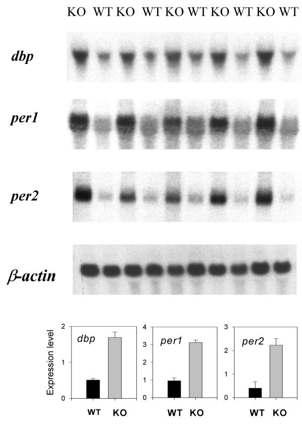
Deletion of the cryptochromes disinhibits the transcriptional activation of CLOCK:BMAL1 and NPAS2:BMAL1 target genes, resulting in increased levels of their transcripts in the SCN, liver, and retina [20,23,25]. We assayed the expression in the brain of three genes that are known targets of cryptochrome mediated transcriptional inhibition: albumin D-binding protein (dbp [26]), period (per)1, and per2 [20,23,27], in the middle of the daily light period. Confirming the earlier studies in other tissues, mRNA levels for all three genes were higher in the brains of cry1,2-/- mice compared to wild type mice (Figure 4). This relative increase was highly significant for all three genes (dbp, per1: 3.3-fold; per2: 5.6-fold; P < 0.0003, unpaired t-tests, n = 5/genotype).
Figure 4.
Whole-brain mRNA levels for dbp, per1, and per2 in cry1,2-/- and wild type mice. Levels of all three genes are elevated in cry1,2-/- mice (KO) relative to wild type (WT) controls at ZT6 when dbp, per1, and per2 mRNAs are lowest in the forebrain of wild type mice [37]. In the lower three panels mean (± 1 SEM) expression levels are depicted. β-actin expression was used as an internal standard.
Sleep deprivation-induced differences in circadian gene expression
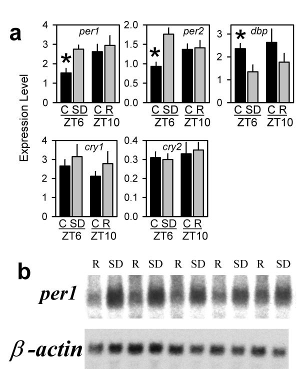
Messenger RNA levels for per1,2, cry1,2, and dbp were quantified by RT-PCR in three brain areas (cerebral cortex, basal forebrain, and hypothalamus) from C57BL/6 mice that were sacrificed immediately after 6 h of SD or after 4 h of recovery sleep (ZT10). Significant differences in expression were observed only in the cortex (Figure 5A) and not in hypothalamus or basal forebrain (data not shown). Both per1 and per2 mRNA levels were higher immediately after SD compared to controls, cry1,2 expression did not change, and dbp mRNA decreased significantly (Figure 5A). After 4 h of recovery sleep, per1,2 and dbp expression returned to the normal levels for that time of day (ZT10; Figure 5A). We also measured the expression of five other circadian genes: bmal1, clock, npas2, per3, and casein kinase-1-ε (csnk1e) in the cortex; the levels of these five mRNAs were not affected by SD (data not shown).
Figure 5.
Sleep deprivation alters mRNA levels of per1, per2, and dbp. (A) RT-PCR analysis of the expression of five 'clock'-genes in the mouse cortex across four experimental conditions [C = control; R = recovery sleep; SD = sleep deprived; ZT = Zeitgeber time (i.e., 6 or 10 h after light-onset)]. g3pdh expression was used as an internal standard. Bars depict mean ± 1 SEM. Asterisks denote significant differences (P < 0.05) between the experimental and corresponding control group (Student-Newman-Keuls post-hoc tests; 1-way ANOVA factor 'condition': P < 0.05, for per1,2, and dbp only; n = 7/condition) (B) per1 mRNA falls significantly in rat cortex during a 2-h recovery period (R) subsequent to 6 h SD ending at ZT6 (P < 0.05, t-test). Northern analysis was performed on cortex of five sleep-deprived rats and five rats that were allowed 2 h of recovery sleep (ZT8).
As confirmation of our results in the mouse, we determined the expression of per2 and per1 in the rat by RT-PCR and Northern analysis, respectively. In a similar experimental paradigm, rats were sleep deprived for 6 h and then either sacrificed immediately after the SD (ZT6), or after 2 h of recovery sleep (ZT8; n = 5/group). The cortex-specific increase in per2 mRNA was confirmed by the RT-PCR analysis and, as in the mouse, per2 expression returned to basal levels after a period of recovery sleep (data not shown). Northern analysis confirmed that recovery sleep was associated with a decline in per1 mRNA relative to the level of expression reached at the end of the SD (Figure 5B).
Discussion
Upon release into constant darkness cry1,2-/- mice immediately become arrhythmic at the behavioral level [22,23], at the level of SCN electrophysiology [28], and at the cellular/molecular level [23]. Of the available mouse models for circadian dysfunction, only per1,2 double mutant mice [29], bmal1 knockout mice [30], and mice with an ablation of the SCN [31] show a similarly dramatic phenotype. Thus, cry1,2-/- mice appear to be a suitable model for studies of the regulation of sleep in the absence of an intact circadian clock.
Under light / dark (LD) conditions, running wheel activity patterns and, as we show here, the distribution of sleep in cry1,2-/- mice still exhibit diurnal variation. LD cycles can influence the expression of sleep by entraining the circadian pacemaker that drives the diurnal rhythm of sleep and/or by directly affecting the expression of sleep, thereby 'masking' the influence of the pacemaker on sleep. Masking seems to be the mechanism by which light drives these rhythms under LD conditions in cry1,2-/- mice [22], since the daily modulation of NREMS that occurs in cry1,2-/- mice under a light/dark cycle immediately disappears upon placement in constant darkness ([22], Figure 3). At the molecular level, per2 expression (but not that of per1) is rhythmic in the SCN of cry1,2-/- mice under LD conditions. Upon release into constant dark conditions, per2 rhythmicity disappears concomitant with the immediate loss of behavioral rhythmicity, suggesting a role for 'light-driven' per2 expression in generating behavioral rhythms [23].
The most striking and unexpected finding of the current study is that, under baseline conditions, cry1,2-/- mice exhibit all the hallmarks of high NREMS pressure, including more consolidated NREMS, increased NREMS time, and higher levels of EEG delta power relative to wild type mice that were attained immediately after NREMS onset. The failure of cry1,2-/- mice to exhibit a robust increase in any of these measures after 6 h SD is consistent with the interpretation that these mice are already under high NREMS pressure during baseline conditions. Determination of the time constants that most accurately describe the dynamics of NREMS delta power revealed that during wake, the propensity for high NREMS delta power increases during wake in cry1,2-/- mice at a faster rate than in wild type mice. This could help explain why NREMS delta power is chronically high in cry1,2-/- mice. The coincidence of high NREMS time and chronically high delta power in cry1,2-/- mice is all the more striking when one considers that during NREMS, the drive for NREMS should dissipate and result in lower delta power [14,32].
These findings in cry1,2-/- mice contrast with the findings of sleep studies in animals that are rendered arrhythmic by lesioning of the SCN. In nocturnal rodents, lesioning the SCN results in more fragmented sleep, with lower EEG delta power, but leaving the daily sleep time unchanged [6-8,31]. Lesioning the SCN in a diurnal primate, the squirrel monkey, did result in an increase in NREMS time, but sleep was more fragmented, with a higher proportion of 'light' NREMS [5]; i.e., with lower overall levels of EEG delta power. Furthermore, the homeostatic response to sleep deprivation does not seem to be altered in SCN-lesioned rodents [6-8]. Thus, the sleep characteristics of cry1,2-/- mice do not support the concept of cry1,2-/- mice as simply a genetic model for ablation of the circadian clock in the SCN. Together, these unexpected results are compatible with a role for cryptochromes in the homeostatic regulation of sleep in addition to their role in generating circadian rhythms.
Recent observations, including the current report, suggest a complex interrelationship between homeostatic and circadian influences on sleep at the molecular level. Deletion of the cycle gene in Drosophila produces flies that have an exaggerated homeostatic response to rest deprivation in addition to their lack of circadian rhythmicity [16]. In a striking parallel to our current results, flies with a mutation in the cryptochrome gene also exhibit increased rest time as well as a reduced compensatory response to rest deprivation (P. Shaw, personal communication). The clock mutation in mice, which has a profound effect on circadian rhythmicity [33], decreases NREMS time and consolidation under baseline conditions [34]. The clock sleep phenotype is the inverse of the sleep characteristics we report here for the cry1,2-/- mice and is thus consistent with CLOCK and CRY1,2 being positive and negative transcriptional regulators, respectively. Albumin D-binding protein (Dbp) is a transcription factor whose expression is under the direct transcriptional control of CLOCK:BMAL1 [26]. Deletion of the dbp gene, which results in a shortening of the circadian period [35], also results in decreased sleep consolidation and NREMS delta power [36].
We assume that the effects on sleep we observed in cry1,2-/- mice are a result of a lack of cryptochrome-dependent inhibition of the transcriptional activation provided by the bHLH-PAS heterodimers CLOCK:BMAL1 and NPAS2:BMAL1 [19,20,37], although cryptochromes also play a role in stabilizing and nuclear sequestration of PER proteins [38], and in photoreception [39]. Lack of cryptochromes results in increased mRNA levels of CLOCK/NPAS2:BMAL1 target genes, including the circadian genes per1 and per2 [20,23]. The expression of these two genes is viewed as a state variable of the molecular circadian clock or a marker of CLOCK/NPAS2:BMAL1-induced transcription, although at least per1 transcription can also be (rapidly) induced by light [40,41], through a CREB-dependent signaling pathway [42-44]. The observation of high brain levels of per1,2 transcripts under baseline conditions in cry1,2-/- mice raises the possibility that these or other CLOCK/NPAS2:BMAL1 target genes are involved in the homeostatic regulation of sleep. The observation of elevated per gene expression in the cortex of sleep-deprived rats and mice (Figure 5) supports this hypothesis.
The increase in per expression after the sleep deprivation was specific to the cerebral cortex, although it cannot be ruled out, based on the present study, that circadian gene expression changes with sleep-wake history in specific nuclei within the other two regions examined; i.e., the hypothalamus and basal forebrain. A surprisingly small number (~0.5%) of the ~10,000 genes screened by mRNA differential display and cDNA microarrays in the cortex to date change their expression with sleep deprivation [45]. It is intriguing and encouraging that, of the initial genes we assayed, three changed their expression with sleep deprivation (per1, per2, dbp), none of which were identified in the aforementioned screens. cry1,2 expression at the mRNA level did not change with sleep deprivation in wild type mice. This observation does not necessarily obviate a direct role for CRY proteins in mediating a response to sleep deprivation. CRY poteins may play a role at steady state levels or there may be post-translational changes in the functioning of CRY proteins in association with sleep deprivation, such as phosphorylation state, ubiquitination [38], or intracellular localization [46] of the protein, all of which are regulated dynamically, at least in vitro. In the liver, CRY protein oscillations are not necessary for circadian oscillations of target transcripts, as CRY proteins are present in excess of PER and oscillations in the latter produce rhythmicity [46]. A similar situation might exist in the cortex.
The high per levels in cry1,2-/- mice and the low per levels in clock mutant mice [26,47] correlate with their contrasting sleep phenotype (see above; [34]). In this context, the sleep abnormalities in dbp-/- mice might also be related to a reduction in per expression since, at least in vitro, DBP can amplify the CLOCK:BMAL1-induced transcription of per [48], but it is not known whether per transcript levels are altered in dbp-/- mice. Apart from the present observations in sleep-deprived rats and mice, several other reports confirm that cortical levels of per expression in wild type animals are high at times when sleep drive is high, irrespective of the phase at which the circadian expression of per peaks in the SCN. Thus, in both nocturnal and diurnal species, per expression in the cortex is maximal in conjunction with the major waking episode [37,49,50]. Under conditions where the phase (methamphetamine administration, restricted feeding) or distribution (circadian splitting) of locomotor activity is altered, per expression in the cortex parallels the overt rhythm of wakefulness, whereas the circadian oscillation of per gene expression in the SCN remains unaffected [49,51,52]. Thus, in contrast to its role in the SCN, PER protein in the cortex is not a component of a self-sustaining circadian oscillator [53]. Instead, per expression in the cortex seems to the follow sleep-wake history, consistent with the hypothesis that it is related to homeostatic regulation of sleep. However, in the current study and those cited above, the expression of per genes was studied at the mRNA level. PER protein level may be affected differentially from that of per mRNA and levels of the PER protein are reduced in cry1,2-/- mice due to reduced stability of PER proteins in the absence of heterodimerizing CRY partners [19,38,46].
Conclusions
In the discussion, we have focused on per1,2 mRNAs as transcriptional targets of cry1,2 because the expression patterns of these circadian genes have been widely described and because their transcriptional control by CRY proteins and by CLOCK/NPAS2:BMAL1 has been well established. At least 90 genes are regulated in a similar fashion by NPAS2:BMAL1 [37] and the identity of the target genes critical for the NREMS phenotype in cry1,2-/- mice remains to be determined. In the absence of cry1,2, the stability and overall level of PER proteins, particularly in the nucleus of the cell, is reduced [19,38,46]. It would therefore be interesting to observe sleep in per1,2 single and double mutant mice, the latter of which have been subjected to behavioral observation [29] but not to sleep EEG studies. per1 single knockout mice and mice expressing a non-functional PER2 protein both exhibit subtle differences from wild type in the homeostatic rebound after SD [54], an observation compatible with the hypothesis that these genes are correlates of sleep homeostasis. In addition, measurement of the effects of sleep deprivation on the expression of cry1,2 and per1,2 genes at the protein level will provide critical information. Finally, from a functional perspective, it is interesting that CLOCK/NPAS2:BMAL1 transcriptional activity is sensitive to redox state [24]. This transcriptional activity might thus provide a link between neuronal activity and an energy regulatory function for NREMS, as has been suggested previously [55].
Methods
Sleep studies
Mice were generated by mating cry1 and cry2 single knockout mice, both of mixed background (ca. 3/4 C57BL/6 – 1/4 129/Sv; [23]) to generate double heterozygotes that were interbred to generate double knockouts (cry1,2-/-) and wild type controls (cry1,2+/+). Eight wild type and 6 cry1,2-/- male mice were surgically prepared for EEG and electromyographic (EMG) recordings as described previously [56]. Following two weeks of post-surgical recovery, mice were isolated for recordings in sound-attenuated chambers. The experiments were conducted under an LD12:12 cycle (lights-on; i.e., Zeitgeber Time ZT0, at 0600 h). Twenty-four hour baseline recordings, starting at ZT0, were followed by a 6 h sleep deprivation (SD) starting at ZT0. The sleep deprivations in this experiment and the other two experiments (see below) were performed by the introduction of novel objects into the cage or by gentle handling. In addition to the SD experiment, animals were subjected to one day of baseline recording in constant darkness separated from the SD experiment by 72 hours. All experimental procedures complied with institutional and NIH guidelines.
Digitized EEG and integrated EMG were stored in 10-s epochs and classified as NREMS, REM sleep (REMS), or wakefulness by visual inspection. The EEG was subjected to a Fast-Fourier-Transformation yielding power spectra between 0–20 Hz. Delta power was calculated as the average EEG power in the delta (1–4 Hz) frequencies for epochs scored as NREMS. For visual representation, hourly delta-power values (Figure 1) were expressed relative to an individual mean (i.e., 24-h baseline mean) before transforming back to absolute values to capture both the highly reproducible individual time course of delta power and the genotype differences in absolute values [10]. Statistical evaluation of genotype differences were based, however, on the absolute EEG values. Post-SD delta power was compared to baseline during the first hour of spontaneous sleep subsequent to SD, while NREMS amount and bout duration (Table 2) were compared over 12 hours (ZT6-ZT18), in accordance with the distinct dynamics of the compensatory responses of these variables to SD [57]. NREMS bouts were defined as periods of NREMS initiated by three consecutive 10-second epochs of NREMS and terminated by three consecutive epochs not classified as NREMS. EEG delta power changes at transitions between wakefulness and NREMS were measured during those transitions characterized by at least 12 consecutive 10-s epochs of wake followed by at least 18 consecutive 10-epochs of NREMS.
A mathematical algorithm was used to quantify the sleep-wake dependent dynamics of delta power during both baseline and recovery from SD [10]. In this algorithm, delta power decreases during NREMS and increases during wakefulness according to saturating exponential functions, the time constants of which are estimated by minimizing the square of the differences between empirical (delta power) data and the values produced by the mathematical functions. The two time constants (for the increase and decrease of delta power) that resulted in the smallest deviation from empirical values within each individual were used to statistically assess genotype effect on the dynamics of delta power [10].
Northern analysis
Five cry1,2-/- mice and 5 wild type controls were sacrificed in the middle of the light period (ZT6). Brains were rapidly removed and frozen on dry ice. Total RNA was extracted and Northern analysis was performed with 10 μg whole brain total RNA as previously described [58].
Ten Wistar rats were sleep deprived for 6 h beginning at light-onset (ZT0). Five rats were sacrificed at ZT6 at the end of the SD and five were allowed to recover for 2 h following the SD and were sacrificed at ZT8. Rats were monitored by EEG/EMG throughout this period as described [59]. Brains were rapidly removed, dissected and frozen, and Northern analysis performed.
Quantitative real-time Reverse-Transcriptase Polymerase-Chain-Reaction (RT-PCR) analyses
Four experimental groups of male C57BL/6J mice were studied: 1) sleep deprived from light-onset (ZT0) to ZT6; 2) control mice for the SD group; 3) 6 h SD (ZT0-ZT6) followed by 4-h recovery sleep (ZT6-ZT10); and 4) control mice for the recovery group (n = 7 mice/group). Animals were sacrificed by decapitation. Brains were rapidly removed and the cerebral cortex, basal forebrain, and hypothalamus were dissected. After a mid-sagittal cut, the entire cortical tissue was peeled off and separated from the hippocampus and underlying diencephalon. Striatal tissue was also removed. From each animal, quantitative real-time PCR determinations of 5 target genes (cry1,2, per1,2, and dbp) and a reference cDNA (glyceraldehyde-3-phosphate dehydrogenase, g3pdh) were made from the cortex, basal forebrain, and hypothalamus (primer/probe sequences available upon request). For five other targets (bmal1, clock, npas2, per3, and csnk1e), expression was quantified in the cortex only. To confirm the specificity of the nucleotide sequences chosen for the primers and probes and the absence of DNA polymorphisms, BLASTN searches were conducted against the dbEST and nonredundant set of Genbank, EMBL, and DDBJ databases. Dual color fluorescence was detected using an ABI Prism 7700 Sequence Detection System (Perkin-Elmer Corp., Foster City, CA). For each experimental sample, the amount of the target and g3pdh reference was determined from the standard curve (range of 0.2–200 ng total RNA) measured in the same assay. A normalized value was obtained by dividing the target cDNA amount by the g3pdh reference. For details see [59].
Authors' contributions
CPS and AS generated the cry1,2-/- mice and provided some preliminary data on microarray gene expression profiles in the brains of these mice. JPW and DME conceived and implemented sleep studies in cry1,2-/- mice. PF analyzed sleep EEG data and conceived, with BFO, AT, and TSK, the molecular studies. BFO, AT, and TSK performed gene expression assays on brain tissues from mice and rats. JPW and PF drafted the manuscript. All authors read and approved the manuscript.
Acknowledgments
Acknowledgments
We appreciate the generous gifts of cDNA clones from Drs. Ueli Schibler (dbp cDNA), Steven Reppert (per2 cDNA), and Hajime Tei (per1 cDNA). Funding was provided by the following NIH grants: HL64243 (JPW and DME), R01HL/MH59658 and R01MH61755 (AT and TSK), GM31082 (CPS and AS), and HL64148 and DA13349 (BFO and PF).
Contributor Information
Jonathan P Wisor, Email: jwisor@stanford.edu.
Bruce F O'Hara, Email: BFO@stanford.edu.
Akira Terao, Email: akira.terao@sri.com.
Chris P Selby, Email: CSelby@med.unc.edu.
Thomas S Kilduff, Email: thomas.kilduff@sri.com.
Aziz Sancar, Email: Aziz_Sancar@med.unc.edu.
Dale M Edgar, Email: DMEdgar@Hypnion.com.
Paul Franken, Email: PFranken@stanford.edu.
References
- Klein DC, Moore RY, Reppert SM. Suprachiasmatic nucleus: The mind's clock. New York: Oxford University Press. 1991.
- Edgar DM. Functional role of the suprachiasmatic nuclei in the regulation of sleep and wakefulness. In: Guilleminault C, editor. Fatal Familial Insomnia: Inherited prion diseases, sleep and the thalamus. New York: Raven Press, Ltd; 1994. pp. 203–213. [Google Scholar]
- Dijk DJ, Czeisler CA. Contribution of the circadian pacemaker and the sleep homeostat to sleep propensity, sleep structure, electroencephalographic slow waves, and sleep spindle activity in humans. J Neurosci. 1995;15:3526–38. doi: 10.1523/JNEUROSCI.15-05-03526.1995. [DOI] [PMC free article] [PubMed] [Google Scholar]
- Borbely AA, Achermann P. Sleep homeostasis and models of sleep regulation. In: Kryger MH, Roth T, Dement WC, editor. Principles and practice in sleep medicine. 3rd. Philadelphia: W.B. Saunders; 2000. pp. 377–390. [Google Scholar]
- Edgar DM, Dement WC, Fuller CA. Effect of SCN lesions on sleep in squirrel monkeys: evidence for opponent processes in sleep-wake regulation. J Neurosci. 1993;13:1065–79. doi: 10.1523/JNEUROSCI.13-03-01065.1993. [DOI] [PMC free article] [PubMed] [Google Scholar]
- Mistlberger RE, Bergmann BM, Waldenar W, Rechtschaffen A. Recovery sleep following sleep deprivation in intact and suprachiasmatic nuclei-lesioned rats. Sleep. 1983;6:217–33. doi: 10.1093/sleep/6.3.217. [DOI] [PubMed] [Google Scholar]
- Tobler I, Borbely AA, Groos G. The effect of sleep deprivation on sleep in rats with suprachiasmatic lesions. Neurosci Lett. 1983;42:49–54. doi: 10.1016/0304-3940(83)90420-2. [DOI] [PubMed] [Google Scholar]
- Trachsel L, Edgar DM, Seidel WF, Heller HC, Dement WC. Sleep homeostasis in suprachiasmatic nuclei-lesioned rats: effects of sleep deprivation and triazolam administration. Brain Res. 1992;589:253–61. doi: 10.1016/0006-8993(92)91284-L. [DOI] [PubMed] [Google Scholar]
- Wurts SW, Edgar DM. Circadian and homeostatic control of rapid eye movement (REM) sleep: promotion of REM tendency by the suprachiasmatic nucleus. J Neurosci. 2000;20:4300–10. doi: 10.1523/JNEUROSCI.20-11-04300.2000. [DOI] [PMC free article] [PubMed] [Google Scholar]
- Franken P, Chollet D, Tafti M. The homeostatic regulation of sleep need is under genetic control. J Neurosci. 2001;21:2610–21. doi: 10.1523/JNEUROSCI.21-08-02610.2001. [DOI] [PMC free article] [PubMed] [Google Scholar]
- Huber R, Deboer T, Tobler I. Effects of sleep deprivation on sleep and sleep EEG in three mouse strains: empirical data and simulations. Brain Res. 2000;857:8–19. doi: 10.1016/S0006-8993(99)02248-9. [DOI] [PubMed] [Google Scholar]
- Edgar DM, Seidel WF. Modafinil induces wakefulness without intensifying motor activity or subsequent rebound hypersomnolence in the rat. J Pharmacol Exp Ther. 1997;283:757–769. [PubMed] [Google Scholar]
- Franken P, Dijk DJ, Tobler I, Borbely AA. Sleep deprivation in rats: effects on EEG power spectra, vigilance states, and cortical temperature. Am J Physiol. 1991;261:R198–208. doi: 10.1152/ajpregu.1991.261.1.R198. [DOI] [PubMed] [Google Scholar]
- Daan S, Beersma DG, Borbely AA. Timing of human sleep: recovery process gated by a circadian pacemaker. Am J Physiol. 1984;246:R161–83. doi: 10.1152/ajpregu.1984.246.2.R161. [DOI] [PubMed] [Google Scholar]
- Cirelli C. How sleep deprivation affects gene expression in the brain: a review of recent findings. J Appl Physiol. 2002;92:394–400. doi: 10.1152/jappl.2002.92.1.394. [DOI] [PubMed] [Google Scholar]
- Shaw PJ, Tononi G, Greenspan RJ, Robinson DF. Stress response genes protect against lethal effects of sleep deprivation in Drosophila. Nature. 2002;417:287–91. doi: 10.1038/417287a. [DOI] [PubMed] [Google Scholar]
- Shaw PJ, Cirelli C, Greenspan RJ, Tononi G. Correlates of sleep and waking in Drosophila melanogaster. Science. 2000;287:1834–7. doi: 10.1126/science.287.5459.1834. [DOI] [PubMed] [Google Scholar]
- Hendricks JC, Finn SM, Panckeri KA, Chavkin J, Williams JA, Sehgal A, Pack AI. Rest in Drosophila is a sleep-like state. Neuron. 2000;25:129–38. doi: 10.1016/s0896-6273(00)80877-6. [DOI] [PubMed] [Google Scholar]
- Kume K, Zylka MJ, Sriram S, Shearman LP, Weaver DR, Jin X, Maywood ES, Hastings MH, Reppert SM. mCRY1 and mCRY2 are essential components of the negative limb of the circadian clock feedback loop. Cell. 1999;98:193–205. doi: 10.1016/s0092-8674(00)81014-4. [DOI] [PubMed] [Google Scholar]
- Shearman LP, Sriram S, Weaver DR, Maywood ES, Chaves I, Zheng B, Kume K, Lee CC, van der Horst GT, Hastings MH, et al. Interacting molecular loops in the mammalian circadian clock. Science. 2000;288:1013–9. doi: 10.1126/science.288.5468.1013. [DOI] [PubMed] [Google Scholar]
- Reppert SM, Weaver DR. Molecular analysis of mammalian circadian rhythms. Annu Rev Physiol. 2001;63:647–76. doi: 10.1146/annurev.physiol.63.1.647. [DOI] [PubMed] [Google Scholar]
- van der Horst GT, Muijtjens M, Kobayashi K, Takano R, Kanno S, Takao M, de Wit J, Verkerk A, Eker AP, van Leenen D, et al. Mammalian Cry1 and Cry2 are essential for maintenance of circadian rhythms. Nature. 1999;398:627–30. doi: 10.1038/19323. [DOI] [PubMed] [Google Scholar]
- Vitaterna MH, Selby CP, Todo T, Niwa H, Thompson C, Fruechte EM, Hitomi K, Thresher RJ, Ishikawa T, Miyazaki J, et al. Differential regulation of mammalian period genes and circadian rhythmicity by cryptochromes 1 and 2. Proc Natl Acad Sci U S A. 1999;96:12114–9. doi: 10.1073/pnas.96.21.12114. [DOI] [PMC free article] [PubMed] [Google Scholar]
- Rutter J, Reick M, Wu LC, McKnight SL. Regulation of clock and NPAS2 DNA binding by the redox state of NAD cofactors. Science. 2001;293:510–4. doi: 10.1126/science.1060698. [DOI] [PubMed] [Google Scholar]
- Okamura H, Miyake S, Sumi Y, Yamaguchi S, Yasui A, Muijtjens M, Hoeijmakers JH, van der Horst GT. Photic induction of mPer1 and mPer2 in cry-deficient mice lacking a biological clock. Science. 1999;286:2531–4. doi: 10.1126/science.286.5449.2531. [DOI] [PubMed] [Google Scholar]
- Ripperger JA, Shearman LP, Reppert SM, Schibler U. CLOCK, an essential pacemaker component, controls expression of the circadian transcription factor DBP. Genes Dev. 2000;14:679–89. [PMC free article] [PubMed] [Google Scholar]
- Griffin EA, Jr, Staknis D, Weitz CJ. Light-independent role of CRY1 and CRY2 in the mammalian circadian clock. Science. 1999;286:768–71. doi: 10.1126/science.286.5440.768. [DOI] [PubMed] [Google Scholar]
- Albus H, Bonnefont X, Chaves I, Yasui A, Doczy J, van der Horst GT, Meijer JH. Cryptochrome-deficient mice lack circadian electrical activity in the suprachiasmatic nuclei. Current biology : CB. 2002;12:1130–3. doi: 10.1016/S0960-9822(02)00923-5. [DOI] [PubMed] [Google Scholar]
- Zheng B, Albrecht U, Kaasik K, Sage M, Lu W, Vaishnav S, Li Q, Sun ZS, Eichele G, Bradley A, et al. Nonredundant roles of the mPer1 and mPer2 genes in the mammalian circadian clock. Cell. 2001;105:683–94. doi: 10.1016/S0092-8674(01)00380-4. [DOI] [PubMed] [Google Scholar]
- Bunger MK, Wilsbacher LD, Moran SM, Clendenin C, Radcliffe LA, Hogenesch JB, Simon MC, Takahashi JS, Bradfield CA. Mop3 is an essential component of the master circadian pacemaker in mammals. Cell. 2000;103:1009–17. doi: 10.1016/s0092-8674(00)00205-1. [DOI] [PMC free article] [PubMed] [Google Scholar]
- Ibuka N, Nihonmatsu I, Sekiguchi S. Sleep-wakefulness rhythms in mice after suprachiasmatic nucleus lesions. Waking Sleeping. 1980;4:167–73. [PubMed] [Google Scholar]
- Borbely AA. A two process model of sleep regulation. Hum Neurobiol. 1982;1:195–204. [PubMed] [Google Scholar]
- Vitaterna MH, King DP, Chang AM, Kornhauser JM, Lowrey PL, McDonald JD, Dove WF, Pinto LH, Turek FW, Takahashi JS. Mutagenesis and mapping of a mouse gene, Clock, essential for circadian behavior. Science. 1994;264:719–725. doi: 10.1126/science.8171325. [DOI] [PMC free article] [PubMed] [Google Scholar]
- Naylor E, Bergmann BM, Krauski K, Zee PC, Takahashi JS, Vitaterna MH, Turek FW. The circadian clock mutation alters sleep homeostasis in the mouse. J Neurosci. 2000;20:8138–43. doi: 10.1523/JNEUROSCI.20-21-08138.2000. [DOI] [PMC free article] [PubMed] [Google Scholar]
- Lopez-Molina L, Conquet F, Dubois-Dauphin M, Schibler U. The DBP gene is expressed according to a circadian rhythm in the suprachiasmatic nucleus and influences circadian behavior. Embo J. 1997;16:6762–71. doi: 10.1093/emboj/16.22.6762. [DOI] [PMC free article] [PubMed] [Google Scholar]
- Franken P, Lopez-Molina L, Marcacci L, Schibler U, Tafti M. The transcription factor DBP affects circadian sleep consolidation and rhythmic EEG activity. J Neurosci. 2000;20:617–25. doi: 10.1523/JNEUROSCI.20-02-00617.2000. [DOI] [PMC free article] [PubMed] [Google Scholar]
- Reick M, Garcia JA, Dudley C, McKnight SL. NPAS2: an analog of clock operative in the mammalian forebrain. Science. 2001;293:506–9. doi: 10.1126/science.1060699. [DOI] [PubMed] [Google Scholar]
- Yagita K, Tamanini F, Yasuda M, Hoeijmakers JH, van Der Horst GT, Okamura H. Nucleocytoplasmic shuttling and mCRY-dependent inhibition of ubiquitylation of the mPER2 clock protein. Embo J. 2002;21:1301–1314. doi: 10.1093/emboj/21.6.1301. [DOI] [PMC free article] [PubMed] [Google Scholar]
- Selby C, Thompson C, Schmitz T, Van Gelder R, S A. Functional redundancy of cryptochrome and classical photoreceptors for nonvisual ocular photoreception in mice. Proc Natl Acad Sci, USA. 2000. [DOI] [PMC free article] [PubMed]
- Albrecht U, Sun ZS, Eichele G, Lee CC. A differential response of two putative mammalian circadian regulators, mper1 and mper2, to light. Cell. 1997;91:1055–1064. doi: 10.1016/s0092-8674(00)80495-x. [DOI] [PubMed] [Google Scholar]
- Shigeyoshi Y, Taguchi K, Yamamoto S, Takekida S, Yan L, Tei H, Moriya T, Shibata S, Loros JJ, Dunlap JC, et al. Light-induced resetting of a mammalian circadian clock is associated with rapid induction of the mPer1 transcript. Cell. 1997;91:1043–1053. doi: 10.1016/s0092-8674(00)80494-8. [DOI] [PubMed] [Google Scholar]
- Yokota S, Yamamoto M, Moriya T, Akiyama M, Fukunaga K, Miyamoto E, Shibata S. Involvement of calcium-calmodulin protein kinase but not mitogen-activated protein kinase in light-induced phase delays and Per gene expression in the suprachiasmatic nucleus of the hamster. J Neurochem. 2001;77:618–27. doi: 10.1046/j.1471-4159.2001.00270.x. [DOI] [PubMed] [Google Scholar]
- Tischkau SA, Mitchell JW, Tyan SH, Buchanan GF, Gillette MU. CREB-dependent activation of Per1 is required for light-induced signaling in the suprachiasmatic nucleus circadian clock. J Biol Chem. 2002. [DOI] [PubMed]
- Gau D, Lemberger T, von Gall C, Kretz O, Le Minh N, Gass P, Schmid W, Schibler U, Korf HW, Schutz G. Phosphorylation of CREB Ser142 regulates light-induced phase shifts of the circadian clock. Neuron. 2002;34:245–53. doi: 10.1016/s0896-6273(02)00656-6. [DOI] [PubMed] [Google Scholar]
- Cirelli C, Tononi G. Gene expression in the brain across the sleep-waking cycle. Brain Res. 2000;885:303–21. doi: 10.1016/S0006-8993(00)03008-0. [DOI] [PubMed] [Google Scholar]
- Lee C, Etchegaray JP, Cagampang FR, Loudon AS, Reppert SM. Posttranslational mechanisms regulate the mammalian circadian clock. Cell. 2001;107:855–67. doi: 10.1016/s0092-8674(01)00610-9. [DOI] [PubMed] [Google Scholar]
- Oishi K, Fukui H, Ishida N. Rhythmic expression of BMAL1 mRNA is altered in Clock mutant mice: differential regulation in the suprachiasmatic nucleus and peripheral tissues. Biochem Biophys Res Commun. 2000;268:164–71. doi: 10.1006/bbrc.1999.2054. [DOI] [PubMed] [Google Scholar]
- Yamaguchi S, Mitsui S, Yan L, Yagita K, Miyake S, Okamura H. Role of DBP in the circadian oscillatory mechanism. Mol Cell Biol. 2000;20:4773–81. doi: 10.1128/MCB.20.13.4773-4781.2000. [DOI] [PMC free article] [PubMed] [Google Scholar]
- Abe H, Honma S, Namihira M, Masubuchi S, Honma K. Behavioural rhythm splitting in the CS mouse is related to clock gene expression outside the suprachiasmatic nucleus. Eur J Neurosci. 2001;14:1121–8. doi: 10.1046/j.0953-816x.2001.01732.x. [DOI] [PubMed] [Google Scholar]
- Mrosovsky N, Edelstein K, Hastings MH, Maywood ES. Cycle of period gene expression in a diurnal mammal (Spermophilus tridecemlineatus): implications for nonphotic phase shifting. J Biol Rhythms. 2001;16:471–8. doi: 10.1177/074873001129002141. [DOI] [PubMed] [Google Scholar]
- Masubuchi S, Honma S, Abe H, Ishizaki K, Namihira M, Ikeda M, Honma K. Clock genes outside the suprachiasmatic nucleus involved in manifestation of locomotor activity rhythm in rats. Eur J Neurosci. 2000;12:4206–14. doi: 10.1046/j.1460-9568.2000.01313.x. [DOI] [PubMed] [Google Scholar]
- Wakamatsu H, Yoshinobu Y, Aida R, Moriya T, Akiyama M, Shibata S. Restricted-feeding-induced anticipatory activity rhythm is associated with a phase-shift of the expression of mPer1 and mPer2 mRNA in the cerebral cortex and hippocampus but not in the suprachiasmatic nucleus of mice. Eur J Neurosci. 2001;13:1190–6. doi: 10.1046/j.0953-816x.2001.01483.x. [DOI] [PubMed] [Google Scholar]
- Abe M, Herzog ED, Yamazaki S, Straume M, Tei H, Sakaki Y, Menaker M, Block GD. Circadian rhythms in isolated brain regions. J Neurosci. 2002;22:350–6. doi: 10.1523/JNEUROSCI.22-01-00350.2002. [DOI] [PMC free article] [PubMed] [Google Scholar]
- Kopp C, Albrecht U, Zheng B, Tobler I. Homeostatic sleep regulation is preserved in mPer1 and mPer2 mutant mice. Eur J Neurosci. 2002;16:1099–106. doi: 10.1046/j.1460-9568.2002.02156.x. [DOI] [PubMed] [Google Scholar]
- Benington JH, Heller HC. Restoration of brain energy metabolism as the function of sleep. Prog Neurobiol. 1995;45:347–360. doi: 10.1016/0301-0082(94)00057-O. [DOI] [PubMed] [Google Scholar]
- Edgar DM, Kilduff TS, Martin CE, Dement WC. Influence of running wheel activity on free-running sleep/wake and drinking circadian rhythms in mice. Physiol Behav. 1991;50:373–378. doi: 10.1016/0031-9384(91)90080-8. [DOI] [PubMed] [Google Scholar]
- Franken P, Malafosse A, Tafti M. Genetic determinants of sleep regulation in inbred mice. Sleep. 1999;22:155–69. [PubMed] [Google Scholar]
- O'Hara BF, Watson FL, Srere HK, Kumar H, Wiler SW, Welch SK, Bitting L, Heller HC, Kilduff TS. Gene expression in the brain across the hibernation cycle. J Neurosci. 1999;19:3781–90. doi: 10.1523/JNEUROSCI.19-10-03781.1999. [DOI] [PMC free article] [PubMed] [Google Scholar]
- Terao A, Peyron C, Ding J, Wurts SW, Edgar DM, Heller HC, Kilduff TS. Prepro-hypocretin (prepro-orexin) expression is unaffected by short-term sleep deprivation in rats and mice. Sleep. 2000;23:867–74. [PubMed] [Google Scholar]


