Journal of Virology logoLink to Journal of Virology
letter
. 2003 Mar;77(6):3888–3889. doi: 10.1128/JVI.77.6.3888-3889.2003

Endogenous Virus and Hepatitis C Virus-Like Particle Budding in BHK-21 Cells

Emmanuelle Blanchard 1, Denys Brand 1, Philippe Roingeard 1,*
PMCID: PMC149545  PMID: 12610167

A recent paper by H. J. Ezelle and coworkers reported the generation of hepatitis C virus (HCV)-like particles by using a recombinant vesicular stomatitis virus (VSV) vector (4). The authors inserted the contiguous HCV Core, E1, and E2 coding region into the VSV genome, and recombinant VSV producing the three HCV structural proteins were used to infect the BHK-21 cell line. With this strategy, the authors claimed to have obtained the complete budding of HCV-like particles, visualized by transmission electron microscopy (TEM) in cytoplasmic vacuoles derived from the rough endoplasmic reticulum (ER). In their Fig. 3 (panels C and D) they indicate “HCV-like virions,” described as 40- to 80-nm particles exhibiting an electron-dense core with an envelope, fully released into the ER lumen (4). Unfortunately, we believe that these particles represent the endogenous viruses of BHK-21 cells known as intracisternal R-type particles that have been widely described elsewhere (3, 6, 7).

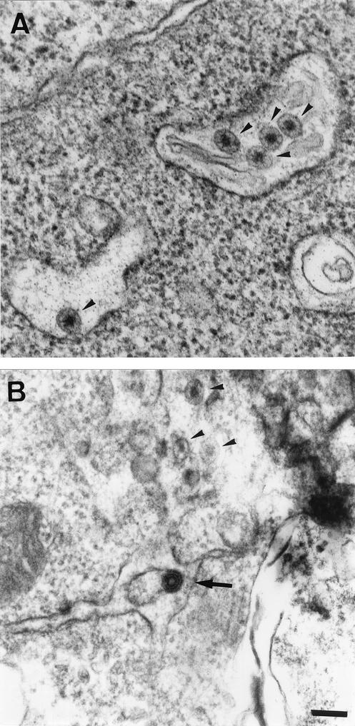
Our group is experienced in the transfection of BHK-21 cells with various constructs encoding viral structural proteins (1, 2, 5). We have frequently observed these intracisternal R-type particles in untransfected BHK-21 studied by TEM (arrowheads in Fig. 1A). In our most recent study, we used a recombinant Semliki forest virus (SFV) replicon to express, in BHK-21 cells, the genes encoding HCV structural proteins (1). The self-assembly of HCV proteins at the ER membrane was associated with the budding of HCV-like particles towards the ER lumen. These HCV-like particles could not be confused with the particles endogenous to BHK-21, as seen in Fig. 1B. HCV-like virions consist of a core-like particle, 30 to 35 nm in diameter, surrounded by an electron-dense ER-derived envelope, yielding a much darker particle with a total diameter of 50 to 60 nm (Fig. 1B, arrow). In our study, these HCV-like particles appeared to display abortive budding in BHK-21 cells. Indeed, few particles were fully released from the ER membrane (1). This is consistent with the low levels of HCV structural proteins detected in the transfected cell supernatant. A similar absence of HCV structural protein secretion was reported by H. J. Ezelle and coworkers in studies of BHK-21 cells infected with recombinant VSV (4).

FIG. 1.

FIG. 1.

Electron micrographs of ultrathin sections of untransfected BHK-21 cells (A) and BHK-21 cells electroporated with an SFV vector encoding the HCV Core, E1, and E2 structural proteins (B). The bar in panel B (for both panels A and B) represents 100 nm. The arrowheads in panels A and B indicate the endogenous viruses known as intracisternal R-type particles, which are frequently encountered in the ER lumen of BHK-21 cells. The arrow in panel B indicates an HCV-like particle budding towards the ER lumen.

Despite the inefficiency of particle secretion, both the SFV and VSV expression systems may be valuable tools for studies of virus assembly mechanisms and virus-host cell interactions. Nevertheless, we would like to emphasize that no system in which the complete budding of HCV-like particles is observed has yet been developed.

REFERENCES

  • 1.Blanchard, E., D. Brand, S. Trassard, A. Goudeau, and P. Roingeard. 2002. Hepatitis C virus-like particle morphogenesis. J. Virol. 76:4073-4079. [DOI] [PMC free article] [PubMed] [Google Scholar]
  • 2.Brand, D., F. Lemiale, G. Thibault, B. Verrier, S. Lebigot, P. Roingeard, L. Buzelay, and F. Barin. 2000. Antigenic properties of recombinant envelope glycoproteins derived from T-cell-line adapted isolates or primary human immunodeficiency virus isolates and their relationship to immunogenicity. Virology 271:350-362. [DOI] [PubMed] [Google Scholar]
  • 3.Compans, R. W., K. V. Holmes, S. Dales, and P. W. Choppin. 1966. An electron microscopic study of moderate and virulent virus-cell interactions of the parainfluenza virus SV5. Virology 30:411-426. [DOI] [PubMed] [Google Scholar]
  • 4.Ezelle, H. J., D. Markovic, and G. N. Barber. 2002. Generation of hepatitis C virus-like particles by use of a recombinant vesicular stomatitis virus vector. J. Virol. 76:12325-12334. [DOI] [PMC free article] [PubMed] [Google Scholar]
  • 5.Hourioux, C., D. Brand, P.-Y. Sizaret, F. Lemiale, S. Lebigot, F. Barin, and P. Roingeard. 2000. Identification of the glycoprotein 41TM cytoplasmic tail domains of human immunodeficiency virus type 1 that interact with Pr 55 gag particles. AIDS Res. Hum. Retrovir. 16:1141-1147. [DOI] [PubMed] [Google Scholar]
  • 6.Shipman, C., Jr., G. C. Vander Weide, and B. I. Ma. 1969. Prevalence of type R virus-like particles in clones of BHK-21 cells. Virology 38:707-710. [DOI] [PubMed] [Google Scholar]
  • 7.Wang, G., M. J. Mulligan, D. N. Baldwin, and M. L. Linial. 1999. Endogenous virus of BHK-21 cells complicates electron microscopy studies of foamy virus maturation. J. Virol. 73:8917. [DOI] [PMC free article] [PubMed] [Google Scholar]
J Virol. 2003 Mar;77(6):3888–3889.

Authors' Reply

Heather J Ezelle 1, Dubrovka Markovic 1, Glen N Barber 1,*

In our recent manuscript, we demonstrated that recombinant vesicular stomatitis virus (VSV) expressing the HCV structural proteins Core, E1, and E2 (VSV-HCV-C/E1/E2) is capable of facilitating the autoassembly of HCV-like particles (HCV-LPs) and generating an HCV-specific immune response (8).

Blanchard et al. have raised a concern regarding the occurrence of type R virus-like particles in BHK-21 cells. In our electron microscopy studies of BHK-21 cells, we did not readily detect type R particles in VSV-infected or uninfected cells. Though Blanchard et al. demonstrate the occurrence of type R particles in their cells, it has been documented that particle abundance can fluctuate among BHK-21 clones as well as in response to multiple agents, including passaging (5, 10, 13). In addition, the HCV-LPs identified in our study were apparent only in VSV-HCV-C/E1/E2-infected cells, not uninfected or VSV-infected control cells, and were measured at 40 to 80 nm. It is worth noting that the available literature indicates that type R particles have been reported to be 100 nm and to have radial projections emanating from their core (1, 7, 15). Nevertheless, since such issues can potentially arise with any one cell line, we naturally complemented studies related to HCV-LP assembly by using a number of cell lines, including those of liver origin.

The point was also made that budding of HCV-LPs in Blanchard et al.'s Semliki forest virus (SFV) system is abortive (3). As stated in our article and shown in Fig. 2B therein, HCV structural proteins can be detected in the medium of VSV-HCV-C/E1/E2 infected cells, which could indicate correct release of HCV-LPs or, alternatively, release as a result of some cellular lysis (8). While this issue is presently being clarified, evidence of budding of HCV-LPs has apparently been reported by Baumert et al., Clayton et al., and Xiang et al. with recombinant baculovirus expression systems (2, 6, 16).

Finally, the question of HCV-LP morphology was also raised. We have extensively analyzed the literature and found that a consensus characterization of electron density and lucency of hepatitis C virions has not been clearly established (3, 4, 6, 9, 11, 12, 14, 16). Among these reports are the demonstration of HCV particles with morphologies similar to our findings (6, 9, 11, 12).

In summary, HCV-LP assembly by using SFV or VSV expression systems may be useful not only for studies of virus assembly and virus-host cell interactions but also as tools for vaccine and therapeutic development.

REFERENCES

  • 1.Albu, E., and K. V. Holmes. 1973. Isolation and preliminary characterization of the RNA-containing R-type, virus-like particle of BHK-21 cells. J. Virol. 12:1164-1172. [DOI] [PMC free article] [PubMed] [Google Scholar]
  • 2.Baumert, T. F., S. Ito, D. T. Wong, and T. J. Liang. 1998. Hepatitis C virus structural proteins assemble into viruslike particles in insect cells. J. Virol. 72:3827-3836. [DOI] [PMC free article] [PubMed] [Google Scholar]
  • 3.Blanchard, E., D. Brand, S. Trassard, A. Goudeau, and P. Roingeard. 2002. Hepatitis C virus-like particle morphogenesis. J. Virol. 76:4073-4079. [DOI] [PMC free article] [PubMed] [Google Scholar]
  • 4.Bosman, C., M. B. Valli, L. Bertolini, A. Serafino, R. Boldrini, M. Marcellini, and G. Carloni. 1998. Detection of virus-like particles in liver biopsies from HCV-infected patients. Res. Virol. 149:311-314. [DOI] [PubMed] [Google Scholar]
  • 5.Cesarini, J. P., and C. de Micco. 1972. Studies on type-H virus-like particles in hamster: their role in oncogenesis. Int. J. Cancer 10:174-185. [DOI] [PubMed] [Google Scholar]
  • 6.Clayton, R. F., A. Owsianka, J. Aitken, S. Graham, D. Bhella, and A. H. Patel. 2002. Analysis of antigenicity and topology of E2 glycoprotein present on recombinant hepatitis C virus-like particles. J. Virol. 76:7672-7682. [DOI] [PMC free article] [PubMed] [Google Scholar]
  • 7.Compans, R. W., K. V. Holmes, S. Dales, and P. W. Choppin. 1966. An electron microscopic study of moderate and virulent virus-cell interactions of the parainfluenza virus SV5. Virology 30:411-426. [DOI] [PubMed] [Google Scholar]
  • 8.Ezelle, H. J., D. Markovic, and G. N. Barber. 2002. Generation of hepatitis C virus-like particles by use of a recombinant vesicular stomatitis virus vector. J. Virol. 76:12325-12334. [DOI] [PMC free article] [PubMed] [Google Scholar]
  • 9.Kaito, M., S. Watanabe, K. Tsukiyama-Kohara, K. Yamaguchi, Y. Kobayashi, M. Konishi, M. Yokoi, S. Ishida, S. Suzuki, and M. Kohara. 1994. Hepatitis C virus particle detected by immunoelectron microscopic study. J. Gen. Virol. 75(Pt. 7):1755-1760. [DOI] [PubMed] [Google Scholar]
  • 10.Kuff, E. L., and K. K. Lueders. 1988. The intracisternal A-particle gene family: structure and functional aspects. Adv. Cancer Res. 51:183-276. [DOI] [PubMed] [Google Scholar]
  • 11.Serafino, A., M. B. Valli, A. Alessandrini, A. Ponzetto, G. Carloni, and L. Bertolini. 1997. Ultrastructural observations of viral particles within hepatitis C virus-infected human B lymphoblastoid cell line. Res. Virol. 148:153-159. [DOI] [PubMed] [Google Scholar]
  • 12.Shimizu, Y. K., S. M. Feinstone, M. Kohara, R. H. Purcell, and H. Yoshikura. 1996. Hepatitis C virus: detection of intracellular virus particles by electron microscopy. Hepatology 23:205-209. [DOI] [PubMed] [Google Scholar]
  • 13.Shipman, C., Jr., G. C. Vander Weide, and B. I. Ma. 1969. Prevalence of type R virus-like particles in clones of BHK-21 cells. Virology 38:707-710. [DOI] [PubMed] [Google Scholar]
  • 14.Trestard, A., Y. Bacq, L. Buzelay, F. Dubois, F. Barin, A. Goudeau, and P. Roingeard. 1998. Ultrastructural and physicochemical characterization of the hepatitis C virus recovered from the serum of an agammaglobulinemic patient. Arch. Virol. 143:2241-2245. [DOI] [PubMed] [Google Scholar]
  • 15.Wang, G., M. J. Mulligan, D. N. Baldwin, and M. L. Linial. 1999. Endogenous virus of BHK-21 cells complicates electron microscopy studies of foamy virus maturation. J. Virol. 73:8917. [DOI] [PMC free article] [PubMed] [Google Scholar]
  • 16.Xiang, J., S. Wunschmann, S. L. George, D. Klinzman, W. N. Schmidt, D. R. LaBrecque, and J. T. Stapleton. 2002. Recombinant hepatitis C virus-like particles expressed by baculovirus: utility in cell-binding and antibody detection assays. J. Med. Virol. 68:537-543. [DOI] [PubMed] [Google Scholar]

Articles from Journal of Virology are provided here courtesy of American Society for Microbiology (ASM)

RESOURCES