Abstract
We describe the use of two insertion sequence elements (ISFtu1 and ISFtu2) in Francisella tularensis to type strains by restriction fragment length polymorphism (RFLP). The RFLP profiles of 17 epidemiologically unrelated isolates were determined and compared. Our results showed that RFLP profiles can be used to assign F. tularensis strains into five main groups corresponding to strains of F. tularensis subsp. tularensis, F. tularensis strain ATCC 6223, strains of F. tularensis subsp. holarctica, strains of F. tularensis subsp. holarctica from Japan, and F. tularensis subsp. mediaasiatica. The results confirm the genetic identities of these subspecies and also support the suggestion that strains of F. tularensis subsp. holarctica from Japan should be considered members of a separate biovar. These findings should support future studies to determine the genetic differences between strains of F. tularensis at the whole-genome level.
Francisella tularensis is the causative agent of tularemia. Tularemia is a disease affecting many mammalian species including humans, rodents, and lagomorphs (5, 13). The bacterium is a small (0.2 to 0.5 μm by 0.7 to 1.0 μm) gram-negative intracellular pathogen which is thought to be maintained in the environment in animal hosts such as ground squirrels, rabbits, hares, voles, and other rodents. A wide range of arthropod vectors has been implicated in the transmission of tularemia between animal hosts and also in the transmission of disease from animals to humans (13, 14). There is also evidence that the bacterium can persist in watercourses, possibly in association with amoebae (12, 22, 27). Tularemia occurs primarily in the Northern Hemisphere, most frequently in Scandinavia, North America, Japan, and Russia (4, 22, 23). However, the organism has also been isolated in countries in central Europe (3, 17, 28, 35) and recently in Spain, Yugoslavia, and Kosovo (2, 3, 28, 35), indicating that tularemia is more widely distributed than was previously thought.
Tularemia in humans can occur in several forms depending on the route of entry of the bacterium into the host. The most common form of the disease is ulceroglandular tularemia, which usually occurs as a consequence of a bite from an arthropod vector that has previously fed on an infected animal (24, 32). Inhalational tularemia is the most acute form of disease and is associated with the inhalation of contaminated aerosols or dust (7, 10).
The genus Francisella contains three species, F. tularensis, F. novicida, and F. philomiragia. F. philomiragia is relatively rare, often associated with water, and generally considered pathogenic only in immunocompromised individuals (34). F. novicida is considered pathogenic only in immunocompromised humans (8). F. tularensis has been divided into three subspecies: F. tularensis subsp. tularensis (also known as type A) is the most virulent and was thought to be confined to North America, but it has recently been isolated in Europe (8, 11, 25). F. tularensis subsp. holarctica (also known as type B) is less virulent and is found mainly in Europe and Asia (8, 26). F. tularensis subsp. mediaasiatica has only been isolated from locations in Central Asia (26, 29) and is also considered to be of relatively low virulence (25).
The importance of highly discriminatory typing systems for F. tularensis cannot be understated. Not only can they provide information regarding the epidemiology of F. tularensis, but they also permit some degree of genetic characterization, which may aid in taxonomic classification. This information may be vital in determining factors that are contributing to the apparent spread of tularemia in central Europe and to the occurrence of the most virulent strains outside of North America.
Recent studies have focused upon molecular discrimination between the different subspecies and strains of F. tularensis. These studies indicate a remarkable homogeneity within the species. The capabilities of PCR methods that use enterobacterial repetitive intragenic consensus (ERIC) sequence, repetitive extragenic palindromic (REP) sequence, or various randomly amplified polymorphic DNA (RAPD) primers have proved to be limited to typing at the subspecies level, but they are not capable of discriminating individual strains (6, 16). Other molecular discrimination studies have used the data arising from the genome sequence of F. tularensis subsp. tularensis strain SchuS4. For example, short sequence repeats arranged in tandem have been identified in the SchuS4 genome, and molecular typing methods based on interstrain variations in copy number at various such short sequence repeat loci have been developed (9, 15). The use of short sequence repeats for typing of individual strains provides the highest level of discrimination reported to date. One drawback of these highly discriminatory methods is that they provide information about the short sequence repeat loci only and cannot detect other sequence variations such as large insertions, deletions, or rearrangements at the whole-genome level.
Although the complete genome sequence of F. tularensis subsp. tularensis strain SchuS4 will soon be determined (18), the organization of this genome relative to those of other strains of F. tularensis subsp. tularensis is unknown. Additionally, the relationships between F. tularensis subsp. tularensis, F. tularensis subsp. holarctica, and F. tularensis subsp. mediaasiatica strains are unclear. It therefore follows that the relationships between attenuated and nonattenuated strains are unknown. Typing methods based upon elements occurring throughout the genome not only could provide insight into the relationships between these strains but could also permit epidemiological surveillance as well. The latter point is particularly pertinent in light of the recently reported occurrence of strains of the highly virulent species F. tularensis subsp. tularensis outside of North America (11) and the ever-increasing threat of bioterrorism.
The scope of this study is to provide a new tool to aid in the understanding of F. tularensis and the relatedness of its subspecies and to enable comparisons of virulent and attenuated strains to be made. Here we describe a method, not previously applied to F. tularensis, which could aid in the typing and hence strain identification of F. tularensis. The methodology is based upon traditional restriction fragment length polymorphism (RFLP) and involves identification of specific subpopulations of the genomic DNA containing insertion sequence (IS) elements. Typing systems based on IS elements have previously proven to be highly discriminative for typing of several bacterial species including Mycobacterium tuberculosis and Yersinia pestis, both of which are considered to be genetically conserved (19, 30, 31). This methodology may contribute to the understanding and identification of F. tularensis and its subspecies, which are of importance with the new focus on F. tularensis as a possible bioterrorist weapon.
MATERIALS AND METHODS
General enzymes and chemicals.
Unless otherwise stated, enzymes for the manipulation of DNA, nucleotides, and reagents for the detection of the digoxigenin (DIG)-labeled probe were obtained from Roche Diagnostics Limited (Lewes, United Kingdom); chemicals were obtained from Sigma Chemical Co. (Poole, United Kingdom), and culture media were obtained from Oxoid Limited (Basingstoke, United Kingdom).
Identification of ISs in the F. tularensis genome.
The genome of F. tularensis subsp. tularensis strain SchuS4 is being sequenced. We used the REPuter program developed at the University of Bliefeld, Bielefeld, Germany, to identify repeated elements such as ISs in the present genome sequence data (20).
Strains and plasmids.
The F. tularensis strains used in this study are not epidemiologically related, and their sources are shown in Table 1. Plasmids containing an IS element cloned into plasmid pUC18 were selected from a shotgun library used for genome sequencing of strain SchuS4.
TABLE 1.
ISFtu1 and ISFtu2 RFLPs of F. tularensis isolates
| Species and lane no.a | Origin | Strain designation | Alternative strain designation | ISFtu1 RFLP patternb
|
ISFtu2 RFLP pattern
|
||||
|---|---|---|---|---|---|---|---|---|---|
| PstI | Bg/II | SwaI | PstI | Bg/II | SwaI | ||||
| F. tularensis subsp. tularensis | |||||||||
| 3 | Human ulcer, 1941, Ohio | FSC237 | Schu-S4 | 1 | I | i | A | a | α |
| 4 | Mite, 1998, Slovakia | FSC199 | SE-221-38 | 1 | I∗ | i | A | a | α |
| 5 | Human lymph node, 1920, Utah | ATCC 6223 | FSC230, B-38 | 2 | II | ii | B | b | β |
| 6 | Tick, 1935, British Columbia, Canada | FSC041 | Vavenby | 1∗ | I∗∗ | i | A | a | α |
| F. tularensis subsp. holarctica | |||||||||
| 7 | Live vaccine strain, NDBR lot 11 | LVS | Live vaccine strain | 3 | III | iii | C | c | χ |
| 8 | Hare, Telemark, Norway, 1963 | FSC092 | HN63 | 3∗ | IV | iii | D | c | χ |
| 9 | Human lymph node, 1926, Japan | FSC017 | Jap-S2 | 4 | V | iv | E | d | δ |
| 10 | Ticks, 1957, Japan | FSC075 | Jama | 4 | V∗ | iv | E | d | δ |
| 11 | Beaver, 1976, Montana | FSC035 | B423A | 3∗ | IV | iii | D | c | χ |
| 12 | Human, 1998, Ljusdal, Sweden | FSC200 | 3∗ | III | iii | C∗ | c | χ | |
| 13 | Hare, 1952, France | FSC025 | Chateauroux | 3∗ | III∗ | iii | D | c | χ∗ |
| 14 | Human, 1981, Ljusdal, Sweden | FSC108 | R 45 | 3∗ | III | iii | C∗ | c | χ∗ |
| 15 | Human Blood, 1996, Raahe, Finland | FSC250 | 1532 | 3∗ | III | iii | C∗ | c | χ |
| 16 | Human Ulcer, 1984, Norway | FSC097 | 83933/84 | 3∗ | III | iii | C∗ | c | χ |
| 17 | Water, 1990, Odessa region, Ukraine | FSC124 | 14588 | 3∗ | III | iii | C∗ | c | χ |
| F. tularensis subspecies mediaasiatica | |||||||||
| 18 | Miday gerbil, 1965, Central Asia | FSC147 | 543 | 5 | VI | v | F | e | ɛ |
| 19 | Tick, 1982, Central Asia, former USSR | FSC148 | 240 | 5∗ | VI | v | F | e | ɛ |
Identical RFLP patterns have each been designated the same arbitrary letter or number code; subtle differences (one or two bands) in an otherwise identical pattern are indicated by asterisks. The restriction enzymes used for digestion of genomic F. tularensis DNA were PstI, BglII, and SwaI.
Experimental approach of the IS element-based RFLP.
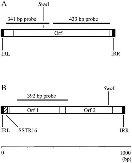
Restriction endonuclease digestion of genomic F. tularensis DNA was performed by two basic strategies. First, we used enzymes with no recognition sites within the IS elements (BglII and PstI). Second, an enzyme which cuts within the IS elements (SwaI cleaves ISFtu1 and ISFtu2) was used to maximize the possibility of determining the copy number of these IS elements in the isolates investigated in this study and, hence, to aid in their discrimination. The positions of the probe sequences in relation to the restriction sites in the IS elements are shown in Fig. 1. A 341-bp DNA fragment was used to hybridize to ISFtu2 from PstI- and BglII-digested genomic DNA. A 433-bp fragment was used to hybridize to ISFtu2 from SwaI-digested genomic DNA. A 392-bp DNA fragment was used to hybridize to ISFtu1 from BglII-, PstI-, and SwaI-digested genomic DNA (Fig. 1).
FIG. 1.
Organizations of ISs ISFtu2 (A) and ISFtu1 (B). Terminal inverted repeat sequences to the left (IRL) and right (IRR) are depicted as closed bars; an internal SSTR16 in ISFtu1 is depicted as a hatched bar. The locations of open reading frames (Orfs), the DNA probes used, and the recognition site for the restriction enzyme SwaI are indicated.
Amplification of probes.
Probes were amplified by PCR. Reaction mixtures contained PCR buffer (1.5 mM MgCl2), deoxynucleoside triphosphates (200 μM), 20 pmol of each primer (MWG AG Biotech, Ebersberg, Germany), and 0.2 U of Taq DNA polymerase in a final volume of 20 μl. One microliter of template DNA was used per reaction (1:10 dilution of plasmid preparation; plasmid midi preparation; Qiagen, Crawley, United Kingdom). DNA amplification was performed on a Gene Amplification System 9600 (PE Applied Biosystems Ltd., Warrington, United Kingdom). For the amplification of ISFtu1 (392 bp), primers 952Fa (5′-GAAAGCTTATTGTCACACCCCATCTA-3′) and 952Ra (5′-GACGGAGTTCGAGCTGAGTAAGTTT-3′) were used, with PCR conditions of 1 cycle of 94°C for 5 min; 30 cycles of 94°C (30 s), 67°C (30 s), and 72°C (30 s); and 1 cycle of 72°C for 7 min. For the amplification of ISFtu2 (341 bp), primers 866Fa (5′-CATGTGCTCTTGCTATTGTTGA-3′) and 866 Ra (5′-ACCGCTGTTATTTAAGATTTGAAA-3′) were used, with PCR conditions of 1 cycle of 94°C for 5 min; 30 cycles of 94°C (30 s), 60°C (30 s), and 72°C (30 s); and 1 cycle of 72°C for 7 min. When SwaI-digested DNA was probed, an alternative ISFtu2-specific probe (433 bp) was amplified by using primers New866F (5′-ATTCATCGTAACCAAATAAAAGTA-3′) and New866R (5′-GCTACGGGATATGATAAAGATGATAAC-3′), with PCR conditions of 1 cycle of 94°C for 5 min; 30 cycles of 94°C (30 s), 58°C (30 s), and 72°C (30 s); and 1 cycle of 72°C for 7 min. The concentrations of the amplified probes were determined by obtaining optical density readings at 260 nm with a 4054 UV/visible spectrophotometer (Amersham Biosciences UK Ltd). The probe was aliquoted and stored at −20°C.
Growth of F. tularensis and genomic DNA extraction.
The bacterial strains were cultured on blood cysteine glucose agar plates supplemented with 10 ml of 10% (wt/vol) histidine per liter at 37°C for 1 day. Colonies were harvested from the plates with a sterile loop, suspended in phosphate-buffered saline (pH 7.2), and incubated overnight at 4°C. The bacteria were then collected by centrifugation (3,000 × g, 10 min) and resuspended in 15 ml of lysing solution (10 mM NaCl, 20 mM Tris-HCl [pH 8] containing 1 mM EDTA and 0.5% [wt/vol] sodium dodecyl sulfate [SDS]). Proteinase K was added to a final concentration 100 mg/ml, and the mixture was incubated for 12 h at 50°C. Three milliliters of 5 M sodium perchlorate was then added to the cleared lysate, followed by incubation at room temperature for 1 h. The lysate was extracted twice with an equal volume of phenol-chloroform-isoamyl alcohol (25:24:1), and the DNA in the aqueous phase was precipitated by the addition of 2 volumes of ice-cold 95% ethanol. The DNA was spooled onto a sterile plastic loop and resuspended in distilled H2O.
Genomic DNA restriction endonuclease digestion and separation.
Genomic DNA samples were ethanol precipitated and resuspended in H2O, and their concentrations were determined by obtaining optical density measurements at 260 nm. Typically, 2 μg of DNA was digested with 10 to 20 U of restriction endonuclease (SwaI, BglII, or PstI) in a final volume of 15 μl overnight at 37°C. The digested DNA was separated in a 0.7% (wt/vol) agarose-TAE (Tris-acetate-EDTA) gel (with 0.5 μg of ethidium bromide per ml) with 1× TAE buffer at 100 V. DNA standards consisting of DIG-labeled HindIII-digested bacteriophage λ (molecular weight marker II; Roche Diagnostics Limited) were included as size markers (5 to 10 ng per lane). Five microliters of the plasmid-cloned IS element was used as a positive control.
Southern transfer and hybridization.
After DNA separation the gels were gently agitated for 45 min in denaturing solution (0.5 M NaOH, 1.5 M NaCl). Following denaturation, the gels were gently agitated for 30 min and then for 15 min in neutralizing buffer (1 M Tris-HCl [pH 8.0], 1.5 M NaCl). The DNA was transferred to a positively charged nylon membrane by capillary action with 10× SSC (diluted from 20× SSC [1× SSC is 0.15 M NaCl plus 0.015 M sodium citrate]; Sigma Chemical Co.). After DNA transfer, the membranes were air dried for 30 min and baked at 120°C for 30 min. Prehybridization and hybridization of the membranes were performed at 37°C in DIG Easy Hyb. The probe was made single stranded by boiling for 8 min and being placed on ice until use. The probe was used at a concentration of 20 or 25 ng of DIG Easy Hyb per ml.
Detection of bound probe.
The membranes were washed twice for 5 min each time at room temperature in 2× SSC-0.1% SDS, followed by two washes at 68°C in 0.1× SSC containing 0.1% SDS for 15 min.
The DIG Wash and Block Buffer set was used for the detection of the DIG label. The hybridized probe was visualized indirectly by using an antibody directed against the DIG label (anti-DIG alkaline phosphatase, Fab fragments). The membrane was washed briefly in 1× wash buffer at room temperature and was then blocked for 45 min at room temperature in 1.5× blocking solution (made up in malic acid buffer). The membranes were incubated with antibody at a dilution of 1:10,000 in 1.5× blocking solution for 30 min at room temperature. Unbound antibody was removed by two 15-min washes at room temperature in 1× wash buffer.
The membranes were equilibrated for 2 min in detection buffer. Twenty to 30 drops (1 ml) of the substrate disodium 3-(4-methoxyspiro{1,2-dioxetane-3,2′-(5′-chloro)tricyclo[3.3.1.13,7]decan}-4-yl) phenyl phosphate was applied to a transparent polyethylene folder. The membranes were incubated at room temperature for 10 min and for a further 15 min at 37°C. The membranes were then exposed to Lumi-film chemiluminescent detection film.
Interpretation of banding patterns.
In this study, the F. tularensis strains with identical banding patterns have been assigned the same arbitrary letter or number code (e.g., A, II, or δ). RFLP patterns that resembled a particular banding pattern but that had a one- or two-band difference from the pattern are denoted by addition of an asterisk (e.g., A∗, II∗, or δ∗) (Table 1).
Nucleotide sequence accession numbers.
The ISFtu2 region of F. tularensis subsp. tularensis strain SchuS4 has been assigned GenBank accession no. AY101577. The ISFtu1 region of F. tularensis subsp. tularensis strain SchuS4 has previously been assigned GenBank accession no. AF357005.
RESULTS
Identification of ISs in F. tularensis.
Analysis of the available genome sequence from F. tularensis subsp. tularensis strain SchuS4 identified two repeated sequences with a typical IS element structure of 952 and 865 bp, respectively. Both IS elements were flanked by terminal inverted repeats and contained putative transposase genes.
The 952-bp IS element was designated ISFtu1 according to the suggested nomenclature and showed similarities to members of the IS630 family of bacterial ISs (21). The available F. tularensis strain SchuS4 genome sequence indicated that 40 copies were present, each flanked by a 14-bp terminal inverted repeat. As described previously, this element contains an internal 16-bp short sequence tandem repeat (SSTR16) adjacent to one of the terminal inverted repeats (15). The various ISFtu1 sequences have 2 to 18 copies of a 16-bp SSTR (GenBank accession no. AF357005). The two putative transposase genes in ISFtu1 are 126 and 118 amino acids, respectively.
The 865-bp IS element was designated ISFtu2 and was placed into the IS5 family of bacterial ISs (21). An analysis of the available genome sequence indicated that 10 copies of this IS element are present in F. tularensis subsp. tularensis strain SchuS4. This IS element was flanked by 19-bp terminal inverted repeats and had an open reading frame encoding a putative transposase of 247 amino acids. A search of the sequences in the GenBank database identified homology to several bacterial putative transposase genes including one from Vibrio salmonicida (GenBank accession no. AJ277063; E value, 10−33; 81 of 218 identical amino acids).
RFLP analysis of F. tularensis with an ISFtu1-specific probe.
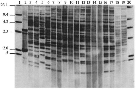
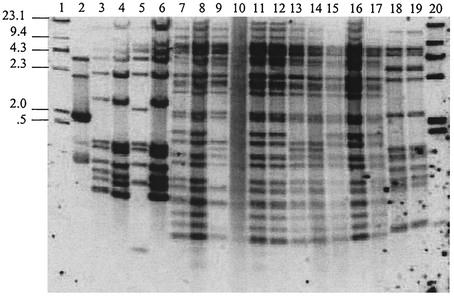
Genomic DNAs from a range of isolates of F. tularensis were digested with the PstI or BglII restriction endonuclease, Southern blotted, and hybridized with the labeled ISFtu1-specific probe (Table 1). Due to the apparent high copy number of this IS it was difficult to obtain discrete probe-reactive bands. When the DNAs of all of the isolates of F. tularensis subsp. tularensis tested were digested with BglII, they had unique, although related, RFLP profiles (Fig. 2). Only the attenuated strain, ATCC 6223, showed a profile which had a low level of similarity to the profiles of other strains in the same subspecies (F. tularensis subsp. tularensis). The profiles obtained after PstI digestion of the DNAs from isolates of F. tularensis subsp. tularensis showed a lower level of discrimination within this subspecies (data not shown), and some isolates had identical patterns. The isolates tested were grouped on the basis of the differences and similarities in probe-reactive bands (Table 1). Three main patterns were identified among the F. tularensis subsp. holarctica isolates after their BglII-digested DNAs were probed with the ISFtu1-specific probe, and two main patterns were identified after PstI digestion of these DNAs. Isolates of F. tularensis subsp. holarctica originating from Japan displayed a distinct profile after digestion of their DNAs with either enzyme. In addition, BglII digestion of these DNAs resulted in a single band difference between the two Japanese isolates investigated. DNAs from F. tularensis subsp. mediaasiatica isolates also had distinct RFLP profiles after digestion with BglII or PstI, and they were probed with an ISFtu1-specific probe. BglII digests did, in addition, permit discrimination of the F. tularensis subsp. mediaasiatica isolates tested in this study.
FIG. 2.
Southern blot of BglII-digested DNAs from 17 strains of F. tularensis hybridized with an ISFtu1-specific probe. Lanes 1 and 20, molecular size marker (sizes are indicated on the left in kilobase pairs); lane 2, plasmid-borne IS element; lanes 3 to 6, F. tularensis subsp. tularensis (FSC 237, FSC 199, ATCC 6223, and FSC 041, respectively); lanes 7 to 17, F. tularensis subsp. holarctica (LVS, FSC 092, FSC 017, FSC 075, FSC 035, FSC 200, FSC 025, FSC 108, FSC 250, FSC 097, and FSC 124, respectively); lanes 18 and 19, F. tularensis subsp. mediaasiatica (FSC 147 and FSC 148, respectively).
RFLP analysis of F. tularensis with an ISFtu2-specific probe.
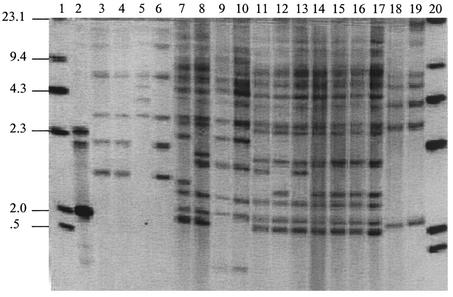
Genomic DNAs from a range of isolates of F. tularensis were digested with the BglII (data not shown) or PstI enzyme, Southern blotted, and hybridized with a labeled ISFtu2-specific probe. The results indicated that clearer discrimination between isolates was possible after PstI digestion of DNA (Fig. 3). However, on comparison with the patterns of hybridization obtained with the ISFtu1-specific probe, poorer discrimination of individual isolates was possible with the ISFtu2-specific probe (Table 1). When the DNAs were digested with PstI and probed with the ISFtu2-specific probe, some of the probe-reactive bands were common to all of the isolates tested. However, some bands were unique either to individual strains or to groups of isolates. The isolates tested were grouped on the basis of the differences and similarities in probe-reactive bands (Table 1). With the exception of attenuated strain ATCC 6223, all of the F. tularensis subsp. tularensis isolates tested had identical profiles when their DNAs were digested with either PstI or BglII. Both of the isolates of F. tularensis subsp. mediaasiatica tested also had identical and distinctive RFLP profiles when their DNAs had been digested with either PstI or BglII. When the DNAs from isolates of F. tularensis subsp. holarctica were digested with BglII, all of the isolates with the exception of the isolates from Japan showed identical profiles. The isolates from Japan shared a distinct profile. PstI digestion revealed three distinct patterns among the F. tularensis subsp. holarctica isolates, with the isolates originating from Japan sharing a unique profile.
FIG. 3.
Southern blot of PstI-digested DNAs from 17 strains of F. tularensis hybridized with an ISFtu2-specific probe. Lanes 1 and 20, molecular size markers (sizes are indicated on the left in kilobase pairs); lane 2, plasmid-borne IS element; lanes 3 to 6, F. tularensis subsp. tularensis (FSC 237, FSC 199, ATCC 6223, and FSC 041, respectively); lanes 7 to 17, F. tularensis subsp. holarctica (LVS, FSC 092, FSC 017, FSC 075, FSC 035, FSC 200, FSC 025, FSC 108, FSC 250, FSC 097, and FSC 124, respectively); lanes 18 and 19, F. tularensis subsp. mediaasiatica (FSC 147 and FSC 148, respectively).
RFLP analysis of F. tularensis DNA with an IS element-cutting enzyme.
The restriction endonuclease SwaI cuts once within the ISFtu2 sequence (at 329 bp) and once in the ISFtu1 sequence (at 829 bp in an ISFtu1 sequence with a 16-bp SSTR occurring in two copies) (Fig. 1). Only fragments containing the 5′ sequence of ISFtu2 or the 3′ sequence of ISFtu1 hybridize with the appropriate IS element-specific probe after SwaI digestion of DNA. This permits the detection of single copies of the IS elements and, subsequently, the determination of the approximate copy number of IS elements in individual isolates.
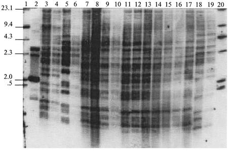
When DNAs from isolates of F. tularensis subsp. tularensis, F. tularensis subsp. holarctica, and F. tularensis subsp. mediaasiatica were digested with SwaI and hybridized with ISFtu1, approximately 40 DNA probe-reactive fragments were present in all of the isolates tested (Fig. 4). When similar experiments were performed with the ISFtu2-specific probe, 12 probe-reactive fragments were detected in F. tularensis subsp. tularensis. In the strains of F. tularensis subsp. holarctica and F. tularensis subsp. mediaasiatica tested, there were approximately 30 to 35 and 13 probe-reactive fragments, respectively (Fig. 5).
FIG. 4.
Southern blot of SwaI-digested DNAs from 17 strains of F. tularensis hybridized with an ISFtu1-specific probe. Lanes 1 and 20, molecular size marker (sizes are indicated on the left in kilobase pairs); lane 2, plasmid-borne IS element; lanes 3 to 6, F. tularensis subsp. tularensis (FSC 237, FSC 199, ATCC 6223, and FSC 041, respectively); lanes 7 to 17, F. tularensis subsp. holarctica (LVS, FSC 092, FSC 017, FSC 075, FSC 035, FSC 200, FSC 025, FSC 108, FSC 250, FSC 097, and FSC 124, respectively); lanes 18 and 19, F. tularensis subsp. mediaasiatica (FSC 147 and FSC 148, respectively).
FIG. 5.
Southern blot of SwaI-digested DNAs from 17 strains of F. tularensis hybridized with an ISFtu2-specific probe. Lanes 1 and 20, molecular size marker (sizes are indicated on the left in kilobase pairs); lane 2, plasmid-borne IS element; lanes 3 to 6, F. tularensis subsp. tularensis (FSC 237, FSC 199, ATCC 6223, and FSC 041, respectively); lanes 7 to 17, F. tularensis subsp. holarctica (LVS, FSC 092, FSC 017, FSC 075, FSC 035, FSC 200, FSC 025, FSC 108, FSC 250, FSC 097, and FSC 124, respectively); lanes 18 and 19, F. tularensis subsp. mediaasiatica (FSC 147 and FSC 148, respectively).
The patterns of hybridization of SwaI-digested DNA fragments (Fig. 4 and 5) revealed that even though single copies of IS elements can be identified, there was a striking similarity of hybridization profiles between the DNAs from different strains within each subspecies. These groupings are detailed in Table 1.
When DNAs from different strains of F. tularensis subsp. tularensis were digested with SwaI and hybridized with the ISFtu1- and ISFtu2-specific probes, all of the profiles except that for strain ATCC 6223 formed a single subgroup. DNAs from different strains of F. tularensis subsp. holarctica showed limited diversity, and digested DNAs from strains FSC 025 and FSC 108, which had been hybridized with the ISFtu2-specific probe, formed a distinct grouping (Fig. 5). Digested DNAs from the two Japanese isolates of F. tularensis subsp. holarctica showed a profile distinct from those obtained with other isolates of F. tularensis subsp. holarctica with both the ISFtu1- and ISFtu2-specific probes (Fig. 4 and 5). Digested DNAs from the F. tularensis subsp. mediaasiatica isolates, which were hybridized with either the ISFtu1- or ISFtu2-specific probe, showed profiles which were distinct from the profiles obtained with DNAs from the other isolates of F. tularensis tested in this study.
DISCUSSION
There are three subspecies of F. tularensis. The subspecies F. tularensis subsp. tularensis, F. tularensis subsp. holarctica, and F. tularensis subsp. mediaasiatica all pose significant threats as human pathogens (8, 11, 25, 26, 29).
This is the first report of a typing methodology that uses the ISs found in F. tularensis. The nucleotide sequence of ISFtu1 has been described previously (15). In this study we have used the genome sequence of F. tularensis subsp. tularensis strain SchuS4 to identify a novel IS element which we have termed ISFtu2. The RFLP typing method reported here exploits probes derived from the sequences of both ISFtu1 and ISFtu2.
Other DNA-based typing methods have exploited sequences that are not derived from F. tularensis. The use of primers consisting of the REP sequence, the ERIC sequence, or RAPD has permitted the discrimination of subspecies of F. tularensis and even individual isolates (6). However, an analysis of the SchuS4 genome sequence has revealed no evidence of REP or ERIC sequences (16).
With the availability of F. tularensis strain SchuS4 genome sequence data, we considered that typing methodologies could be based upon known sequence elements within the genome. A typing system based upon SSTRs could, in theory, offer a highly discriminatory typing system due to their vulnerability to mutation (33). Two studies based upon such a system have been reported (9, 15). In the report of Johansson et al. (15), eight discrete SSTR loci were identified from the F. tularensis subsp. tularensis strain SchuS4 genome sequence. Two of the loci (SSTR9 and SSTR16) containing unique SSTR elements showed high degrees of allelic variability between strains. The PCR products from both SSTR loci were highly discriminatory when they were used in combination, giving a Simpson's index of diversity of 0.97. This permits discrimination of individual strains. The report of Farlow et al. (9) described six variable number tandem repeat loci and a previously identified 30-bp region of heterogeneity (15) that distinguishes members of the subspecies F. tularensis subsp. holarctica. The polymorphisms at these loci were used to resolve 56 isolates into 39 unique types. This demonstrates higher-level relationships, which are consistent with the present subspecies classification.
In this study an alternative view of the organization of the genomes from different strains of F. tularensis is offered. Entire genomes can be compared with each other on the basis of the numbers and distributions of IS elements. IS elements are mobile DNA elements that can transpose themselves into diverse sites within the genome. They are maintained in bacterial populations as they replicate and transpose independently to the chromosomal DNA. Hence, they are subject to continuous variation during the history of a bacterial clone. Therefore, RFLP based upon IS elements could reveal much about the epidemiology and evolutionary genetics of bacterial pathogens (31, 33).
The most important finding of our study is that despite the diverse geographical origins of the strains tested, the distributions of the IS elements are generally stable among isolates of each subspecies. On the basis of the RFLP patterns of the isolated DNAs, the F. tularensis isolates tested fell into one of five main groups, namely, F. tularensis subsp. tularensis, the attenuated strain F. tularensis subsp. tularensis ATCC 6223, F. tularensis subsp. holarctica, Japanese isolates of F. tularensis subsp. holarctica, and F. tularensis subsp. mediaasiatica. This ability to group isolates on the basis of IS element distribution is in contrast to the results found with recently emerged pathogens such as M. tuberculosis and Y. pestis. Typing of isolates of these species, which is based on the IS elements IS6110 and IS100, respectively, showed that these species are genetically diverse in terms of both IS element distribution and copy number (1, 19, 30). Our results suggest that the F. tularensis IS elements are not frequently involved in genome rearrangements. Alternatively, the homogeneity may reflect a slow rate of evolution of the bacterium due to a long mean generation time, possibly because of a dormant phase in an ecological niche between epidemics in mammalian hosts.
The potential for using IS element-based RFLP as a typing method for the discrimination of individual isolates of F. tularensis is somewhat limited. However, our analysis of IS element copy number and RFLP profiles did permit some groups within subspecies to be identified. Significantly, our results demonstrated that the Japanese isolates of F. tularensis subsp. holarctica tested could be discriminated from the other F. tularensis subsp. holarctica isolates tested in this study. It has been reported that the Japanese isolates of F. tularensis subsp. holarctica have distinct differences from other isolates of the subspecies in terms of their genetic makeup (26, 29). The Japanese isolates consistently grouped separately from the other isolates of F. tularensis subsp. holarctica used in this study. Therefore, our results seem to support the suggestion that Japanese isolates are a distinct subgroup that has sometimes been termed F. tularensis subsp. holarctica biovar japonica (26, 29).
Similarly, our results provide new insight into the genetic organization of DNA from isolates of F. tularensis subsp. mediaasiatica in comparison with the DNA of other isolates of F. tularensis. The 16S rDNA genotype of F. tularensis subsp. mediaasiatica has previously been shown to be most similar to the 16S rDNA genotype of F. tularensis subsp. tularensis (29), and isolates of both of these subspecies show citrulline ureidase activity (26, 29). Our findings show that the copy numbers of ISFtu1 and ISFtu2 in the F. tularensis subsp. mediaasiatica isolates tested are most similar to the copy numbers (but not the distributions) of these IS elements in isolates of F. tularensis subsp. tularensis. This finding may indicate that these subspecies have relatively similar evolutionary histories. In support of this suggestion, a whole-genome microarray analysis of F. tularensis isolates has revealed that although isolates of F. tularensis subsp. mediaasiatica possess a moderate virulence, their DNA is most similar to the DNA from isolates of highly virulent F. tularensis subsp. tularensis (M. Broekhuijsen, P. Larsson, A. Johansson, et al., submitted for publication).
Although both of the attenuated isolates evaluated in this study (strains ATCC 6223 and LVS) had unique profiles, our analysis has not revealed any consistent differences between the RFLP profiles of virulent and attenuated isolates of F. tularensis. Our finding that the profile of the DNA of isolate ATCC 6223 consistently differs from those of the DNAs of other strains of F. tularensis subsp. tularensis corroborates the findings of previous studies in which a microarray (Broekhuijsen et al., submitted), pulsed-field gel electrophoresis (A. Johansson and A. Sjöstedt, unpublished results), or RAPD PCR typing (16) was used to analyze the genomic makeup. DNA from F. tularensis LVS digested with PstI had a profile distinct from those of the other F. tularensis subsp. holarctica isolates tested, but the IS element copy number appeared to be similar to that in other F. tularensis subsp. holarctica isolates. Similar IS element copy numbers may indicate that IS elements ISFtu1 and ISFtu2 may not contribute to the attenuated status of this strain. Conversely, the unique banding patterns of LVS may suggest that the IS elements caused adjacent deletions within the genome, and these could account for the attenuation of LVS.
Acknowledgments
This work was supported by DynPort Vaccine Company LLC, Frederick, Md., and the Joint Vaccine Acquisiton Program, Frederick, Md.
REFERENCES
- 1.Achtman, M., K. Zurth, G. Morelli, G. Torrea, A. Guiyoule, and E. Carniel. 1999. Yersinia pestis, the cause of plague, is a recently emerged clone of Yersinia pseudotuberculosis. Proc. Natl. Acad. Sci. USA 96:14043-14048. [DOI] [PMC free article] [PubMed] [Google Scholar]
- 2.Anonymous. 2000. Tularemia, Kosovo. Wkly. Epidemiol. Rec. 75:133-134. [PubMed] [Google Scholar]
- 3.Bachiller Luque, P., J. L. P. Castrillon, M. M. Luquero, F. J. M. Martin, J. D. L. L. Lopez-Areal, P. P. Pascual, M. A. Mazon, and V. H. Guilarte. 1998. Preliminary report of an epidemic tularemia outbreak in Valladolid. Rev. Clin. Esp. 198:789-793. [PubMed] [Google Scholar]
- 4.Boyce, J. M. 1975. Recent trends in the epidemiology of tularemia in the United States. J. Infect. Dis. 131:197-199. [DOI] [PubMed] [Google Scholar]
- 5.Craven, R. B., and A. M. Barnes. 1991. Plague and tularemia. Infect. Dis. Clin. N. Am. 5:65-175. [PubMed] [Google Scholar]
- 6.de la Puente-Redondo, V. A., N. G. D. Blanco, C. B. Gutierrez-Martin, F. J. Garcia-Pena, and E. F. R. Ferri. 2000. Comparison of different PCR approaches for typing of Francisella tularensis strains. J. Clin. Microbiol. 38:1016-1022. [DOI] [PMC free article] [PubMed] [Google Scholar]
- 7.Dennis, D. T., T. V. Inglesby, D. A. Henderson, J. G. Bartlett, M. S. Ascher, E. Eitzen, A. D. Fine, A. M. Friedlander, J. Hauer, M. Layton, S. R. Lillibridge, J. E. McDade, M. T. Osterholm, T. O'Toole, G. Parker, T. M. Perl, P. K. Russell, K. Tonat, et al. 2001. Tularemia as a biological weapon: medical and public health management. JAMA 285:2763-2773. [DOI] [PubMed] [Google Scholar]
- 8.Eigelsbach, H. T., and V. G. McGann. 1984. Genus Francisella Dorofe'ev 1947, 176AL, p. 394-399. In N. R. Krieg and J. G. Holt (ed.), Bergey's manual of systematic bacteriology, vol. 1. The Williams & Wilkins Co., Baltimore, Md.
- 9.Farlow, J., K. L. Smith, J. Wong, M. Abrams, M. Lytle, and P. Keim. 2001. Francisella tularensis strain typing using multiple-locus, variable number tandem repeat analysis. J. Clin. Microbiol. 39:3186-3192. [DOI] [PMC free article] [PubMed] [Google Scholar]
- 10.Gill, V., and B. A. Cunha. 1997. Tularemia pneumonia. Semin. Resp. Infect. 12:61-67. [PubMed] [Google Scholar]
- 11.Gurycova, D. 1998. First isolation of Francisella tularensis subsp. tularensis in Europe. Eur. J. Epidemiol. 14:797-802. [DOI] [PubMed] [Google Scholar]
- 12.Gustafsson, K. 1989. Growth and survival of four strains of Francisella tularensis in a rich medium preconditioned with Acanthamoeba palestinensis. Can. J. Microbiol. 35:1100-1104. [DOI] [PubMed] [Google Scholar]
- 13.Hopla, C. E. 1974. The ecology of tularemia. Adv. Vet. Sci. Comp. Med. 18:25-53. [PubMed] [Google Scholar]
- 14.Hubalek, Z., W. Sixl, and J. Halouzka. 1998. Francisella tularensis in Dermacentor reticulatus ticks from the Czech Republic and Austria. Wien. Klin. Wochenschr. 110:909-910. [PubMed] [Google Scholar]
- 15.Johansson, A., I. Göransson, P. Larsson, and A. Sjöstedt. 2001. Extensive allelic variation among Francisella tularensis strains in a short-sequence tandem repeat region. J. Clin. Microbiol. 39:3140-3146. [DOI] [PMC free article] [PubMed] [Google Scholar]
- 16.Johansson, A., A. Ibrahim, I. Göransson, U. Eriksson, D. Gurycova, J. E. Clarridge, and A. Sjöstedt. 2000. Evaluation of PCR-based methods for discrimination of Francisella species and subspecies and development of a specific PCR that distinguishes the two major subspecies of Francisella tularensis. J. Clin. Microbiol. 38:4180-4185. [DOI] [PMC free article] [PubMed] [Google Scholar]
- 17.Jusatz, H. J. 1952. Tularemia in Europe, 1926-1951, p. 7-16. In E. Rodenwaldt (ed.), Welt-Suchen atlas, vol. 1. Falk-Verlag, Hamburg, Germany.
- 18.Karlsson, J., R. G. Prior, K. Williams, L. Lindler, K. A. Brown, N. Chatwell, K. Hjalmarsson, N. Loman, K. A. Mack, M. Pallen, M. Popek, G. Sandstöm, A. Sjöstedt, T. Svensson, I. Tamas, S. G. E. Andersson, B. W. Wren, P. C. F. Oyston, and R. W. Titball. 2000. Sequencing of the Francisella tularensis strain Schu 4 genome reveals the shikimate and purine metabolic pathways, targets for the construction of a rationally attenuated auxotrophic vaccine. Microbiol. Comp. Gen. 5:25-39. [DOI] [PubMed] [Google Scholar]
- 19.Kremer, K., D. van Soolingen, R. Frothingham, W. H. Haas, P. W. Hermans, C. Martin, P. Palittapongarnpim, B. B. Plikaytis, L. W. Riley, M. A. Yakrus, J. M. Musser, and J. D. van Embden. 1999. Comparison of methods based on different molecular epidemiological markers for typing of Mycobacterium tuberculosis complex strains: interlaboratory study of discriminatory power and reproducibility. J. Clin. Microbiol. 37:2607-2618. [DOI] [PMC free article] [PubMed] [Google Scholar]
- 20.Kurtz, S., and C. Schleiermacher. 1999. REPuter: fast computation of maximal repeats in complete genomes. Bioinformatics 15:426-427. [DOI] [PubMed] [Google Scholar]
- 21.Mahillon, J., and M. Chandler. 1998. Insertion sequences. Microbiol. Mol. Biol. Rev. 62:725-774. [DOI] [PMC free article] [PubMed] [Google Scholar]
- 22.Morner, T. 1992. The ecology of tularemia. Rev. Sci. Tech. Off. Int. Epizoot. 11:1123-1130. [PubMed] [Google Scholar]
- 23.Ohara, Y., T. Sato, H. Fujita, T. Ueno, and M. Homma. 1991. Clinical manifestations of tularemia in Japan—analysis of 1355 cases observed between 1924 and 1987. Infection 19:14-17. [DOI] [PubMed] [Google Scholar]
- 24.Ohara, Y., T. Sato, and M. Homma. 1998. Arthropod-borne tularemia in Japan: clinical analysis of 1374 cases observed between 1924 and 1996. J. Med. Entomol. 35:471-473. [DOI] [PubMed] [Google Scholar]
- 25.Olsufjev, N. G., and I. S. Meshcheryakova. 1982. Infraspecific taxonomy of tularemia agent Francisella tularensis McCoy and Chapin. J. Hyg. Epidemiol. Microbiol. Immunol. 26:291-299. [PubMed] [Google Scholar]
- 26.Olsufjev, N. G., and I. S. Meshcheryakova. 1983. Subspecific taxonomy of Francisella tularensis. Int. J. Syst. Bacteriol. 33:872-874. [Google Scholar]
- 27.Parker, R. R., E. A. Steinhaus, G. M. Kohls, and W. L. Jellison. 1951. National Institutes of Health bulletin, p. 7-33. National Institutes of Health, Bethesda, Md. [PubMed]
- 28.Reintjes, R., I. Dedushaj, A. Gjini, T. R. Jorgensen, B. Cotter, A. Lieftucht, F. D'Ancona, D. T. Dennis, M. A. Kosoy, G. Mulliqi-Osmani, R. Grunow, A. Kalaveshi, L. Gashi, and I. Humolli. 2002. Tularemia outbreak investigation in Kosovo: case control and environmental studies. Emerg. Infect. Dis. 8:69-73. [DOI] [PMC free article] [PubMed] [Google Scholar]
- 29.Sandström, G., A. Sjöstedt, M. Forsman, N. V. Pavlovich, and B. N. Mishan'kin. 1992. Characterization and classification of strains of Francisella tularensis isolated in the central Asian focus of the Soviet Union and in Japan. J. Clin. Microbiol. 30:172-175. [DOI] [PMC free article] [PubMed] [Google Scholar]
- 30.Sreevatsan, S., X. Pan, K. E. Stockbauer, N. D. Connell, B. N. Kreiswirth, T. S. Whittam, and J. M. Musser. 1997. Restricted structural gene polymorphism in the Mycobacterium tuberculosis complex indicates evolutionarily recent global dissemination. Proc. Natl. Acad. Sci. USA 94:9869-9874. [DOI] [PMC free article] [PubMed] [Google Scholar]
- 31.Stanley, J., and N. Saunders. 1996. DNA insertion sequences and the molecular epidemiology of Salmonella and Mycobacterium. J. Med. Microbiol. 45:236-251. [DOI] [PubMed] [Google Scholar]
- 32.Tärnvik, A., M. Ericsson, I. Golovliov, G. Sandstrom, and A. Sjöstedt. 1996. Orchestration of the protective immune response to intracellular bacteria: Francisella tularensis as a model organism. FEMS Immunol. Med. Microbiol. 13:221-225. [DOI] [PubMed] [Google Scholar]
- 33.van Belkum, A. 1999. The role of short sequence repeats in epidemiologic typing. Curr. Opin. Microbiol. 2:306-311. [DOI] [PubMed] [Google Scholar]
- 34.Wenger, J. D., D. G. Hollis, R. E. Weaver, C. N. Baker, G. R. Brown, D. J. Brenner, and C. V. Broome. 1989. Infection caused by Francisella philomiragia (formerly Yersinia philomiragia). A newly recognised human pathogen. Ann. Intern. Med. 110:888-892. [DOI] [PubMed] [Google Scholar]
- 35.Wicki, R., P. Sauter, C. Mettler, A. Natsch, T. Enzler, N. Pusterla, P. Kuhnert, G. Egli, M. Bernasconi, R. Lienhard, H. Lutz, and C. M. Leutenegger. 2000. Swiss Army survey in Switzerland to determine the prevalence of Francisella tularensis, members of the Ehrlichia phagocytophila genogroup, Borrelia burgdorferi sensu lato, and tick-borne encephalitis virus in ticks. Eur. J. Clin. Microbiol. Infect. Dis. 19:427-432. [DOI] [PubMed] [Google Scholar]


