Abstract
Genetic analysis of wild-type Crimean-Congo hemorrhagic fever (CCHF) virus strains recovered in the European part of Russia was performed. Reverse transcriptase PCR followed by direct sequencing was used to recover partial sequences of the CCHF virus medium (M) genome segment (M segment) from four pools of Hyalomma marginatum ticks and six human patients. Phylogenetic analysis of the M-segment sequences from Russian strains revealed a close relatedness of the strains (nucleotide sequence diversity, ≤5.0%). The strains differed significantly from CCHF viruses from other regions of the world (nucleotide sequence diversity, 10.3 to 20.4%), suggesting that CCHF virus strains recovered in the European part of Russia form a distinct group.
Crimean-Congo hemorrhagic fever (CCHF) virus causes a hemorrhagic and toxic syndrome disease in humans and high mortality rates of up to 50%. The infection was first discovered in the Crimean region of Russia and is now registered in many regions of the world (1, 2, 3). Humans become infected through bites of the Ixodid tick, by contact with a patient with CCHF during the acute phase of infection, or by contact with blood or tissues from viremic livestock. Hyalomma ticks are one of the major vectors of CCHF virus (3).
Sporadic cases of the CCHF are registered in European Russia each year. During 2000 and 2001, 133 primary cases of CCHF were identified in European Russia, with a fatality rate of 9.0% (6). However, the genetic diversity of CCHF virus in this geographic region has been unclear. Only the CCHF virus small (S) genome segment (S segment) sequence from one virus strain (strain Drosdov), isolated in 1967 in the Astrahan region of Russia (1), was genetically characterized in 1997 (GenBank accession number U88412).
CCHF viruses from different regions of the world have shown considerable molecular heterogeneity in their S genome segments (4, 5, 8). Very few data on the RNA of the medium (M) genome segments (M segments) are available; until now, only four complete M-segment sequences of CCHF viruses from China and Nigeria have been characterized (7). In our study, partial sequences of the M segments from 10 CCHF virus strains recovered in European Russia were determined and analyzed.
Primary blood samples from patients with CCHF, tissues samples from patients with fatal cases, and Hyalomma marginatum ticks were collected in the southern regions of European Russia during 2000 and 2001. The primary samples were screened for the presence of viral antigens by an enzyme antigen immunoassay. Four CCHF virus-positive tissue pools from H. marginatum ticks, blood samples from CCHF patients, and five positive tissue samples from patients with fatal cases were collected in three administrative regions of European Russia (Stavropol, Rostov, and Volgograd) and were tested by reverse transcriptase (RT) PCR.
Total RNA was extracted from selected tissues and blood by using the RNAeasy kit (Qiagen GmbH, Hilden, Germany) according to the recommendations of the supplier. RNA was converted to cDNA and amplified with the Access RT-PCR kit (Promega, Madison, Wis.) according to the recommendations of the manufacturer. Primers M-U (5′-CCC AGA TCT GTT TCA TTG TCA CCR GTT CAA TC-3′, where R is G or A) and M-L (5′-CCC GTC GAC GCA GAC AGC CAR CAT GAC A-3′) were used to amplify a 366-bp section that is located in the central region of the M segment and that corresponds to nucleotide (nt) positions 2586 to 2951 of CCHF virus strain BA66019 (7). Amplified bands of the predicted size were demonstrated in all samples investigated. Both strands of the amplicons were sequenced and resulted in a target sequence of 323 bp (nt positions 2607 to 2932).
The sequences obtained were aligned with the corresponding M-segment section of known CCHF viruses (African and Asian strains) by using the BLAST utility (National Center for Biotechnology Information, Bethesda, Md.). Comparative analysis of the CCHF virus M-segment sequences (Table 1) included four partial sequences from H. marginatum ticks, six sequences from CCHF patients in European Russia, one sequence from a Nigerian tick, and two sequences from Chinese CCHF patients (7). All sequences derived from the Russian H. marginatum ticks and CCHF patients in this study were closely related to each other (nucleotide sequence diversity, <5.0%; amino acid sequence diversity, <2.8%) and showed clear geographic clustering; the nucleotide sequence diversity between strains from the same locality did not exceed 0.6% (data not shown). All sequences could be divided into two groups: sequences originating from strains collected near the Tsimlyanskoye artificial sea in the Volgograd and Rostov regions (the VLG-ROS group, which had a nucleotide sequence diversity of 0.9 to 1.9% and which included strains TI28017, TI28019, HU29176, HU29901, HU29662, and HU29664) and sequences derived from strains from the Stavropol region and the southern part of the Rostov region (the STV-ROS group, which had nucleotide sequence diversity of 0.0 to 1.5% and which included strains TI27960, HU29219, and HU29901). The nucleotide sequence diversity between the two groups ranged from 3.7 to 5.0%.
TABLE 1.
M-segment nucleotidea and amino acid sequence differences among Russian CCHF virus strains and between Russian CCHF virus strains and those from other countries
| Groupb | Sequencec | % Difference
|
||||
|---|---|---|---|---|---|---|
| Russian STV-ROS group | Russian VLG-ROS group | Nigerian strain 10200 | Chinese strain 88166 | Chinese strain 66019 | ||
| Russian | nt | 0.9-1.9 | 3.7-5.0 | 19.8-20.1 | 17.6-18.6 | 20.1-20.4 |
| STV-ROS | aa | 0.0-0.9 | 1.9-2.8 | 13.1-14.0 | 10.3-11.2 | 14.0-14.9 |
| Russian | nt | 0.0-1.5 | 19.8-21.1 | 16.1-17.6 | 19.2-20.7 | |
| VLG-ROS | aa | 0.0-1.9 | 14.0-15.9 | 10.3-12.1 | 13.1-14.0 | |
Bases 2607 to 2929.
The VLG-ROS group includes strains TI28017, TI28019, HU29175, HU29176, TI29323, HU29662, and HU29664; the STV-ROS group includes strains TI27960, HU29219, and HU29901.
nt, nucleotide sequence; aa, amino acid sequence.
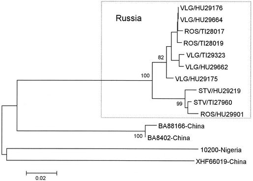
The nucleotide sequence divergences between the CCHF virus strains from Russia and related viral strains were as follows: 19.8 to 21.1% for CCHF virus strain IbAr10200 from a Nigerian tick, 16.1 to 18.6% for CCHF virus strain BA 88166 from a Chinese patient, and 19.2 to 20.7% for CCHF virus strain 66019 from a Chinese patient. These results indicate that, on the basis of sequence divergence, Russian CCHF viruses are most closely related to each other and form a distinct group. The phylogenetic tree generated by neighbor-joining analysis showed a geographic clustering of the sequences of CCHF viruses from European Russia. These constituted a well-supported branch that could be further divided into two lineages, VLG-ROS and STV-ROS (Fig. 1). Thus, our analysis provides strong evidence for the circulation of closely related genetic variants of CCHF virus in the southern part of European Russia.
FIG. 1.
Phylogenetic tree based on partial sequences of the M segment (nt positions 2607 to 2932). The sequences of CCHF viruses from Russia and other countries were analyzed by a neighbor-joining method with Kimura two-parameter distances by using MEGA software (version 2.1). Bootstrap confidence limits were based on 1,000 replicates; only those values that exceed 70% are shown. GenBank accession numbers for CCHF virus sequences from Russia are AF401647 to AF401650 and AY093620 to AY093625; the GenBank accession numbers of other sequences are AF338470 for strain BA88166, AB069674 for strain BA8402, AF350448 for strain XHFV66019, and U39455 for strain IbAr10200. Samples from Russia are designated with the following nomenclature: VLG, Volgograd region; ROS, Rostov region; STV, Stavropol region; TI, tick sample; HU, human sample. For example, STV/HU29219 is human sample 29219 obtained from the Stavropol region.
The potential roles of migratory birds and the movement of infected livestock (and ticks) in the spread of the virus over distant geographical areas have been studied (3, 4, 8). On the basis of the detection of genetically closely related viruses in the United Arab Emirates and Somalia, it has been suggested that animals imported from Somalia were the source of infection in the United Arab Emirates (8). The study of genetic variants of CCHF virus in the area between the Don and Volga Rivers, with a maximal distance of 400 km between localities, was one more reason to evaluate a migratory hypothesis. Our results are consistent with a point-source outbreak in the region studied and did not support a migratory hypothesis.
Nucleotide sequence accession numbers.
The sequences of the Russian isolates have been submitted to GenBank and have received accession numbers AF401647 to AF401650 and AY093620 to AY093625.
Acknowledgments
This work was supported by the CTR program (in the United States) through the International Science and Technology Center (grant 1291-2p) and by the Ministry of Public Health of the Russian Federation.
REFERENCES
- 1.Aristova, V. A., L. V. Kolobukhina, M. Y. Shchelkanov, and D. K. Lvov. 2001. Ecology and clinical features of Crimean-Congo hemorrhagic fever in Russia and neighboring countries. Vopr. Virusol. 45:7-15. (In Russian.)
- 2.Chumakov, M. P. 1947. A new virus disease—Crimean hemorrhagic fever. Nov. Med. 4:9-11. [Google Scholar]
- 3.Hoogstraal, H. 1979. The epidemiology of tick-borne Crimean-Congo hemorrhagic fever in Asia, Europe and Africa. J. Med. Entomol. 15:307-417. [DOI] [PubMed] [Google Scholar]
- 4.Khan, A. S., G. O. Maupin, P. E. Rollin, A. M. Noor, H. H. Shurie, A. G. Shalabi, et al. 1997. An outbreak of Crimean—Congo hemorrhagic fever in the United Arab Emirates, 1994-1995. Am. J. Trop. Med. Hyg. 57:519-525. [DOI] [PubMed] [Google Scholar]
- 5.Mariott, A. C., and P. A. Nuttall. 1992. Comparison of the S RNA segments and nucleoprotein sequences of Crimean-Congo hemorrhagic fever, Hazara and Dugbe viruses. Virology 109:795-799. [DOI] [PubMed] [Google Scholar]
- 6.Onishenko, G. G. 2001. On the epidemic situation and morbidity of natural focal infections in the Russian Federation and measures of their prophylaxis. Zh. Mikrobiol. 3:22-28.11550553 [Google Scholar]
- 7.Papa, A., B. Ma, S. Kouidou, Q. Tang, C. Hang, and A. Antoniadis. 2002. Genetic characterization of the M RNA segment of Crimean Congo hemorrhagic fever virus strains, China. Emerg. Infect. Dis. 8:50-53. [DOI] [PMC free article] [PubMed] [Google Scholar]
- 8.Rodriguez, L. L., G. O. Maupin, T. G. Ksiazek, O. E. Rollin, A. S. Khan, T. F. Schwarz, R. S. Lofts, J. F. Smith, A. M. Noor, C. J. Peters, and S. T. Nichol. 1997. Molecular investigation of a multisource outbreak of Crimean-Congo hemorrhagic fever in the United Arab Emirates. Am. J. Trop. Med. Hyg. 57:512-518. [DOI] [PubMed] [Google Scholar]


