Abstract
A central function of memory is to permit an organism to distinguish between stimuli that have been previously encountered and those that are novel. Although the medial temporal lobe (which includes the hippocampus and surrounding perirhinal, parahippocampal, and entorhinal cortices) is known to be crucial for recognition memory, controversy remains regarding how the specific subregions within the medial temporal lobe contribute to recognition. We used event-related functional MRI to examine the relation between activation in distinct medial temporal lobe subregions during memory formation and the ability (i) to later recognize an item as previously encountered (item recognition) and (ii) to later recollect specific contextual details about the prior encounter (source recollection). Encoding activation in hippocampus and in posterior parahippocampal cortex predicted later source recollection, but was uncorrelated with item recognition. In contrast, encoding activation in perirhinal cortex predicted later item recognition, but not subsequent source recollection. These outcomes suggest that the subregions within the medial temporal lobe subserve distinct, but complementary, learning mechanisms.
One of the most influential observations in memory research has been the profound inability of patients with bilateral medial temporal lobe (MTL) damage to consciously remember events that have occurred since the neural insult (1). This deficit, which is specific to declarative or explicit memory (2, 3), appears both during attempts to recall past events as well as when recognition of previously encountered stimuli is required. Subsequent investigations of the memory impairment in human amnesia, as well as in animal models, have led to advances in understanding MTL contributions to the encoding, retrieval, and consolidation of declarative memories (refs. 2, 4, and 5, but see ref. 6). However, although MTL consists of multiple structures (the hippocampus and surrounding entorhinal, perirhinal, and parahippocampal cortices), considerable debate surrounds the precise characterization of the mnemonic processes subserved by these various subregions. One point of controversy centers on the question of whether the hippocampus and perirhinal cortex make similar or distinct contributions to recognition (7–11).
At the anatomical level, consideration of the architecture raises the possibility that MTL subregions, while interacting, may mediate different computations. Consistent with its role in cross-modal memory and object identification (12), perirhinal cortex receives extensive input from visual association areas in the ventral visual stream and about a third of its input from polymodal or nonvisual unimodal cortices (13). In humans, perirhinal cortex may receive input from semantic association cortices (14–16). By contrast, parahippocampal cortex receives input mainly from visuospatial association areas in the dorsal visual stream as well as from other structures including dorsolateral prefrontal and retrosplenial cortices (17, 18). Perirhinal and parahippocampal cortices provide about two-thirds of the input to entorhinal cortex, which, in turn, provides the majority of the input to hippocampus (19). Because it receives input from much of the surrounding cortex, the hippocampus is well situated to bind disparate event features into an integrated conjunctive representation.
At the functional level, some animal lesion and electrophysiological data suggest that the subregions within MTL differentially contribute to recognition (9). For example, relative to hippocampal insult, lesions of perirhinal cortex in monkeys yield a more severe impairment in object recognition performance (20–22). Complementary data indicate that recognition deficits after selective hippocampal damage, if present, can be modest (refs. 23 and 24, but see refs. 25 and 26). Across-study comparisons further suggest that the extent of perirhinal, but not hippocampal, damage positively correlates with the magnitude of recognition impairment (ref. 27, but see ref. 28), implicating perirhinal cortex as critical for recognition. Moreover, electrophysiological measures indicate that hippocampal neurons differentially code for associative relations rather than for individual items, whereas perirhinal neurons code for item familiarity and recency (i.e., context-independent responses that signal prior item encounter) (9, 29, 30).
In humans, damage that is thought to be selective to the hippocampus yields item and associative recognition deficits (31–34). However, an emerging body of data suggests that damage limited to the hippocampus may differentially impair contextual recollection relative to item familiarity, and thus recognition deficits are seen when recollection of contextual details contributes to recognition performance in neurologically intact controls (32, 35). In contrast, recognition performance after hippocampal insult can be comparable to that of control subjects when the utility of contextual recollection is minimized for the controls (32). Such intact performance may depend on item memory, or stimulus familiarity, which can be differentially spared after early- (33) and late-onset (32, 36) selective hippocampal damage. Thus, structures beyond the hippocampus may subserve item memory, with some theorists specifically implicating perirhinal cortex as central for item familiarity (9). Unfortunately, the role of perirhinal cortex in recognition memory has yet to be directly addressed in humans because damage limited to perirhinal cortex is extremely rare (but see ref. 37).
Although the preceding evidence points to functional differentiation within MTL, other data raise questions regarding whether the hippocampus and perirhinal cortex make distinct contributions to recognition (25, 26, 38–40). For example, some animal lesion data indicate that recognition can be impaired after selective hippocampal damage (25, 26, 40), and findings from humans with damage thought to be limited to the hippocampus raise the possibility that item familiarity and contextual recollection may be similarly impaired (31, 34, 41). Such outcomes are consistent with the perspective that subregions within MTL interact to contribute to recognition memory, with hippocampal computations mediating both item memory and contextual recollection.
Given the centrality of this issue for understanding the functional architecture of MTL, the present study used event-related functional MRI (fMRI) during episodic encoding to test whether perirhinal cortex, parahippocampal cortex, and hippocampus support distinct forms of learning and thus different aspects of recognition memory. Although prior neuroimaging studies of recognition memory have revealed activation in the hippocampus (42–44), it remains unclear whether hippocampal activation during recognition reflects a specific role in associative memory (45, 46) and contextual recollection (47) or may also mark hippocampal contributions to item familiarity (31). Moreover, to date, little has been gleaned from neuroimaging regarding the functional contributions of human perirhinal cortex to recognition (48). The present study was specifically designed to permit identification of (i) structures whose activation at encoding correlates with later recognition that an item was previously encountered irrespective of whether item recognition is accompanied by recollection, and (ii) structures whose activation at encoding correlates with later recollection of the contextual details (i.e., the source; ref. 49) associated with the item's encounter.
Methods
Subjects.
Four female and 10 male right-handed, native-English speakers (aged 18–26 yr) were paid $50 for their participation. Informed consent was obtained in a manner approved by the institutional review boards at Massachusetts Institute of Technology and Massachusetts General Hospital.
Behavioral Procedures.
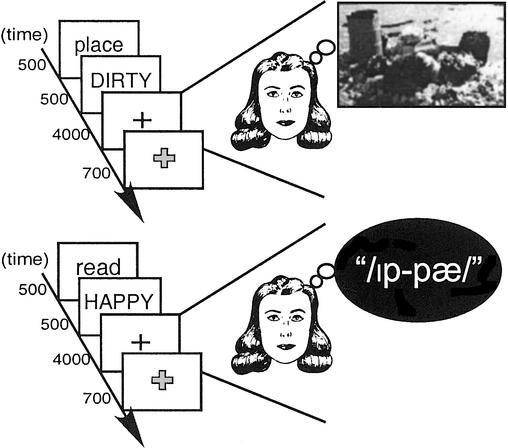
Across eight fMRI encoding scans, 200 visually presented adjectives were encoded through mental imagery (“Image” task) and 200 through orthographic-to-phonological transformation (“Read” task) (Fig. 1). On each trial, a 500-ms cue (place/read) signaled the encoding task to be performed on an adjective that was then visually presented for 500 ms. During Image trials, subjects generated a mental image of a spatial scene (i.e., a “place”) described by the adjective (e.g., for DIRTY, the subject might imagine a garbage dump). During Read trials, subjects covertly pronounced the word backwards (e.g., HAPPY might be pronounced/ip-pæ/). After a 4,000-ms fixation period, during which subjects performed the indicated task, the fixation cross changed color signaling subjects to indicate their level of task success by pressing one of four buttons: 1 = unsuccessful, 2 = partially successful, 3 = succeeded with effort, 4 = succeeded with ease. To ensure that subsequent memory effects do not reflect differential task success, analyses were restricted to successful trials. Items were counterbalanced across conditions. The order of conditions was determined by using a sequencing program designed to maximize the efficiency of the event-related design (50). Conditions were “jittered” by using variable duration (2–18 s) fixation null events.
Figure 1.
Encoding conditions performed during fMRI scanning are illustrated.
Subsequent recognition memory for studied items and recollection of the source (Image or Read) associated with each item were indexed by a two-step memory test administered ≈20 h after encoding. During this self-paced (nonscanned) test, all words studied during fMRI scanning as well as 400 unstudied distractors (Novel) were presented. Subjects first indicated whether they recognized the word as having been studied by responding “old” or “new.” When responding “old,” subjects then indicated which encoding task was performed with the item (i.e., Image or Read). Thus, measures of item recognition (recognized vs. forgotten) and source recollection (source correct vs. source incorrect) were obtained for each studied word. These measures were used to analyze the fMRI encoding data according to subsequent item and source memory.
fMRI Procedures.
Functional data were acquired by using a gradient-echo echo-planar sequence (1.5T Siemens Sonata, repetition time = 2 s, echo time = 40 ms, 21 axial slices, 3.125 × 3.125 × 5 mm, 1-mm skip, 210 volumes per run). High-resolution T1-weighted (MP-RAGE) structural images were collected for anatomical visualization. A bite-bar minimized head motion. Visual stimuli were projected onto a screen viewed through a mirror; responses were collected by using a magnet-compatible response pad.
Data were analyzed with SPM99 (Wellcome Department of Cognitive Neurology, London), with standard preprocessing procedures (for details see ref. 46). Structural and functional images were normalized to anatomical and EPI templates based on the MNI305 stereotaxic space. Images were resampled into 3-mm cubic voxels and spatially smoothed with an 8-mm full-width half maximum isotropic Gaussian kernel.
Statistical analyses were performed by using the general linear model. Trials were modeled by using a canonical hemodynamic response function and its temporal derivative. Effects were estimated by using a subject-specific fixed-effects model, with session-specific effects and low-frequency signal components treated as confounds. Linear contrasts yielded subject-specific estimates that were entered into second-level random-effects analyses. Regions consisting of at least five contiguous voxels that exceeded an uncorrected threshold of P < 0.001 were considered reliable.
To assess activation during the two encoding tasks, Image and Read trials were contrasted with fixation and with each other. Prior imaging data have revealed parahippocampal cortical activation during scene imagery (51) and hippocampal activation during associative encoding (45, 46). Thus, MTL regions were expected to be particularly responsive during Image trials because this condition required goal-directed semantic retrieval and generation of an associated spatial mental image. As discussed below, this prediction was confirmed. By contrast, the Read condition failed to yield above-baseline MTL activation; this may reflect the limited associative demands of the read task or the presence of phonological encoding computations during both fixation and Read trials. Accordingly, subsequent memory analyses (see below) were restricted to Image-encoded trials.
Neural correlates of subsequent memory were assessed in a priori predicted regions of interest (ROIs) identified in the group-level analyses of Image encoding (i.e., Image > Read and Image > Fixation contrasts). ROIs included all significant voxels within 6 mm of each maximum. For each subject, signal was calculated by selectively averaging data with respect to peristimulus time per condition. The conditions consisted of (i) trials associated with later item recognition and correct source recollection (Item + Source), (ii) trials associated with later item recognition but recollection failure (Item Only), and (iii) trials associated with later item forgetting (Forgotten). The resulting data were subjected to mixed-effect ANOVA, treating subsequent memory and peristimulus time as repeated measures and subjects as a random effect. Interaction analyses assessed between-region differences in the pattern of peak hemodynamic response across subsequent memory outcomes.
Results
Subsequent Recognition Performance.
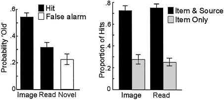
Item recognition, collapsed across correct and incorrect source recollection, differed depending on the task performed at encoding [F(2,26) = 52.99; P < 0.001], with recognition being superior for Imaged than for Read words [F(1,13) = 51.56; P < 0.001]; the hit rate for Read items was greater than the false alarm rate [F(1,13) = 7.84; P < 0.02] (Fig. 2 Left). For correctly recognized words (i.e., “hits”), source recollection was similar for Imaged and Read items (F < 1; Fig. 2 Right). For false alarms, there was a modest, but reliable (P < 0.05), bias to guess Read (0.63; chance is 0.50). To ensure that Item Only trials reflected “true” item memory and not guessing, the probability of recognizing an Image-encoded trial without source recollection (Item Only) was computed, correcting for the opportunity to guess. The resulting probability of item recognition without recollection was higher than the false alarm rate [0.30 vs. 0.15, respectively; t(13) = 7.82, P < 0.0001].
Figure 2.
Subsequent memory is plotted according to overall item recognition and source recollection. (Left) Item recognition was superior after Image than after Read encoding; Read encoding was higher than the false alarm rate. (Right) For recognized items, source recollection after Image and Read trials was comparable.
fMRI Task Effects.
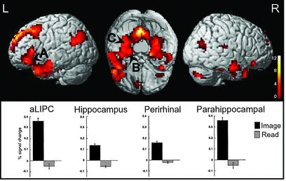
fMRI analyses revealed greater MTL activation in hippocampus, perirhinal, and parahippocampal cortices during Image relative to Read encoding and during Image encoding relative to the fixation; MTL activation during Read encoding did not fall above baseline (Fig. 3). These results complement prior demonstrations of parahippocampal cortical activation during scene imagery (51), and extend such effects to hippocampus and perirhinal cortex. Moreover, engagement of the hippocampus during the Image task, but not during the Read task, is consistent with the putative role of the hippocampus in associative processing (45, 46), as associative computations were required to generate an image based on the semantics associated with the adjective. Image encoding also engaged the anterior extent of left inferior prefrontal cortex [aLIPC; ≈Brodmann's area (BA) 47/45; Fig. 3] and bilateral retrosplenial cortices. The selective response of aLIPC to the Image, but not the Read, condition is consistent with the hypothesis that aLIPC differentially mediates controlled semantic retrieval (52, 53).
Figure 3.
Statistical maps displayed on a canonical 3D brain reveals regions demonstrating greater activation during Image than during Read trials. Critically, MTL and prefrontal regions demonstrated a greater response during Image compared with Read encoding. Regions included anterior left inferior frontal cortex (A: −54, 24, 3, ≈BA 47/45), left hippocampus (B: −33, −21, −21), left perirhinal cortex (B: −36, −12, −33, ≈BA 35/36), and left parahippocampal cortex (C: −33, −39, −18, ≈BA 37); peak responses did not fall above baseline during Read encoding. Other regions engaged during Image encoding included right MTL and bilateral retrosplenial cortices (complete coordinates are available on request). Note that activations are displayed on left- and right-hemisphere views, as well as on a cerebellumless ventral view (occipital lobe facing top).
Both encoding tasks elicited activation in the posterior extent of left inferior prefrontal cortex, extending into premotor cortex (MNI coordinates: −45, 14, 21 and −45, 10, 25; ≈BA 44/6). Relative to Image encoding and to baseline, Read encoding also differentially activated left premotor (≈BA 6), right inferior prefrontal (≈BA 47/45), bilateral posterior parietal (≈BA 7/40), and left lateral occipito-temporal (≈BA 37/21) cortices.
fMRI Subsequent Memory Effects.
The behavioral measures of subsequent item recognition and source recollection were used to back-sort the fMRI encoding events into imaged trials that were later recognized and accompanied by source recollection (Item + Source), recognized but not accompanied by source recollection (Item Only), or forgotten. The subsequent memory analysis revealed that the magnitude of encoding activation in the hippocampus, parahippocampal cortex, and perirhinal cortex correlated with different forms of subsequent memory (Fig. 4). There were three critical outcomes that bear on our understanding of MTL contributions to recognition memory.
Figure 4.
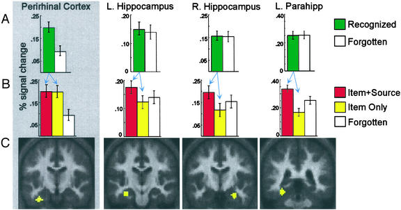
Peak signal change during Image encoding revealed qualitatively different activation patterns in left perirhinal cortex (−36, −12, −33), bilateral hippocampus (−33, −21, −21 and 30, −9, −24), and left parahippocampal cortex (−33, −39, −18). (A) Encoding activation, sorted according to whether the item was later recognized (irrespective of source recollection outcome) or forgotten, revealed dissociations between perirhinal cortex and each hippocampal region and between perirhinal and parahippocampal cortices. Perirhinal cortex demonstrated greater activation for items later recognized relative to those later forgotten, whereas neither hippocampus nor parahippocampal cortex differed according to item memory. (B) Time course analyses revealed a main effect of source memory outcome (Item + Source > Item Only) in right and left hippocampus and parahippocampal cortex, but not in perirhinal cortex. As displayed, peak signal change paralleled this pattern. (C) MTL regions of interest rendered on group-averaged structural images; anatomical localization of these ROIs to perirhinal cortex, hippocampus, and parahippocampal cortex, respectively, was confirmed at the individual-subject level.
First, when encoding trials were segregated based on whether the item was later recognized (collapsed across Item + Source and Item Only) or forgotten, activation in left perirhinal cortex predicted subsequent item recognition, whereas activation in bilateral hippocampus and parahippocampal cortices was similar irrespective of item recognition outcome (Fig. 4A). Specifically, activation in perirhinal cortex was greater during trials for which items were later recognized than during those later forgotten [F(1,13) = 15.21, P < 0.005], whereas neither hippocampal nor parahippocampal cortical activation differed according to subsequent item recognition (F < 1). Critically, the activation pattern in perirhinal cortex dissociated from that in each hippocampal region [Memory × Region, F(1,13) > 6.19, P < 0.05)], as well as from that in parahippocampal cortex [Memory × Region, F(1,13) = 11.90, P < 0.01]. In contrast, the patterns of activation in hippocampus and in parahippocampal cortex were qualitatively similar (F < 1.0).
Second, the dissociation between perirhinal encoding responses and the encoding responses in hippocampus and in parahippocampal cortex held even when comparing the two trial types (Item Only and Forgotten) that shared an absence of source recollection but differed with respect to item recognition (perirhinal/hippocampus: F(1,13) > 4.56, P ≤ 0.05; perirhinal/parahippocampal cortex: F(1,13) = 10.87, P < 0.01; Fig. 4B). Thus, perirhinal cortex demonstrated greater activation during encoding that yielded subsequent item recognition in the absence of source recollection (Item Only) relative to subsequent item forgetting. In contrast, neither hippocampus nor parahippocampal cortex demonstrated greater activation during Item Only relative to Forgotten trials (Fig. 4B), a critical observation that bears on theories of hippocampal contributions to recognition.
Third, when encoding events were segregated into those later recognized with (Item + Source) or without (Item Only) source recollection, activation in bilateral hippocampus and left parahippocampal cortex, but not in perirhinal cortex, predicted later recollection [right and left hippocampus: F(1,13) = 11.43, P < 0.005 and F(1,13) = 3.66, P < 0.08; parahippocampal cortex: F(1,13) = 5.78, P < 0.01; perirhinal cortex: F(1,13) = 1.37, P > 0.25] (Fig. 4B). Thus, perirhinal activation correlated with subsequent item recognition, but not source recollection. In contrast, hippocampal and parahippocampal activation did not track later item recognition, but rather correlated with whether recognition was accompanied by successful or unsuccessful contextual recollection.
Subsequent memory effects during Image encoding were also observed beyond MTL. Neural correlates of subsequent source recollection were observed in bilateral retrosplenial cortices (6, −51, 3 and −3, −57, 12; ≈BA 29/30). These regions demonstrated greater activation during Item + Source relative to Item Only trials [F(1,13) ≥ 5.48, P < 0.05], and no reliable difference between Item Only and Forgotten events [F(1,13) < 2.15, P > 0.15]. Similar effects were observed in two aLIPC regions [(−54, 24, 3 and −48, 24, −9; ≈BA 47/45): Item + Source > Item Only (F(1,13) > 5.19, P < 0.05) and Forgotten>Item Only (−54, 24, 3: F(1,13) = 4.93, P = 0.05) or Forgotten ≅ Item Only (−48, 24, −9: F(1, 13) = 1.56, P > 0.20)].
Discussion
Considerable debate has focused on characterizing the contributions of anatomically distinct MTL subregions to recognition memory. The present fMRI data revealed that hippocampal and parahippocampal cortical computations during encoding are functionally dissociable from those subserved by perirhinal cortex. Engagement of hippocampal and parahippocampal computations during learning correlated with later source recollection, but not with subsequent item recognition. In contrast, encoding activation in perirhinal cortex correlated with whether the studied item would be subsequently recognized, but failed to predict whether item recognition would be accompanied by source recollection. This functional dissociation between MTL subregions provides compelling evidence that human hippocampal and perirhinal structures code for distinct properties of an episodic event. Thus, although anatomically interconnected, MTL subregions appear to mediate different, yet complementary, learning mechanisms in the service of declarative memory.
The hippocampus has been posited to differentially support conjunctive learning mechanisms that are important for contextual recollection, whereas, for individual episodes, perirhinal cortex has been hypothesized to subserve item memory or stimulus familiarity (9, 54, 55). Several prior neuroimaging studies have revealed increased hippocampal activation during associative encoding (45, 46, 56). However, few studies have examined the specific contributions of MTL encoding mechanisms to recognition (57–60), and none have observed hippocampal correlates specific to later recollection. In the present study, hippocampal encoding responses predicted whether subjects would be able to subsequently recollect the source associated with an item's prior encounter, consistent with the putative role of hippocampus in conjunctive learning that supports later pattern completion. Moreover, and of equal importance, we observed that hippocampal activation did not correlate with item recognition relative to recognition failure, and this was the case even when source recollection outcome was held constant (Item Only vs. Forgotten trials). This pattern indicates that hippocampal activation does not track item recognition per se, but rather is specifically associated with encoding computations that support subsequent contextual recollection.
A selective role for hippocampus in recollection-based recognition was recently suggested by an fMRI study of episodic retrieval (47). Eldridge and colleagues used subjective measures of recollection (“remembering”) and familiarity (“knowing”), and observed greater hippocampal activation during recognition accompanied by remembering than by knowing, whereas activation during knowing did not differ from that to “misses” and “correct rejections.” To the extent that know responses reflect item familiarity, this pattern suggests that hippocampus does not contribute to item recognition. However, an alternative interpretation can readily account for these findings without recourse to positing a selective role of hippocampus for recollection (31). Specifically, because misses and correct rejections are by definition less familiar at test than are known items, the processing of these test probes may have elicited additional hippocampal encoding processes (61). If so, then it remains possible that hippocampus also supports item recognition, but that this familiarity signal, which would be expressed as a difference between know and miss/correct rejection trials, was obscured by the conflation of encoding and retrieval processes. Moreover, remember responses may themselves engender reencoding of the rich contextual details accompanying recollection of a specific study episode. Thus, retrieval-based indices of MTL function likely reflect an amalgam of encoding and retrieval computations.
The present fMRI indices of MTL function, by contrast, were acquired during encoding and thus are not susceptible to the ambiguities associated with studies of retrieval. As a result, the present observation that hippocampal encoding activation correlates with subsequent contextual recollection, but not with subsequent item recognition, provides more definitive evidence that hippocampal computations differentially support recollection rather than item familiarity. These neuroimaging findings complement data from selective hippocampal lesions in humans (refs. 32, 33, and 36, but see refs. 31 and 34).
Of even greater importance, the present data provide the first observation that, in humans, perirhinal activation during encoding correlates with later item recognition and not with subsequent source recollection. That is, in contrast to hippocampus and parahippocampal cortex, perirhinal encoding activation differentiated between the learning of items that were later recognized (Item + Source and Item Only) relative to those that were later forgotten. To the extent that Item Only trials reflect item memory in the absence of recollection, then the similar perirhinal response to Item + Source and Item Only events indicates that perirhinal computations do not support the rapid formation of conjunctions between an item and the contextual details accompanying the item's encounter. Rather, for individual episodes, perirhinal responses appear important for the acquisition of traces that support later item recognition. This important outcome is agnostic with respect to whether perirhinal cortex may also mediate the gradual abstraction of conjunctive regularities that are present across multiple study episodes (54, 55, 62).
Positive correlations between subsequent free recall and perirhinal encoding activation were recently observed using fMRI (63), and similar correlations were demonstrated in rhinal cortex using intracranial electroencephalography (64). These prior observations do not decisively implicate perirhinal cortex in learning that supports recollection because neither study included measures of item-based memory. Indeed, in the present study, the pattern of perirhinal cortical activation indicates that trials associated with later recollection (Item + Source) elicit greater perirhinal activation relative to the average of trials associated with recollection failure (Item Only and Forgotten), which is a similar analysis to that performed in these prior studies of free recall. Crucially, by differentiating subsequent memory outcomes according to both item memory and source recollection, the present study provides compelling evidence that, when item memory was held constant, perirhinal activation was insensitive to recollection outcome (Item + Source vs. Item Only). Of equal significance, this pattern in perirhinal cortex dissociated from that in hippocampus and parahippocampal cortex, thus revealing that human perirhinal cortex, rather than hippocampus, correlates with item recognition (9, 65).
Although our findings are in accord with the perspective that hippocampus, parahippocampal cortex, and perirhinal cortex subserve complementary learning mechanisms (9, 65), an alternative interpretation for the observed dissociations within MTL remains possible. In particular, the patterns of activity in parahippocampal and perirhinal cortices may reflect the nature of the inputs to these regions rather than dissociable encoding computations. Given that parahippocampal cortex is known to be engaged during scene imagery (51), it is possible that parahippocampal computations are correlated with later source recollection for the Image task precisely because the criterial information required to make an accurate source decision is spatial. From this perspective, perirhinal cortical computations may also mediate conjunctive learning that supports recollection, but the recollected knowledge may constitute semantic or lexical properties associated with each item (14–16). These nonspatial semantic features, though not assisting in arriving at the correct source decision, may nevertheless serve as the basis for correct item recognition. Thus, from this perspective, both Item + Source and Item Only trials constitute recollected events, with both entailing semantic/lexical recollection and only the Item + Source trials also including spatial recollection.
To the extent that this alternative interpretation better accounts for the present data, as opposed to a functional distinction along item familiarity and source recollection, then this interpretation would predict that other structures that are central to semantic elaboration during encoding would parallel the pattern observed in perirhinal cortex. Anterior LIPC has been associated with semantic and lexical processing (66–68), and the present study revealed selective aLIPC activation during Image relative to Read encoding trials. Importantly, the pattern of encoding activation in aLIPC paralleled that in hippocampus and parahippocampal cortex, rather than that in perirhinal cortex. This result suggests that semantic computations were differentially engaged during, and contributed to subsequent source recollection for, the Item + Source trials, whereas semantic recollection was not associated with Item Only events (69). Thus, we believe it unlikely that the activation in perirhinal cortex reflects the role of perirhinal computations for subsequent recollection of event-specific semantic/lexical features. Rather, the perirhinal response, taken together with the broader pattern of results in our study, is consistent with a role for perirhinal cortex in supporting subsequent item familiarity.
Moreover, it is important to note that, although a semantic/spatial interpretation may be appealing when considering the present perirhinal and parahippocampal dissociation, this perspective does not offer an account for the observed dissociation between hippocampal and perirhinal responses. The hippocampus receives strong direct (70) and indirect (71, 72) input from both perirhinal and parahippocampal cortices. Thus, if activation in perirhinal and parahippocampal cortices primarily reflects the “content” of information received and subsequently recollected, one would expect hippocampal activation to reflect a pattern consistent with graded recollection. That is, hippocampus should have demonstrated greater activation during trials yielding subsequent spatial and semantic recollection (Item + Source) than during trials yielding semantic recollection (Item Only), which in turn should have been greater than during trials subsequently forgotten. However, contrary to this prediction, the pattern of hippocampal activation paralleled that seen in parahippocampal cortex (i.e., Item + Source > Item Only ≤ Forgotten). Thus, although it remains possible that parahippocampal processes supporting later source recollection are linked to its role in spatial scene encoding, our data demonstrate that hippocampal processing, though privy to both perirhinal and parahippocampal inputs, is differentially correlated with later source recollection, whereas perirhinal processing is differentially correlated with later item memory.
MTL is known to be crucial for mediating recognition, though the specific roles subserved by the subcomponents of this circuit remain controversial. Evidence for functional differentiation within MTL has been observed in animal lesion and electrophysiological studies (8, 9). By contrast, efforts to understand the function of specific MTL structures in humans have proven complex. Some studies of patients with lesions thought to be selective to the hippocampus suggest that hippocampus may differentially mediate contextual recollection relative to item familiarity (32, 33, 36), whereas other data suggest that selective hippocampal insult similarly impairs recollection and familiarity (31). These across study differences may reflect difficult to detect, but important, differences in lesion focus or methodological issues related to indexing recollection and familiarity. The present fMRI study of MTL encoding function in healthy humans provides important evidence suggesting that the subregions within human MTL mediate distinct, but complementary, learning mechanisms, and thus make unique contributions to recognition memory.
Acknowledgments
We gratefully acknowledge D. Schacter's contributions through theoretical discussions and are most appreciative of his consideration. We thank R. Henson for the cerebellum-free 3D anatomical referent; M. Hutson and R. Kurnick for assistance with data collection; R. Poldrack for development of analysis tools; and I. Dobbins, R. Poldrack, C. Ranganath, L. Squire, and W. Suzuki for insightful comments on an earlier draft of this manuscript. This work was supported by the National Science Foundation (0133126), the National Institute of Mental Health (MH60941 and MH12793), the McKnight Endowment Fund for Neuroscience, and P. Newton.
Abbreviations
- aLIPC
anterior left inferior prefrontal cortex
- fMRI
functional MRI
- BA
Brodmann's area
- MTL
medial temporal lobe
References
- 1.Scoville W B, Milner B. J Neurol Neurosurg Psychiatry. 1957;20:11–21. doi: 10.1136/jnnp.20.1.11. [DOI] [PMC free article] [PubMed] [Google Scholar]
- 2.Squire L R. Psychol Rev. 1992;99:195–231. doi: 10.1037/0033-295x.99.2.195. [DOI] [PubMed] [Google Scholar]
- 3.Schacter D L, Wagner A D, Buckner R L. In: The Oxford Handbook of Memory. Tulving E, Craik F I M, editors. New York: Oxford Univ. Press; 2000. pp. 627–643. [Google Scholar]
- 4.Cohen N J, Eichenbaum H E. Memory, Amnesia, and the Hippocampal System. Cambridge, MA: MIT Press; 1993. [Google Scholar]
- 5.McGaugh J L. Science. 2000;287:248–251. doi: 10.1126/science.287.5451.248. [DOI] [PubMed] [Google Scholar]
- 6.Nadel L, Samsonovich A, Ryan L, Moscovitch M. Hippocampus. 2000;10:352–368. doi: 10.1002/1098-1063(2000)10:4<352::AID-HIPO2>3.0.CO;2-D. [DOI] [PubMed] [Google Scholar]
- 7.Gaffan D. Exp Brain Res. 1994;99:411–422. doi: 10.1007/BF00228977. [DOI] [PubMed] [Google Scholar]
- 8.Eichenbaum H. Nat Rev Neurosci. 2000;1:41–50. doi: 10.1038/35036213. [DOI] [PubMed] [Google Scholar]
- 9.Brown M W, Aggleton J P. Nat Rev Neurosci. 2001;2:51–61. doi: 10.1038/35049064. [DOI] [PubMed] [Google Scholar]
- 10.Squire L R. C R Acad Sci Ser III. 1998;321:153–156. doi: 10.1016/s0764-4469(97)89814-9. [DOI] [PubMed] [Google Scholar]
- 11.Verfaellie M, Keane M M. In: Neuropsyhology of Memory. Squire L R, Schacter D L, editors. New York: Guilford; 2002. pp. 35–46. [Google Scholar]
- 12.Murray E A, Bussey T J, Hampton R R, Saksida L M. Ann NY Acad Sci. 2000;911:166–174. doi: 10.1111/j.1749-6632.2000.tb06725.x. [DOI] [PubMed] [Google Scholar]
- 13.Suzuki W A. Semin Neurosci. 1996;8:3–12. [Google Scholar]
- 14.Ricci P T, Zelkowicz B J, Nebes R D, Meltzer C C, Mintun M A, Becker J T. NeuroImage. 1999;9:88–96. doi: 10.1006/nimg.1998.0386. [DOI] [PubMed] [Google Scholar]
- 15.Newman A J, Pancheva R, Ozawa K, Neville H J, Ullman M T. J Psycholinguist Res. 2001;30:339–364. doi: 10.1023/a:1010499119393. [DOI] [PubMed] [Google Scholar]
- 16.Fernandez G, Klaver P, Fell J, Grunwald T, Elger C E. Hippocampus. 2002;12:514–519. doi: 10.1002/hipo.10050. [DOI] [PubMed] [Google Scholar]
- 17.Suzuki W A, Amaral D G. J Comp Neurol. 1994;350:497–533. doi: 10.1002/cne.903500402. [DOI] [PubMed] [Google Scholar]
- 18.Goldman-Rakic P S, Selemon L D, Schwartz M L. Neuroscience. 1984;12:719–743. doi: 10.1016/0306-4522(84)90166-0. [DOI] [PubMed] [Google Scholar]
- 19.Witter M P, Amaral D G. J Comp Neurol. 1991;307:437–459. doi: 10.1002/cne.903070308. [DOI] [PubMed] [Google Scholar]
- 20.Zola-Morgan S, Squire L R, Amaral D G, Suzuki W A. J Neurosci. 1989;9:4355–4370. doi: 10.1523/JNEUROSCI.09-12-04355.1989. [DOI] [PMC free article] [PubMed] [Google Scholar]
- 21.Meunier M, Bachevalier J, Mishkin M, Murray E A. J Neurosci. 1993;13:5418–5432. doi: 10.1523/JNEUROSCI.13-12-05418.1993. [DOI] [PMC free article] [PubMed] [Google Scholar]
- 22.Murray E A, Mishkin M. J Neurosci. 1986;6:1991–2003. doi: 10.1523/JNEUROSCI.06-07-01991.1986. [DOI] [PMC free article] [PubMed] [Google Scholar]
- 23.Meunier M, Hadfield W, Bachevalier J, Murray E A. J Neurophysiol. 1996;75:1190–1205. doi: 10.1152/jn.1996.75.3.1190. [DOI] [PubMed] [Google Scholar]
- 24.Murray E A, Mishkin M. J Neurosci. 1998;18:6568–6582. doi: 10.1523/JNEUROSCI.18-16-06568.1998. [DOI] [PMC free article] [PubMed] [Google Scholar]
- 25.Beason-Held L L, Rosene D L, Killiany R J, Moss M B. Hippocampus. 1999;9:562–574. doi: 10.1002/(SICI)1098-1063(1999)9:5<562::AID-HIPO10>3.0.CO;2-X. [DOI] [PubMed] [Google Scholar]
- 26.Alvarez P, Zola-Morgan S, Squire L R. J Neurosci. 1995;15:3796–3807. doi: 10.1523/JNEUROSCI.15-05-03796.1995. [DOI] [PMC free article] [PubMed] [Google Scholar]
- 27.Baxter M G, Murray E A. Hippocampus. 2001;11:61–71. doi: 10.1002/1098-1063(2001)11:1<61::AID-HIPO1021>3.0.CO;2-Z. [DOI] [PubMed] [Google Scholar]
- 28.Zola S M, Squire L R. Hippocampus. 2001;11:92–98. doi: 10.1002/hipo.1027. [DOI] [PubMed] [Google Scholar]
- 29.Sobotka S, Ringo J L. Exp Brain Res. 1993;96:28–38. doi: 10.1007/BF00230436. [DOI] [PubMed] [Google Scholar]
- 30.Brown M W, Wilson F A, Riches I P. Brain Res. 1987;409:158–162. doi: 10.1016/0006-8993(87)90753-0. [DOI] [PubMed] [Google Scholar]
- 31.Manns J R, Hopkins R O, Reed J M, Kitchener E G, Squire L R. Neuron. 2003;37:171–180. doi: 10.1016/s0896-6273(02)01147-9. [DOI] [PubMed] [Google Scholar]
- 32.Holdstock J S, Mayes A R, Roberts N, Cezayirli E, Isaac C L, O'Reilly R C, Norman K A. Hippocampus. 2002;12:341–351. doi: 10.1002/hipo.10011. [DOI] [PubMed] [Google Scholar]
- 33.Baddeley A, Vargha-Khadem F, Mishkin M. J Cognit Neurosci. 2001;13:357–369. doi: 10.1162/08989290151137403. [DOI] [PubMed] [Google Scholar]
- 34.Stark C E, Bayley P J, Squire L R. Learn Mem. 2002;9:238–242. doi: 10.1101/lm.51802. [DOI] [PMC free article] [PubMed] [Google Scholar]
- 35.Mayes A R, Holdstock J S, Isaac C L, Hunkin N M, Roberts N. Hippocampus. 2002;12:325–340. doi: 10.1002/hipo.1111. [DOI] [PubMed] [Google Scholar]
- 36.Yonelinas A P, Kroll N E, Quamme J R, Lazzara M M, Sauve M J, Widaman K F, Knight R T. Nat Neurosci. 2002;5:1236–1241. doi: 10.1038/nn961. [DOI] [PubMed] [Google Scholar]
- 37.Buffalo E A, Reber P J, Squire L R. Hippocampus. 1998;8:330–339. doi: 10.1002/(SICI)1098-1063(1998)8:4<330::AID-HIPO3>3.0.CO;2-L. [DOI] [PubMed] [Google Scholar]
- 38.Reed J M, Squire L R. Behav Neurosci. 1997;111:667–675. doi: 10.1037//0735-7044.111.4.667. [DOI] [PubMed] [Google Scholar]
- 39.Zola S M, Squire L R, Teng E, Stefanacci L, Buffalo E A, Clark R E. J Neurosci. 2000;20:451–463. doi: 10.1523/JNEUROSCI.20-01-00451.2000. [DOI] [PMC free article] [PubMed] [Google Scholar]
- 40.Clark R E, Zola S M, Squire L R. J Neurosci. 2000;20:8853–8860. doi: 10.1523/JNEUROSCI.20-23-08853.2000. [DOI] [PMC free article] [PubMed] [Google Scholar]
- 41.Hopkins R O, Kesner R P, Goldstein M. Brain Cognit. 1995;27:180–201. doi: 10.1006/brcg.1995.1016. [DOI] [PubMed] [Google Scholar]
- 42.Stark C E, Squire L R. Learn Mem. 2001;8:190–197. doi: 10.1101/lm.40701. [DOI] [PMC free article] [PubMed] [Google Scholar]
- 43.Yonelinas A P, Hopfinger J B, Buonocore M H, Kroll N E, Baynes K. NeuroReport. 2001;12:359–363. doi: 10.1097/00001756-200102120-00035. [DOI] [PubMed] [Google Scholar]
- 44.Schacter D L, Wagner A D. Hippocampus. 1999;9:7–24. doi: 10.1002/(SICI)1098-1063(1999)9:1<7::AID-HIPO2>3.0.CO;2-K. [DOI] [PubMed] [Google Scholar]
- 45.Henke K, Weber B, Kneifel S, Wieser H G, Buck A. Proc Natl Acad Sci USA. 1999;96:5884–5889. doi: 10.1073/pnas.96.10.5884. [DOI] [PMC free article] [PubMed] [Google Scholar]
- 46.Davachi L, Wagner A D. J Neurophysiol. 2002;88:982–990. doi: 10.1152/jn.2002.88.2.982. [DOI] [PubMed] [Google Scholar]
- 47.Eldridge L L, Knowlton B J, Furmanski C S, Bookheimer S Y, Engel S A. Nat Neurosci. 2000;3:1149–1152. doi: 10.1038/80671. [DOI] [PubMed] [Google Scholar]
- 48. Henson, R. N. A., Cansino, S., Herron, J. E., Robb, W. G. K. & Rugg, M. D. Hippocampus, in press. [DOI] [PubMed]
- 49.Johnson M K, Hashtroudi S, Lindsay D S. Psychol Bull. 1993;114:3–28. doi: 10.1037/0033-2909.114.1.3. [DOI] [PubMed] [Google Scholar]
- 50.Dale A M. Hum Brain Mapp. 1999;8:109–114. doi: 10.1002/(SICI)1097-0193(1999)8:2/3<109::AID-HBM7>3.0.CO;2-W. [DOI] [PMC free article] [PubMed] [Google Scholar]
- 51.O'Craven K M, Kanwisher N. J Cognit Neurosci. 2000;12:1013–1123. doi: 10.1162/08989290051137549. [DOI] [PubMed] [Google Scholar]
- 52.Petersen S E, Fox P T, Posner M I, Mintun M, Raichle M E. Nature. 1988;331:585–589. doi: 10.1038/331585a0. [DOI] [PubMed] [Google Scholar]
- 53.Wagner A D, Pare-Blagoev E J, Clark J, Poldrack R A. Neuron. 2001;31:329–338. doi: 10.1016/s0896-6273(01)00359-2. [DOI] [PubMed] [Google Scholar]
- 54.O'Reilly R C, Rudy J W. Psychol Rev. 2001;108:311–345. doi: 10.1037/0033-295x.108.2.311. [DOI] [PubMed] [Google Scholar]
- 55. Norman, K. A. & O'Reilly, R. C. Psychol. Rev., in press.
- 56.Sperling R A, Bates J F, Cocchiarella A J, Schacter D L, Rosen B R, Albert M S. Hum Brain Mapp. 2001;14:129–139. doi: 10.1002/hbm.1047. [DOI] [PMC free article] [PubMed] [Google Scholar]
- 57.Henson R N, Rugg M D, Shallice T, Josephs O, Dolan R J. J Neurosci. 1999;19:3962–3972. doi: 10.1523/JNEUROSCI.19-10-03962.1999. [DOI] [PMC free article] [PubMed] [Google Scholar]
- 58.Cansino S, Maquet P, Dolan R J, Rugg M D. Cereb Cortex. 2002;12:1048–1056. doi: 10.1093/cercor/12.10.1048. [DOI] [PubMed] [Google Scholar]
- 59.Brewer J B, Zhao Z, Desmond J E, Glover G H, Gabrieli J D. Science. 1998;281:1185–1187. doi: 10.1126/science.281.5380.1185. [DOI] [PubMed] [Google Scholar]
- 60.Paller K A, Wagner A D. Trends Cognit Sci. 2002;6:93–102. doi: 10.1016/s1364-6613(00)01845-3. [DOI] [PubMed] [Google Scholar]
- 61.Stark C E, Squire L R. J Neurosci. 2000;20:7776–7781. doi: 10.1523/JNEUROSCI.20-20-07776.2000. [DOI] [PMC free article] [PubMed] [Google Scholar]
- 62.Murray E A, Richmond B J. Curr Opin Neurobiol. 2001;11:188–193. doi: 10.1016/s0959-4388(00)00195-1. [DOI] [PubMed] [Google Scholar]
- 63.Strange B A, Otten L J, Josephs O, Rugg M D, Dolan R J. J Neurosci. 2002;15:523–528. doi: 10.1523/JNEUROSCI.22-02-00523.2002. [DOI] [PMC free article] [PubMed] [Google Scholar]
- 64.Fernandez G, Effern A, Grunwald T, Pezer N, Lehnertz K, Dumpelmann M, Van Roost D, Elger C E. Science. 1999;285:1582–1585. doi: 10.1126/science.285.5433.1582. [DOI] [PubMed] [Google Scholar]
- 65.O'Reilly R C, Rudy J W. Hippocampus. 2000;10:389–397. doi: 10.1002/1098-1063(2000)10:4<389::AID-HIPO5>3.0.CO;2-P. [DOI] [PubMed] [Google Scholar]
- 66.Poldrack R A, Wagner A D, Prull M W, Desmond J E, Glover G H, Gabrieli J D. NeuroImage. 1999;10:15–35. doi: 10.1006/nimg.1999.0441. [DOI] [PubMed] [Google Scholar]
- 67.Fiez J A. Hum Brain Mapp. 1997;5:79–83. [PubMed] [Google Scholar]
- 68.Clark D, Wagner A D. Neuropsychologia. 2003;41:304–317. doi: 10.1016/s0028-3932(02)00163-x. [DOI] [PubMed] [Google Scholar]
- 69.Friedman D, Trott C. Neuropsychologia. 2000;38:542–557. doi: 10.1016/s0028-3932(99)00122-0. [DOI] [PubMed] [Google Scholar]
- 70.Suzuki W A, Amaral D G. Neurosci Lett. 1990;115:43–48. doi: 10.1016/0304-3940(90)90515-b. [DOI] [PubMed] [Google Scholar]
- 71.Insausti R, Amaral D G, Cowan W M. J Comp Neurol. 1987;264:356–395. doi: 10.1002/cne.902640306. [DOI] [PubMed] [Google Scholar]
- 72.Suzuki W A, Amaral D G. J Neurosci. 1994;14:1856–1877. doi: 10.1523/JNEUROSCI.14-03-01856.1994. [DOI] [PMC free article] [PubMed] [Google Scholar]


