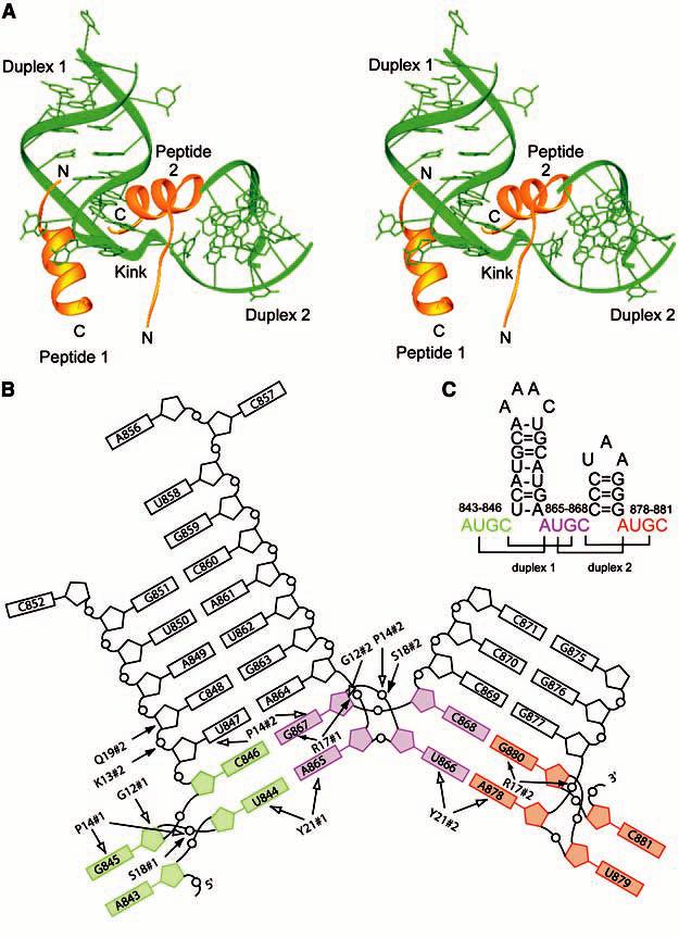
Fig. 1.

(A) Stereoimage of the AMV RNA-CP complex. The RNA is shown in green and the CP26 peptides are shown in gold. (B) Schematic diagram of the AMV RNA and CP26 protein contacts. The AUGC sequences have been color coded. The 5´ AUGC is colored green, the interhelical AUGC sequence is colored purple, and the 3´ AUGC sequence is colored red. Hydrogen-bonding contacts between the RNA and peptide are indicated with filled arrows and van der Waals contacts are indicated by open arrows. #1, peptide 1; #2, peptide 2; G, Gly; K, Lys; P, Pro; Q, Gln; R, Arg; S, Ser; Y, Tyr. (C) Secondary structure of the 39-nucleotide minimal binding domain found at the 3´ end of AMV RNAs, AMV843-881. The AUGC sequences are color coded as in (B). Base pairs are indicated by brackets.
