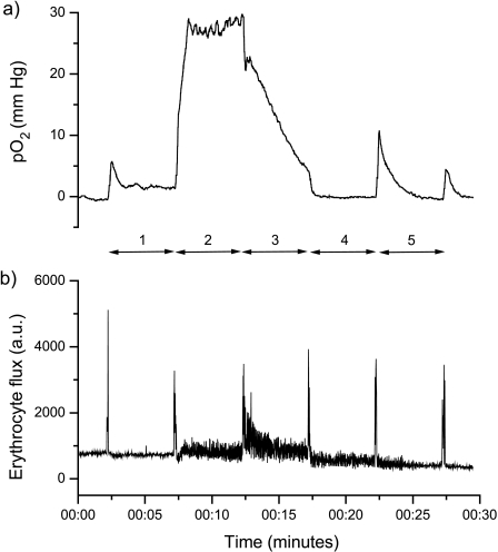
Figure 2.
Traces obtained from (a) one OxyLite pO2 probe, coupled with (b) an OxyFlo LDF probe, from a C6 wild-type glioma. The probe was inserted into the tumor and then with time retracted back through the tumor to measure the pO2 at five locations as indicated. The intense spikes in the LDF trace clearly mark the time of movement of the probe.

