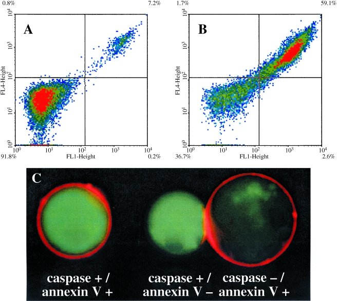
Figure 2.
Labeling of untreated and camptothecin-treated Jurkat T cells with active Cy5.5-annexin V. (A) Untreated cells incubated with active Cy-annexin V and FITC-annexin V. The FL1 channel is for fluorescein whereas FL4 is for Cy5.5. Unlabeled cells are in the left quadrant (91.8%), whereas cells labeled with both annexin Vs are in the upper right quadrant (7.2%). (B) Camptothecin-treated cells incubated with active Cy-annexin V and FITC-annexin V. Double-labeled cells are in the upper right quadrant (59.1%), with unlabeled cells in the lower left quadrant (36.7%). (C) Fluorescence micrographs of camptothecin-treated cells stained with active Cy-annexin V and the caspase probe FITC-VAD-FMK. A membrane-bound NIRF signal from active Cy-annexin V and a cytoplasmic green fluorescence from FITC-VAD-FMK are seen.

