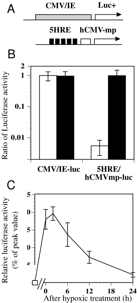
Figure 1.
The luciferase reporter assay using stably transfected cells under aerobic and hypoxic conditions. (A) Schematic diagram of the luciferase reporter plasmids with CMV/IE and 5HRE/hCMVmp expression units. (B) Comparison of the luciferase activity under aerobic (clear columns) and hypoxic (black columns) conditions. Stably transfected clones with the plasmids shown above were exposed to aerobic and hypoxic conditions (0.02% O2) for 6 hours and assayed for luciferase activity. The luciferase activities were normalized to that of a CMV/IE-luc clone under aerobic conditions. The error bars show the standard deviations (SD) of four independent samples. (C) The time course of luciferase activity after reoxygenation following hypoxic treatment of a 5HRE/hCMVmp-luc clone. The cells were treated under hypoxic conditions for 24 hours and then returned to aerobic conditions. The relative luciferase activity to its peak value was shown. Bars, SD of four independent experiments.

