Abstract
The location of the phospholipase C β1-isoform (PLC-β1) in the mouse oocyte and its role in the resumption of meiosis were examined. We used specific monoclonal antibodies to monitor the in vitro dynamics of the subcellular distribution of the enzyme from the release of the oocyte from the follicle until breakdown of the germinal vesicle (GVBD) by Western blotting, electron microscope immunohistochemistry, and confocal microscope immunofluorescence. PLC-β1 became relocated to the oocyte cortex and the nucleoplasm during the G2/M transition, mainly in the hour preceding GVBD. The enzyme was a 150-kDa protein, corresponding to PLC-β1a. Its synthesis in the cytoplasm increased during this period, and it accumulated in the nucleoplasm. GVBD was dramatically inhibited by the microinjection of anti-PLC-β1 monoclonal antibody into the germinal vesicle (GV) only when this accumulation was at its maximum. In contrast, PLC-γ1 was absent from the GV from the time of release from the follicle until 1 h later, and microinjection of anti-PLC-γ1 into the GV did not affect GVBD. Our results demonstrate a relationship between the relocation of PLC-β1 and its role in the first step of meiosis.
INTRODUCTION
In mammalian oocytes, meiosis resumes when the envelope of the germinal vesicle (GV), i.e., the oocyte nucleus, has disappeared. This event, called germinal vesicle breakdown (GVBD), occurs a few hours after, in vivo, the luteinizing hormone (LH) surge or, in vitro, oocyte release from the follicle. The nature of the link between oocyte release and meiotic arrest has not yet been clearly identified. From the time at which the oocyte receives the releasing message until GVBD, many cytological and biochemical events, some known and some unknown, may occur; they include translocation of the GV to the plasma membrane, chromatin reorganization, and changes in the synthesis of cell cycle proteins. Spontaneous calcium oscillations have been observed in the cytoplasm (Carroll and Swann, 1992; Lefèvre et al., 1995), and in the GV (Lefèvre et al., 1995) of the mouse oocyte arrested at the G2 stage of the meiotic cell cycle during the period preceding GVBD. We have demonstrated that changes in nuclear Ca2+ occur via calcium channels associated with type I inositol trisphosphate receptors, and appear to be necessary for the G2/M transition to occur (Pesty et al., 1998).
It is known from other cellular models that the activation of a phosphoinositide-specific phospholipase C (PLC) catalyzes the hydrolysis of phosphatidylinositol 4,5-biphosphate to generate diacylglycerol and inositol 1,4,5-trisphosphate (Berridge, 1997). Recently, it has also been suggested that a similar transduction signal operates in the nucleus of several cell types (Payrastre et al., 1992; Divecha et al., 1993a,b; Asano et al., 1994), and that it is involved in certain cellular events, such as proliferation and differentiation (Zini et al., 1996; Manzoli et al., 1997; Matteucci et al., 1998). The idea of a nuclear phosphatidylinositol cycle is reinforced by the presence of phosphatidylinositol-transfer proteins involved in the transfer of phosphatidylinositol, not synthesized by the nucleus, from an extranuclear site to this organelle (Capitani et al., 1990; D'Santos et al., 1998). It has been proposed that phosphatidylinositol-transfer proteins deliver phosphatidylinositol to lipid kinases to yield phosphatidylinositol 4,5-biphosphate (PIP2) as a substrate for PLC-β (Thomas et al., 1993). It has also been shown that the enzymes responsible for the metabolism of inositol lipids and present in the nuclei of several cell types are the β1- and β2-isoforms of PLC (Martelli et al., 1992; Divecha et al., 1993b; Zini et al., 1993, 1994; Bertagnolo et al., 1997). PLC-β1 has been demonstrated in mouse oocytes by using polymerase chain reaction (Dupont et al., 1996). PLC-β differs from other isozymes γ and δ in that it contains a long COOH-terminal sequence that contributes to its translocation and its association with the nucleus (Kim et al., 1996; Manzoli et al., 1997). However, PLC-γ1 and PLC-δ4 have also been detected in this compartment (Zini et al., 1994; Marmiroli et al., 1994; Liu et al., 1996). Several reports indicate that activation of both PLC (Carnero et al., 1993) and inositol metabolism (Carrasco et al., 1990; Pesty et al., 1998) cause the resumption of meiosis in oocytes of several species.
We have now used immunoblotting and immunochemistry to verify the presence and the location of the PLC-β1 isoform in the mouse oocyte. We also used the microinjection of a specific anti-PLC-β1 monoclonal antibody into the oocytes during in vitro maturation to analyze the role of the enzyme in the resumption of meiosis.
MATERIALS AND METHODS
Oocyte Recovery
The ovaries of 6-wk-old female CD1 mice (Charles River, Saint-Aubin-Lès-Elbeuf, France) were stimulated by injecting 5 IU of pregnant mare's serum gonadotropin (Chronogest; Intervet International, Boxmeer, Holland) The oocytes were collected as previously described (Pesty et al., 1998). The GV-stage oocytes were either used immediately or cultured in M16 medium (Fulton and Whittingham, 1978) supplemented with 15 mg/ml bovine serum albumin (BSA) (fraction V; Sigma, Saint Quentin, Fallavier, France) and maintained at 37°C in a 5% CO2-humidified atmosphere for 30, 60, 90, or 120 min before experiments.
Western Blotting
A total of ∼200 oocytes was used for each Western blot. These oocytes were kept in culture for 30, 60, or 90 min after their release from the follicles and then homogenized by repeated freezing and thawing in 10 μl of homogenization buffer (50 mM Tris-HCl, 75 mM KCl, 50 mM NaF, 10 mM Na2HPO4, 1 mM EDTA, 1 mM 4-(2-aminoethyl)-benzenesulfonyl fluoride), with or without the calpain inhibitors I and II (5 mM; Calbiochem, La Jolla, CA). They were then suspended in 10 μl of Laemmli 2× sample buffer, either directly or after ultracentrifugation at 4°C to separate the subcellular fractions (cytoplasm and nucleoplasm), and boiled for 5 min. The samples were chilled on ice and then electrophoresed on a 7.5% polyacrylamide-0.1% SDS gel at constant voltage (200 V) for ∼45 min. The separated proteins were transferred to a Polyscreen polyvinylidene difluoride membrane (NEN Life Science Products, Boston, MA) in an electrotrans-blot apparatus (Bio-Rad, Cambridge, United Kingdom) by using 100 V for 1.5 h. The membrane was then incubated with blocking buffer (5% skim milk, 0.1% Tween 20 [Merck, Darmstadt, Germany] in Dulbecco's phosphate buffer [PBS; Sigma]) for 1 h with gentle shaking to block nonspecific binding. Two commercially available anti-PLC-β1 monoclonal antibodies (mAbs) were used (mAb1 from Upstate Biotechnology, Lake Placid, NY, and mAb2 from Transduction Laboratories, Lexington, KY). The mAb1 was a mix of monoclonal antibodies that interacted with several domains located at both the N- and C-terminal regions of the β1 antigen (Suh et al., 1988). The mAb2 used was specific for the N-terminal region. The anti-PLC-β1 mAb1 (2 μg/ml) was added first, and the mixture was incubated overnight at 4°C. The membrane was washed four times with 0.1% Tween 20 in PBS for 15 min and incubated in blocking buffer containing horseradish peroxidase-conjugated anti-mouse IgG (1:2000; Amersham; Saclay, France) for 1.5 h. It was washed with 0.1% Tween 20 in PBS four times and specific binding was visualized on X-ray film (Sigma) by using an enhanced chemiluminescence immunoblot kit (Amersham) according to the manufacturer's instructions. The antibody was then removed by washing the membrane in a solution of 62.5 mM Tris-HCl, pH 6.7, 2% SDS, 100 mM β-mercaptoethanol. Then the membrane was incubated with the anti-PLC-β1 mAb2 (0.5 μg/ml). In parallel, Western blot analysis of other pools of 200 oocytes were performed with a mAb against another PLC isoform, anti-PLC-γ1 mAb (0.8 μg/ml; Upstate Biotechnology), or with the second antibody alone as a negative control. Western blot analysis of mouse brain homogenate was also done as a positive control. All these experiments were repeated at least three times.
Confocal Microscope Immunofluorescence
Whole Oocyte.
Oocytes were incubated for 5 min in 0.01% α-chymotrypsin in PBS supplemented with 3% BSA to remove the zona pellucida and then fixed in 2% paraformaldehyde (Sigma) in PBS for 1 h at 37°C. The fixed oocytes were placed in two successive blocking solutions in PBS: 15 min in 3 mg/ml ammonium chloride (NH4Cl; Sigma), followed by 15 min in 0.05% Tween 20 and 1.5% BSA. They were then incubated overnight at 4°C with the anti-PLC-β1 mAb1 diluted in the second blocking solution (0.05 mg/ml), washed three times, and immunostained for 45 min at 37°C with goat anti-mouse IgG secondary antibody, fluorescein isothiocyanate-conjugated (1:50 in the second blocking solution; Jackson ImmunoResearch Laboratories, West Grove, PA). Saponin (0.5%) was added throughout the procedure to ensure membrane permeability. The immunostained oocytes were examined by confocal microscopy (MRC 600; Bio-Rad) with a 40× objective (NPL Fluotar 40/0.70) in a single optical section through the GV or by using the Z-series procedure with a 2-μm step. This procedure was performed on six different pools of oocytes that had been kept in culture for 0, 30, 60, 90, or 120 min
Isolated Nucleus.
Oocytes that had been cultured for 0, 30, 60, or 90 min were mechanically disrupted with a very fine micropipette. Their isolated nuclei were treated as described above for immunofluorescence, and examined by using a 60× objective (PlanApo, 60/0.95; Nikon; Champigny sur Marne, France).
Control Experiments.
Control experiments were performed to confirm the specificity of the labeling. Oocytes were incubated with the second antibody alone or with anti-PLC-γ1 mAb (16 μg/ml).
Electron Microscope Immunocytochemistry
Oocytes were fixed in 2% paraformaldehyde and 0.05% glutaraldehyde (Sigma) in PBS (pH 7.4) for 1 h at 4°C, washed for 30 min in 0.1 M sodium cacodylate, dehydrated in an ethanol series (70 to 100%), and embedded in Unicryl resin (British Biocell International, Cardiff, United Kingdom) at 60°C for 2 d. Ultrathin sections were incubated in 50 mM glycine in PBS for 20 min and then saturated in 5% BSA IgG-free (Sigma) in PBS for 1 h. They were next incubated overnight at 4°C with the anti-PLC-β1 mAb1 (0.03 mg/ml) in PBS-BSA 5%, and then with goat anti-mouse IgG conjugated with 10-nm colloidal gold particles (British Biocell International) diluted 1:10 in PBS-BSA 5% for 1 h at room temperature. The sections were stained with saturated aqueous uranyl acetate for 30 min.
Controls consisted of 1) gold-conjugated goat anti-mouse IgG without the primary antibody, 2) replacement of the primary antibody by purified nonimmune IgG, or 3) incubation with the anti-PLC-γ1 mAb (0.8 μg/ml). The observations were made by using a Phillips 301 electron microscope.
Microinjection Procedures
The anti-PLC-β1 mAb1 (diluted in the microinjection medium: 140 mM KCl, 1 mM MgCl2, 5 mM HEPES, pH 7.2) was microinjected into the GV or the cytoplasm of oocytes that had been cultured for 0, 30, 60, or 90 min. The oocytes were then maintained in M16 medium at 37°C and examined in the light microscope every hour until 4 h after release from the follicle to check for GVBD.
The lifetime of the anti-PLC-β1 mAb1 inside the nucleus was estimated by microinjecting it into the GV at time zero. The oocytes were cultured for a further 90 min and then fixed for immunohistochemical analysis.
As a control, medium alone or medium containing mouse γ-globulins was injected into the nucleus or the cytoplasm of oocytes 30 min after release from the follicle. In parallel, anti-PLC-γ1 mAb was microinjected into either cell compartment at 60 min after follicular release.
Holding pipettes were prepared from borosilicate glass capillaries (GC120-10; Clark Electromedical Instruments, Pangbourne, United Kingdom) by using a horizontal micropipette puller (model 773; Campden Instruments, London, United Kingdom) and a De Fonbrune microforge (Alcatel, Malakoff, France). The microinjections were performed with sterile, ready-made needles (femtotips; Eppendorf, Hamburg, Germany). Oocytes to be microinjected were placed in a 30-μl drop of M2 medium under mineral oil (Sigma) on a cell culture chamber (POC chamber; Helmut Saur, Reutlingen, Germany) and maintained at 37°C (heating stage MS100; Linkam Scientific Instruments, Tadworth, United Kingdom) under an inverted microscope.
Statistical Analysis
The data given are the averages of at least three experiments and are expressed as means ± SE. Values were considered to be statistically different when p was < 0.05 in an ANOVA with a protected least-significant difference Fisher test.
RESULTS
Identification of PLC-β1 in Whole Oocytes and in Cell Subfractions by Immunoblotting (Figure 1)
Figure 1.
Western blot analysis of PLC-β1 and PLC-γ1. Soluble proteins from whole-mouse oocytes (lanes 2A and 1B) or subcellular fractions (cytosol [lanes 3–4A and 2–3B) and nucleoplasm (lanes 5–6A and 4–5B]) were analyzed by 7.5% SDS-PAGE. Approximately 200 oocytes were pooled for each lane. The gels were electroblotted onto Polyscreen polyvinylidene difluoride membranes and the lanes were probed with either anti-PLC-β1 mAb1 and mAb2 or anti-PLC-γ1 mAb. Mouse brain protein (lane 1A) was used as a positive control. Molecular weights are indicated in kDa. The blots selected for A correspond to oocytes treated without calpain inhibitors; the blots in B corresponded to oocytes treated in presence of calpain inhibitors.
Bands of ∼150, 140, and 100 kDa were observed in the mouse brain homogenate, for each of the anti-PLC-β1 mAbs used, as in the rat (Park et al., 1993; Caramelli et al., 1996) and cow (Suh et al., 1988) brains.
Whole-mouse oocyte homogenate contained a 100-kDa protein, a product of the cleavage of the native 150-kDa PLC-β1. The native form was always clearly detectable in whole oocytes when calpain inhibitors were added during extraction. However, the 100-kDa immunoreactive band was still clearly visible, as if the protease was not totally inhibited.
The native form was not detected in either the nucleoplasm or cytoplasm fractions obtained from oocytes not treated with calpain inhibitors, for any anti-PLC-β1 mAb used. In contrast, the native PLC-β1 was clearly detected in the cytoplasm 30 min after release from the follicle when the oocytes were treated with calpain inhibitors, whereas it was almost undetectable in the nuclear fraction. This immunoreactive band was detected in both cell compartments 90 min after release from the follicle, with slightly more in the cytoplasm.
The 100-kDa immunoreactive band was more prominent in the cytoplasm than in the nucleoplasm from the oocytes treated 30 min after release from the follicle when calpain was not inhibited, whereas the situation was reversed in oocytes treated 60 min after release.
We checked the specificity of the mAb1 and mAb2 by performing similar experiments with an antibody against the γ1-isoform. A major 150-kDa band was visible in both cell fractions 60 min after follicular release, but it was very weak in the nucleoplasm.
Confocal Microscope Immunofluorescence in Whole Oocytes and in Isolated Nuclei
Location of PLC-β1 in Whole Oocytes
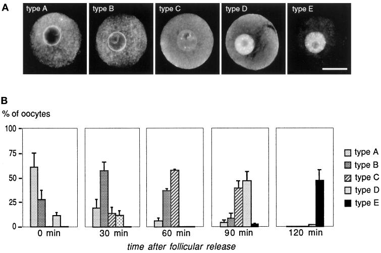
Confocal microscopic examination of whole oocytes immunostained with the anti-PLC-β1 mAb at different times after follicular release to GVBD revealed changes in the location of PLC-β1. There were five basic patterns of staining according to these cell dynamics (Figure 2A). The frequencies at which they were observed were calculated in relation to the time after follicular release (Figure 2B). These five patterns of PLC-β1 distribution are described as follows (Figure 2).
Figure 2.
Pattern of development of anti-PLC-β1 immunostaining before GVBD in the mouse oocyte. (A) Selected photographs show the patterns of anti-PLC-β1 immunostaining observed by confocal microscopy of whole-mouse oocytes analyzed at different times after release. These observations are recorded in a single optical section through the GV. Type A: large spots of dense staining in the cytoplasm, intense labeling around the nuclear membrane; nucleoplasm and plasma membrane not stained. Type B: very bright nuclear membrane and slight labeling in the nucleoplasm and around the nucleolus. Type C: increased labeling of the nucleoplasm with large spots inside the nucleoplasm and a less stained nuclear membrane. Type D: extensive immunoreactivity almost exclusively inside the nucleus and around the cortex; isolated spots of fluorescence scattered in the cytoplasm. Type E: immunofluorescence exclusively in the remaining nucleus area. Bar, 40 μm. (B) Each histogram represents the distribution of the different types of anti-PLC-β1 immunostaining observed for the oocytes fixed at different times after release from the follicles (0, 30, 60, 90, and 120 min). One or two types predominate at each time studied. The mean frequencies are calculated from at least three experiments for each time of observation. The total number of immunostained oocytes are as follows: 0 min, n = 20; 30 min, n = 29; 60 min, n = 19; 90 min, n = 28; and 120 min, n = 30. More than half of the oocytes had progressed further toward the MI stage by 120 min after release, and were not taken into account in the histogram.
Type A showed large spots of dense staining in the cytoplasm and intense labeling around the nuclear membrane, but without staining of the nucleoplasm or the plasma membrane. This intracellular distribution of PLC-β1 was observed in 61.1 ± 15.0% of the oocytes immunostained immediately after release, and in 18.8 ± 9.4, 5.6 ± 3.9, and 4.5 ± 3.2% of those immunostained after 30, 60, and 90 min, respectively.
Type B, characterized by a very bright nuclear membrane and faint labeling in the nucleoplasm and around the nucleolus. The majority (56.6 ± 9.6%) of the oocytes fixed after 30 min in culture had this pattern, whereas only 27.8 ± 10.7, 36.7 ± 2.4, and 8.3 ± 5.9%, respectively, of those immunostained after 0, 60, and 90 min in culture did so.
Type C had increased labeling of the nucleoplasm (large dots were noticeable inside the nucleoplasm), but less staining of the nuclear membrane. A majority of the oocytes (57.8 ± 1.6%) stained 60 min after follicular release had this PLC-β1 distribution. This pattern was observed in 13.4 ± 6.7 and 38.6 ± 8.0% of the oocytes fixed after 30 or 90 min, respectively, whereas it was never observed in those fixed immediately after follicular release.
Type D was identified by an extensive immunoreactivity almost exclusively inside the nucleus (with faint staining of the nuclear membrane) and around the oocyte cortex, with only isolated spots of fluorescence scattered in the cytoplasm. This pattern of PLC-β1 distribution was observed in 46.2 ± 9.1% of the oocytes examined after 90 min in culture, as well as in those that still had their GV after 120 min in culture (only 1.7 ± 1.2% of the cultured oocytes). However, 11.1 ± 4.3, 11.3 ± 5.6, and 0.0%, respectively, of the oocytes observed after 0, 30, or 60 min in culture had this particular staining.
Type E exhibited immunofluorescence only in the remaining nucleus area. This staining occurred only in oocytes that had just broken the nuclear membrane. Although 1.7 ± 1.2% of the oocytes still had type D staining after 120 min in culture, 45.0 ± 10.6% had type E staining. Meiosis had progressed further in the remaining oocytes (>50%); some of them were already at the MI stage.
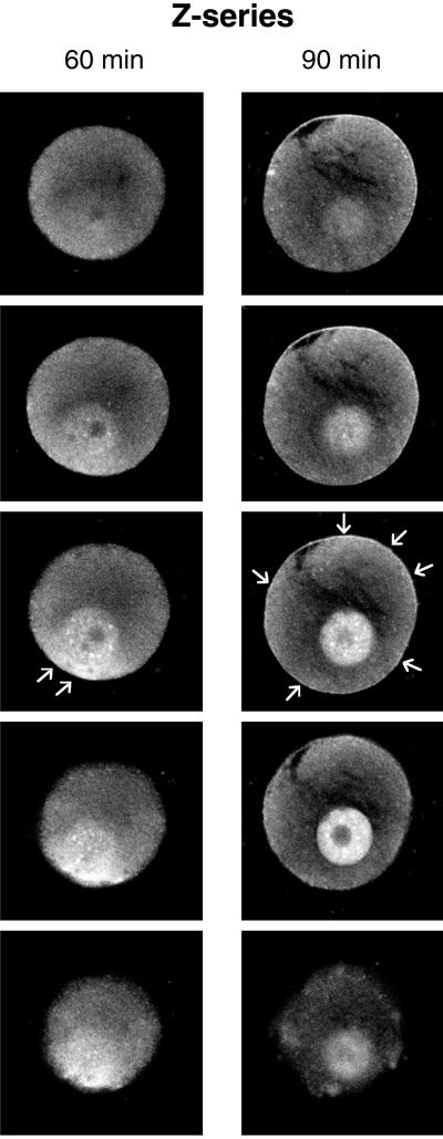
These patterns were all recorded in a single optical section through the GV. However, confocal Z-series studies on the immunostained oocytes revealed new information about the cytoplasmic localization of PLC-β1: it was first translocated to a pole of the oocyte cortex; then, when the immunofluorescence pattern was changing from type C to type D, it was found around the whole cortex (Figure 3).
Figure 3.
Representative Z-series of optical sections of anti-PLC-β1 immunostained oocytes. (A) After 60 min in culture, the cytoplasmic PLC-β1 was located at a pole of the oocyte cortex, near the germinal vesicle (arrows). (B) After 90 min in culture, it was located all around the plasma membrane (arrows). Frames were taken at 2-μm intervals and every third image was selected for each series.
Location of PLC-β1 in Isolated Nuclei
The immunohistochemical studies were performed on isolated nuclei recovered from oocytes maintained in culture for 0, 30, 60, or 90 min (Figure 4). PLC-β1 immunoreactivity appeared only at the nuclear membrane of most of the isolated nuclei cultured for 0 min, whereas the labeling was also seen around the nucleolus of oocytes cultured for 30 min. The labeling increased in the nucleoplasm (with numerous dots) in most of the nuclei isolated from oocytes cultured for 60 or 90 min, whereas the fluorescence of the nuclear membrane decreased.
Figure 4.
Pattern of development of anti-PLC-β1 immunostaining in isolated nuclei. The selected photographs are representative of the development of the pattern of anti-PLC-β1 immunostaining observed by confocal microscopy in isolated nuclei. The nuclei were isolated from oocytes cultured for 0, 30, 60, or 90 min and immediately immunostained with the anti-PLC-β1 mAb. Intense labeling around the nuclear membrane; the nucleoplasm was very slightly stained at time zero. Very bright nuclear membrane and slight labeling in the nucleoplasm and around the nucleolus after 30 min in culture. Large spots in the nucleoplasm and a less stained nuclear membrane after 60 min in culture. Major immunoreactivity with more fluorescent spots after 90 min in culture. Bar, 10 μm.
Location of PLC-γ1 in Whole Oocytes
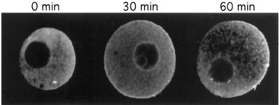
Confocal microscopy of whole oocytes immunostained with the anti-PLC-γ1 mAb and cultured for different times showed the absence of this isoform from the oocyte nucleoplasm, at least until 60 min in culture (Figure 5). Immediately after follicular release, 83.3% of the oocytes (n = 12) showed diffuse immunostaining for PLC-γ1 in the cytoplasm, whereas 68.4% (n = 19) of the oocytes showed staining at one pole of the cortex (Figure 5) after 30 min in culture, and 80.0% (n = 15) after 60 min in culture.
Figure 5.
Pattern of development of anti-PLC-γ1 immunostaining in whole-mouse oocytes. The selected photographs indicate the pattern of development of anti-PLC-γ1 immunostaining observed by confocal microscopy in whole-mouse oocytes analyzed at different times after release. These observations were recorded in a single optical section through the GV. No immunostaining in the nucleus at the three studied times. Diffuse PLC-γ1 immunostaining in the cytoplasm at time zero. The labeling was first intense around the plasma membrane, and then at a pole of the cortex after 30 and 60 min in culture.
Electron Microscope Immunocytochemistry in Whole Oocytes
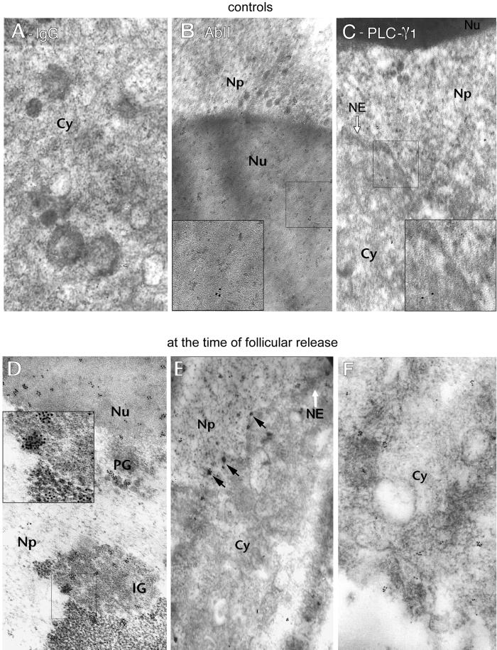
We analyzed the distribution of PLC-β1 in greater detail by electron microscope immunocytochemical analysis of oocytes after 0, 60, and 90 min in culture. To compare gold particle distribution between the zona pellucida, microvilli, cytoplasm, and nucleus we selected only those sections of oocytes in which we could observe at least part of these domains. In all cases, gold particles were almost totally absent from the resin and the zona pellucida. Background labeling was almost negligible in the control specimen incubated with nonimmune pure IgG (Figure 6A) or with only the secondary antibody (Figure 6B). There were very few gold particles in the nucleoplasm of oocytes immunolabeled with the anti-PLC-γ1 mAb after 60 min in culture (Figure 6C).
Figure 6.
Electron microscope immunocytochemistry of oocytes at the time of their release. (A–C) Control samples in which the sections were incubated with nonimmune pure IgG (A), with only the secondary antibody (B). The labeling is almost negligible in both cell compartments. (C) Oocytes treated 60 min after follicular release with the anti-PLC-γ1 mAb. A small number of gold particles are distributed throughout the cytoplasm (Cy) and very few are visible in the nucleoplasm (Np). (D–F) Oocytes treated with the anti-PLC-β1 mAb immediately after release. (D) In the nucleus, the labeling was mainly in the IGs, in the PGs, and in the nucleolus. (E and F) A small number of gold particles are distributed throughout the cytoplasm, but are practically absent from the nucleoplasm. Numerous aggregates (black arrows) are visible along the nuclear envelope (NE). Magnification, 40,000×. The regions selected in small squares are presented as 2× enlarged inserts.
The oocytes treated with the anti-PLC-β1 had a few gold particles aggregated along the nuclear envelope and the cytoplasm (2.6 ± 0.2 particles/μm2 for 2 oocytes studied) (Figure 6, D and F) immediately after release from the follicle. The nucleoplasm was nearly free of particles (1.2 ± 0.2 particles/μm2), except for a few aggregates in perichromatin granules (PGs), over clusters of interchromatin granules (IGs) and in the nucleolus (13.4 ± 0.8 particles/μm2) (Figure 6D).
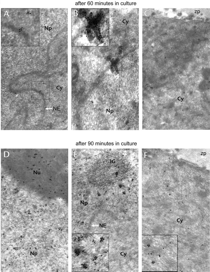
Oocytes treated after 60 min in culture (Figure 7, A–C) had labeling distributed similarly in the cytoplasm (2.8 ± 0.3 particles/μm2) and the nucleus (2.6 ± 0.3 particles/μm2) (5 oocytes studied) and it was also detected in the nucleolus (8.7 ± 1.2 particles/μm2). We also saw labeling that appeared to represent the passage of PLC-β1 through the nuclear envelope, in one case (Figure 7B).
Figure 7.
Electron microscope immunocytochemistry 60 and 90 min after follicular release. (A–C) Oocytes treated with anti-PLC-β1 60 min after follicular release. (A) Gold particles are similarly distributed in the cytoplasm (Cy) and the nucleoplasm (Np); some gold particles are still present on the nuclear envelope (arrow). (B) A large aggregate of gold particles crossing the nuclear envelope. (C) Diffuse staining of the oocyte cortex and cytoplasm; zp, zona pellucida. (D–F) Oocytes treated with anti-PLC-β1 90 min after follicular release. (D) Nucleolus (Nu) is also strongly stained. (E) Gold particles are concentrated in the nucleoplasm and in the IGs; the nuclear envelope (NE) is free of particles. (F) Labeling in the cortex region is noticeable. Magnification, 40,000×. The regions selected in small squares are presented as 2× enlarged inserts.
There were fewer gold particles in the cytoplasm of oocytes treated 90 min after follicular release (Figure 7, D–F) (5.0 ± 0.3 particles/μm2 for the 2 oocytes studied), than in the nucleoplasm (16.8 ± 1.0 particles/μm2) or the nucleolus (14.2 ± 1.9 particles/μm2). The gold particles in the nucleus appeared to be isolated in the nucleoplasm or scattered in aggregates on PGs and IGs.
PLC-β1 and Meiosis Resumption
We analyzed the effects of inhibiting PLC-β1 with the specific anti-PLC-β1 mAb1 on the kinetics of meiosis resumption in relation to the time in culture, and to the cellular compartment into which the mAb was microinjected (cytoplasm or GV) (Figure 8). The microinjected oocytes were then observed under the light microscope every hour until 4 h after follicular release.
Figure 8.
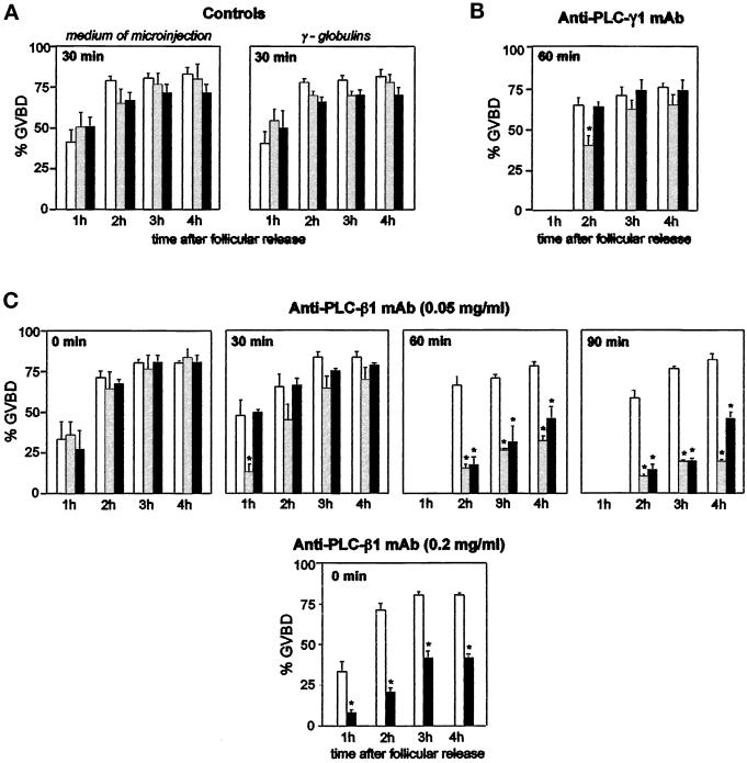
GVBD kinetics. (A) Microinjection into the cytoplasm, or the GV of medium alone or medium containing mouse γ-globulins, did not affect the GVBD process. (B) Inhibition of cytoplasmic or nuclear PLC-γ1 had no effect on GVBD kinetics, except for the first hour in the case of microinjection into the cytoplasm. (C) Inhibition of cytoplasmic or nuclear PLC-β1 had a negative effect upon GVBD kinetics. The anti-PLC-β1 mAb (0.05 or 0.2 mg/ml) was microinjected into the cytoplasm or the GV at t = 0, 30, 60, or 90 min after follicular release. The oocytes were then observed regularly to verify the occurrence of GVBD. The frequencies ± SE of GVBD are calculated at least on three experiments. □, no microinjection; ░⃞, microinjection into the cytoplasm; ▪, microinjection into the nucleus. ∗, statistically significant difference compared with control at p < 0.05.
Controls
Microinjection medium alone or medium containing γ-globulins into the cytoplasm (n = 34 and 27, respectively) or the nucleus (n = 43 and 33, respectively) resulted in a GVBD identical to that of uninjected oocytes (Figure 8A).
Effect of Anti-PLC-γ1
Microinjection of the anti-PLC-γ1 mAb (16 μg/ml) into the cytoplasm (n = 32) 60 min after follicular release slowed meiosis; the GVBD rate 1 h after microinjection was lower (39.3 ± 6.8%) than that for the control oocytes (62.9 ± 4.1%, p < 0.05). In contrast, microinjection of the anti-PLC-γ1 mAb into the GV (n = 41) had no effect on the GVBD rate (Figure 8B).
Effect of Anti-PLC-β1
Microinjection of the anti-PLC-β1 mAb1 (0.05 mg/ml) immediately after follicular release into either the GV (n = 46) or the cytoplasm (n = 43) had no effect on the kinetics of meiosis, which was similar to that of control oocytes (n = 55) (Figure 8C).
The effects on the GVBD of injecting anti-PLC-β1 mAb 30 min after follicular release depended on the cell compartment receiving the mAb. Anti-PLC-β1 mAb injected into the GV had no effect on meiosis (46.9 ± 3.0%, n = 30) compared with control oocytes (44.3 ± 10.5%, n = 92). However, injecting anti-PLC-β1 mAb into the cytoplasm temporarily inhibited the resumption of meiosis; very few oocytes resumed meiosis during the first hour after follicular release compared with controls (11.7 ± 6.3%, n = 32, p < 0.05). During the following hours the delay induced by the mAb disappeared, and the difference between the two groups was no longer discernible.
Meiois was dramatically delayed by injecting anti-PLC-β1 mAb into the cytoplasm (n = 28) or the GV (n = 35) of oocytes cultured for 60 min. The GVBD rate remained significantly lower than in the control (n = 24), even 4 h after follicular release (32.1 ± 4.1 or 45.8 ± 4.6 versus 81.9 ± 4.9%), no matter into which cellular compartment it was microinjected.
Microinjection of anti-PLC-β1 mAb into the cytoplasm (n = 28) or the GV (n = 31) of oocytes cultured for 90 min strongly inhibited the GVBD. This inhibition was maintained until the end of the culture (19.4 ± 1.5 or 45.8 ± 4.6% versus 81.9 ± 4.9%). However, a more concentrated solution of the mAb1 (0.2 mg/ml) dramatically inhibited the GVBD when microinjected into the nucleus (n = 30), immediately after release from the follicle.
Lifetime of mAb in GV during Culture
When anti-PLC-β1 mAb was microinjected into the GV at time zero, PLC-β1 was always immunorevealed after 90 min in culture, and still in the nucleus (5 of 5 tested oocytes). This indicates that the mAb did not diffuse across the nuclear envelope, which was not damaged during culture.
DISCUSSION
We have shown that the subcellular distribution of PLC-β1 changes before the resumption of meiosis, and appears to be correlated with its role in the rupture of the nuclear envelope.
Dynamics of the PLC-β1 Subcellular Location
Our confocal microscope immunofluorescence studies indicate that the subcellular location of PLC-β1a changes before GVBD. The enzyme is located mostly in the cytoplasm and around the nuclear membrane when oocytes are released from the follicle. It is still present on the nuclear membrane 0.5 h later, and beginning to appear in the nucleoplasm and around the nucleolus. Later, although it has disappeared to some extent from the nuclear membrane, it is relocated inside the nucleus. At the same time, it moves from the cytoplasm to a pole of the oocyte, and then all around the cortex. The close link between PLC-β1 and the nucleus is consistent with the fact that this isoform differs from other isozymes in that it contains a long COOH-terminal sequence, which contributes to its translocation and its association with the nucleus (Kim et al., 1996; Manzoli et al., 1997). However, this nuclear location of PLC-β1 is still controversial. The β1-isoform is detected mainly in the nucleus of rat liver cells, according to some studies (Bertagnolo et al., 1995), or in the cytosol (Divecha et al., 1993b). Many reports indicate variations in the subcellular location due to the state of cell differentiation (Martelli et al., 1994; Divecha et al., 1995; Zini et al., 1995). Our conclusions concerning the developing pattern of PLC-β1 nuclear distribution are reinforced by our immunohistochemical data for isolated nuclei, which show a similar increase in the immunostaining in relation to the progression of the meiosis. The PLC-β1 labeling is exclusively in the area of the nucleus just after the disappearance of the nuclear membrane, implying that it is associated with the nuclear matrix rather than the nuclear membrane.
We confirmed the relocation of PLC-β1 to the GV by electron microscope immunocytochemistry. The immunogold detection of PLC-β1 showed that the gold particles were mainly in the IGs and the PGs when the enzyme was concentrated in the nucleus. These granules are ribonucleoprotein structures belonging to the nuclear matrix and involved in mRNA production (Takeuchi et al., 1990; Maraldi et al., 1993; Puvion and Puvion-Dutilleul, 1996; Santella and Kyozuka, 1997). The colocation of PLC-β1 and ribonucleoprotein structures, also observed in PC12 cells (Zini et al., 1994), suggests that the enzyme is involved in the transport and release of transcripts in the mouse oocyte.
Our Western blotting studies in whole-mouse oocytes detected a 150-kDa PLC-β1 corresponding to PLC-β1a (Bahk et al., 1994). This differs from PLC-β1b (140 kDa) by its carboxy-terminal sequence. Only a 100-kDa immunoreactive band appeared when calpain inhibitors were not added, corresponding to a cleavage product of the native enzyme by calpain (Martelli et al., 1992). This Ca2+-dependent protease cleaves PLC-β1 at the linkage between amino acid residues 880–881, generating the 100- and 45-kDa proteins, which correspond to the amino-terminal and carboxy-terminal portions, respectively (Park et al., 1993). Comparative analysis of cytoplasmic and nucleoplasmic fractions by immunoblot, at two different times after follicular release, confirmed the relocation of the PLC-β1 in the GV. The intensity of the catalytically active immunoreactive band of ∼100 kDa (Wu et al., 1993) was stronger in culture after 60 min than after 30 min in the nuclear fractions. This immunoreactive band has been detected by Western blotting in these fractions of several cell types (Martelli et al., 1992; Zini et al., 1994, 1995; Caramelli et al., 1996). The native PLC-β1a was clearly detected in the nuclear fraction 90 min after release from the follicle when calpain inhibitors were used, whereas its amount remained almost constant in the cytoplasm from 30 min. These data suggest increased synthesis in the cytoplasm with accumulation of the native form in the GV in the hour preceding GVBD. The presence of the 100-kDa fragment in the nucleus at a time when the 150-kDa band is poorly detected (i.e., at 30 min) indicates that the native PLC-β1 is already present and cleaved by a residual amount of calpain. The 100-kDa fragment lacking the C-terminal region necessary for nuclear translocation (Kim et al., 1996) seems unable to move from the cytoplasm into the nucleus. And calpain has been found in the GV of the starfish (Santella et al., 1998) and rat (Malcov et al., 1997) oocytes, where it is predominantly present just before GVBD (Santella et al., 1998).
Our results demonstrate that the concentration of native PLC-β1 moves from the cytoplasm and probably the nuclear envelope to the nucleoplasm. These observations and the presence of inositol trisphosphate receptors inside the GV of mouse oocytes (Pesty et al., 1998) demonstrate the existence of a nuclear phosphoinositide cycle, as demonstrated in other cell types (Zini et al., 1994; Mazzotti et al., 1995; Cocco et al., 1996; Manzoli et al., 1996; Vann et al., 1997).
Involvement of PLC-β1 in the Meiotic Resumption Process
The next question is whether the PLC-β1 plays a role in the G2/M phase transition and whether this role depends on its subcellular location. We observed a relationship between the change in the PLC-β1 subcellular location and the inhibition of GVBD by the anti-PLC-β1 mAb microinjected into the GV or the cytoplasm. When the enzyme was immunorevealed at the nuclear membrane but not in the nucleoplasm, the microinjection of less concentrated anti-PLC-β1 mAb had no effect on the resumption of meiosis. This suggests that the PLC-β1 associated with the nuclear membrane is inactive. Microinjection of the mAb into the nucleus inhibits the reinitiation of meiosis as soon as the amount of enzyme in the nucleoplasm increases. The lack of effect of the mAb on GVBD when it is injected at the time of oocyte release from the follicle was surprising, because it remains in this compartment throughout the culture period. However, injection of more mAb into the GV overcame this effect, suggesting that the results obtained with dilute mAb are due to association of the mAb with the PLC-β1 on the nuclear envelope, thereby impeding combination of the mAb with the enzyme translocated later. Like nuclear PLC-β1, the cytoplasmic enzyme appears to be involved in the resumption of meiosis.
This is in agreement with the role of nuclear phospholipase C in mitosis in leukemic cells (Sun et al., 1997) and during meiosis in Xenopus oocytes (Carnero and Lacal, 1993). However, the activation of PLC by G-protein αq subunit is not an absolute requirement for maturation of Xenopus oocytes (Guttridge et al., 1995). Nevertheless, the only fragment detected in the mouse oocyte subcellular fractions not treated with calpain inhibitors was the 100-kDa proteolytic fragment of PLC-β1a. This could be due to an accumulation of calpain in the GV before GVBD, as in the starfish oocytes (Santella et al., 1998). This fragment, which is as catalytically active as the intact enzyme (Rhee et al., 1989; Wu et al., 1993), is not activated by the G-protein αq subunit (Park et al., 1993). It might instead be activated by the G-protein βγ subunits because the PH domain and the sequence just following it in the N-terminal region are essential for interaction with and stimulation by these subunits (Williams, 1999). Thus, the mouse oocyte GVBD process could be regulated by the 100-kDa fragment of PLC-β1a via the G-protein βγ subunits.
This phenomenon of relocation of proteins, such as p34cdc2–cyclin B complex (Ookata et al., 1992) and calpain (Santella et al., 1998), to the nucleus has been observed before GVBD in starfish oocyte. The link between these molecules and meiosis has to be further investigated.
Using the same technical approaches, we have demonstrated that the γ1-isoform of PLC is almost undetectable in the GV when the oocyte is released from the follicle and is not translocated further in this cellular compartment during meiosis, at least until 60 min after the oocyte's release. The γ1-isoform is absent from the oocyte nucleus in other cell models (Divecha et al., 1993b; Bertagnolo et al., 1995; Diakonova et al., 1997). However, as already observed (Dupont et al., 1996), it is present in the cytoplasm with a 150-kDa molecular mass. And GVBD is not affected when the γ1-isoform is inhibited by its specific mAb in the GV. The nature of the behavior of PLC-β1 is highlighted by the different dynamics of the PLC-γ1 that we have used for comparison in this study.
In conclusion, the synthesis and relocation of PLC-β1 to both the oocyte cortex and the nucleus in the hour preceding GVBD appear to be essential for this process to occur. Nuclear and cortical PLC-β1 seem to act synergistically. However, the mechanisms by which the oocyte PLC-β1 is activated remain unknown, as do the Ca2+-dependent phosphorylation cascades triggered by the activation of this enzyme.
ACKNOWLEDGMENTS
We thank Francine Puvion-Dutilleul (Organisation Fonctionnelle du Noyau, UPR 9044, Villejuif, France) for helpful comments on our ultrastructural photographs and Geoff Watts and Owen Parkes for editing the text. The ultrastructural observations were carried out at the “Service de Microscopie Electronique” (Institut Fédératif de Recherche Biologie Intégrative, Centre National de la Recherche Scientifique, Paris VI University). Nathalie Avazeri was supported by a fellowship from the French Organon laboratories.
REFERENCES
- Asano M, Tamiya-Koizumi K, Homma Y, Takenawa T, Nimura Y, Kojima K, Yoshida S. Purification and characterization of nuclear phospholipase C specific for phosphoinositides. J Biol Chem. 1994;269:12360–12366. [PubMed] [Google Scholar]
- Bahk YY, Lee YH, Lee TG, Seo J, Ryu SH, Suh P-G. Two forms of phospholipase C-β1 generated by alternative splicing. J Biol Chem. 1994;269:8240–8245. [PubMed] [Google Scholar]
- Berridge MJ. Elementary and global aspects of calcium signaling. J Physiol. 1997;499:291–306. doi: 10.1113/jphysiol.1997.sp021927. [DOI] [PMC free article] [PubMed] [Google Scholar]
- Bertagnolo V, Marchisio M, Capitani S, Neri LM. Intranuclear translocation of phospholipase C beta2 during HL-60 myeloid differentiation. Biochem Biophys Res Commun. 1997;235:831–837. doi: 10.1006/bbrc.1997.6893. [DOI] [PubMed] [Google Scholar]
- Bertagnolo V, Mazzoni M, Ricci D, Carini C, Neri LM, Previati M, Capitani S. Identification of PI-PLC β1, γ1, and δ1 in rat liver: subcellular distribution and relationship to inositol lipid nuclear signaling. Cellular Signaling. 1995;7:669–678. doi: 10.1016/0898-6568(95)00036-o. [DOI] [PubMed] [Google Scholar]
- Capitani S, Helms B, Mazzoni M, Previati M, Bertagnolo V, Wirtz KW, Manzoli FA. Uptake and phosphorylation of phosphatidylinositol by rat liver nuclei. Role of phosphatidylinositol transfer protein. Biochim Biophys Acta. 1990;1044:193–200. doi: 10.1016/0005-2760(90)90303-f. [DOI] [PubMed] [Google Scholar]
- Caramelli E, Matteucci A, Zini N, Carini C, Guidotti L, Ricci D, Maraldi NM, Capitani S. Nuclear phosphoinositide-specific phospholipase C, phosphatidylinositol 4,5-bisphosphate and protein kinase C during rat spermatogenesis. Eur J Cell Biol. 1996;71:154–164. [PubMed] [Google Scholar]
- Carnero A, Lacal JC. Phospholipase-induced maturation of Xenopus laevisoocytes: mitogenic activity of generated metabolites. J Cell Biochem. 1993;52:440–448. doi: 10.1002/jcb.240520408. [DOI] [PubMed] [Google Scholar]
- Carrasco D, Allende CC, Allende JE. The incorporation of myo-inositol into phosphatidylinositol derivatives is stimulated during hormone-induced meiotic maturation of amphibian oocytes. Exp Cell Res. 1990;191:313–318. doi: 10.1016/0014-4827(90)90020-b. [DOI] [PubMed] [Google Scholar]
- Carroll J, Swann K. Spontaneous cytosolic calcium oscillations driven by inositol trisphosphate occur during in vitromaturation of mouse oocytes. J Biol Chem. 1992;267:11196–11201. [PubMed] [Google Scholar]
- Cocco L, Capitani S, Maraldi NM, Mazzotti G, Barnabei O, Gilmour RS, Manzoli FA. Inositol lipid cycle and autonomous nuclear signaling. Adv Enzyme Regul. 1996;36:101–114. doi: 10.1016/0065-2571(95)00007-0. [DOI] [PubMed] [Google Scholar]
- Diakonova M, Chilov D, Arnaoutov A, Alexeyev V, Nikolsky N, Medvedeva N. Intracellular distribution of phospholipase Cγ1 in cell lines with different levels of transformation. Eur J Cell Biol. 1997;73:360–367. [PubMed] [Google Scholar]
- Divecha N, Banfic H, Irvine RF. Inositides and the nucleus and inositides in the nucleus. Cell. 1993a;74:405–407. doi: 10.1016/0092-8674(93)80041-c. [DOI] [PubMed] [Google Scholar]
- Divecha N, Letcher AJ, Banfic HH, Rhee SG, Irvine RF. Changes in the components of a nuclear inositide cycle during differentiation in murine erythroleukaemia cells. Biochem J. 1995;312:63–67. doi: 10.1042/bj3120063. [DOI] [PMC free article] [PubMed] [Google Scholar]
- Divecha N, Rhee SG, Letcher AJ, Irvine RF. Phosphoinositide signaling enzymes in rat liver nuclei: phosphoinositidase C isoform β1 is specifically, but not predominantly, located in the nucleus. Biochem J. 1993b;289:617–620. doi: 10.1042/bj2890617. [DOI] [PMC free article] [PubMed] [Google Scholar]
- D'Santos CS, Clarke JH, Divecha N. Phospholipid signaling in the nucleus. Een DAG uit het leven van de inositide signalering in de nucleus. Biochem Biophys Acta. 1998;1436:201–232. doi: 10.1016/s0005-2760(98)00146-5. [DOI] [PubMed] [Google Scholar]
- Dupont G, McGuinness OM, Johnson MH, Berridge MJ, Borgese F. Phospholipase C in mouse oocytes: characterization of β and γ isoforms and their possible involvement in sperm-induced Ca2+ spiking. Biochem J. 1996;316:583–591. doi: 10.1042/bj3160583. [DOI] [PMC free article] [PubMed] [Google Scholar]
- Fulton BP, Whittingham DG. Activation of mammalian oocytes by intracellular injection of calcium. Nature. 1978;273:149–151. doi: 10.1038/273149a0. [DOI] [PubMed] [Google Scholar]
- Guttridge KL, Smith LD, Miledi R. Xenopus Gqα subunit activates the phosphatidylinositol pathway in Xenopusoocytes but not consistently induce oocyte maturation. Proc Natl Acad Sci USA. 1995;92:1297–1301. doi: 10.1073/pnas.92.5.1297. [DOI] [PMC free article] [PubMed] [Google Scholar]
- Kim CG, Park D, Rhee SG. The role of carboxyl-terminal basic amino acids in Gqα-dependent activation, particulate association, and nuclear localization of phospholipase C-β1. J Biol Chem. 1996;271:21187–21192. doi: 10.1074/jbc.271.35.21187. [DOI] [PubMed] [Google Scholar]
- Lefèvre B, Pesty A, Testart J. Cytoplasmic and nucleic calcium oscillations in immature mouse oocytes: evidence of wave polarization by confocal imaging. Exp Cell Res. 1995;218:166–173. doi: 10.1006/excr.1995.1144. [DOI] [PubMed] [Google Scholar]
- Liu N, Fukami K, Yu H, Takenawa T. A new phospholipase C δ4 is induced at S-phase of the cell cycle and appears in the nucleus. J Biol Chem. 1996;271:355–360. doi: 10.1074/jbc.271.1.355. [DOI] [PubMed] [Google Scholar]
- Malcov M, Ben-Yossef D, Glaser T, Shalgi R. Changes in calpain during meiosis in the rat egg. Mol Reprod Dev. 1997;48:119–126. doi: 10.1002/(SICI)1098-2795(199709)48:1<119::AID-MRD14>3.0.CO;2-W. [DOI] [PubMed] [Google Scholar]
- Marmiroli S, Ognibene A, Bavelloni A, Sinti C, Cocco L, Maraldi NM. Interleukin 1α stimulates nuclear phospholipase C in human osteosarcoma SaOS-2 cells. J Biol Chem. 1994;269:13–16. [PubMed] [Google Scholar]
- Manzoli L, Billi AM, Rubbini S, Bavelloni A, Faenza I, Gilmour RS, Rhee SG, Cocco L. Essential role for nuclear phospholipase C β1 in insulin-like growth factor I-induced mitogenesis. Cancer Res. 1997;57:2137–2139. [PubMed] [Google Scholar]
- Manzoli L, Gilmour RS, Martelli AM, Billi AM, Cocco L. Nuclear inositol lipid cycle: a new central intermediary in signal transduction. Anticancer Res. 1996;16:3283–3286. [PubMed] [Google Scholar]
- Maraldi NM, Zini N, Santi S, Bavelloni A, Valmori A, Marmiroli S, Ognibene A. Phosphoinositidase C isozymes in SaOS-2 cells: immunocytochemical detection in nuclear and cytoplasmic compartments. Biol Cell. 1993;79:243–250. doi: 10.1016/0248-4900(93)90143-3. [DOI] [PubMed] [Google Scholar]
- Martelli AM, Gilmour RS, Bertagnolo V, Neri LM, Manzoli L, Cocco L. Nuclear localization and signaling activity of phosphoinositidase Cβ in Swiss 3T3 cells. Nature. 1992;358:242–245. doi: 10.1038/358242a0. [DOI] [PubMed] [Google Scholar]
- Martelli AM, Billi AM, Gilmour RS, Neri LM, Manzoli L, Ognibene A, Cocco L. Phosphoinositide signaling in nuclei of Friend cells: phospholipase C β down-regulation is related to cell differentiation. Cancer Res. 1994;54:2536–2540. [PubMed] [Google Scholar]
- Matteucci A, Faenza I, Gilmour RS, Manzoli L, Billi AM, Peruzzi D, Bavelloni A, Rhee SG, Cocco L. Nuclear but not cytoplasmic phospholipase C β1 inhibits differentiation of Erythroleukemia cells. Cancer Res. 1998;58:5057–5060. [PubMed] [Google Scholar]
- Mazzotti G, Zini N, Rizzi E, Rizzoli R, Galanzi A, Ognibene A, Santi S, Matteucci A, Martelli AM, Maraldi NM. Immunocytochemical detection of phosphatidylinositol 4,5-bisphosphate localization sites within the nucleus. J Histochem Cytochem. 1995;43:181–191. doi: 10.1177/43.2.7822774. [DOI] [PubMed] [Google Scholar]
- Ookata K, Hisanaga S, Okano T, Tachibana K, Kishimoto T. Relocation and distinct subcellular localization of p34cdc2-cyclin B complex at meiosis reinitiation in starfish oocytes. EMBO J. 1992;11:1763–1772. doi: 10.1002/j.1460-2075.1992.tb05228.x. [DOI] [PMC free article] [PubMed] [Google Scholar]
- Park D, Jhon D-Y, Lee C-W, Ryu SH, Rhee SG. Removal of the carboxy-terminal region of phospholipase C-β1 by calpain abolishes activation by Gαq. J Biol Chem. 1993;268:3710–3714. [PubMed] [Google Scholar]
- Payrastre B, Nievers M, Boonstra J, Breton M, Verkleij AJ, Van Bergen en Henegouwen PMP. A differential location of phosphoinositide kinases, diacylglycerol kinase, and phospholipase C in the nuclear matrix. J Biol Chem. 1992;267:5078–5084. [PubMed] [Google Scholar]
- Pesty A, Avazeri N, Lefèvre B. Nuclear calcium release by InsP3-receptor channels plays a role in meiosis reinitiation in the mouse oocyte. Cell Calcium. 1998;24:239–251. doi: 10.1016/s0143-4160(98)90048-3. [DOI] [PubMed] [Google Scholar]
- Puvion E, Puvion-Dutilleul F. Ultrastructure of the nucleus in relation to transcription and splicing: roles of perichromatin fibrils and interchromatin granules. Exp Cell Res. 1996;229:217–225. doi: 10.1006/excr.1996.0363. [DOI] [PubMed] [Google Scholar]
- Rhee SG, Suh P-G, Ryu S-H, Lee SY. Studies of inositol phospholipid-specific phospholipase C. Science. 1989;244:546–550. doi: 10.1126/science.2541501. [DOI] [PubMed] [Google Scholar]
- Santella L, Kyozuka K. Association of calmodulin with nuclear structures in starfish oocytes and its role in the resumption of meiosis. Eur J Biochem. 1997;246:602–610. doi: 10.1111/j.1432-1033.1997.t01-1-00602.x. [DOI] [PubMed] [Google Scholar]
- Santella L, Kyozuka K, De Riso L, Carafoli E. Calcium, protease action, and the regulation of the cell cycle. Cell Calcium. 1998;23:123–130. doi: 10.1016/s0143-4160(98)90110-5. [DOI] [PubMed] [Google Scholar]
- Suh P-G, Ryu SH, Choi WC, Lee KY, Rhee SG. Monoclonal antibodies to three phospholipase C isoforms from bovine brain. J Biol Chem. 1988;263:14497–14504. [PubMed] [Google Scholar]
- Sun B, Murray NR, Fields AP. A role for nuclear phosphatidylinositol-specific phospholipase C in the G2/M phase transition. J Biol Chem. 1997;272:26313–26317. doi: 10.1074/jbc.272.42.26313. [DOI] [PubMed] [Google Scholar]
- Takeuchi IK, Sonta S, Takeuchi YK. Perichromatin granule-like granular structures in the nuclei of antral-follicular oocytes of the Chinese hamster. J Electron Microsc. 1990;39:421–425. [PubMed] [Google Scholar]
- Thomas GMH, Cunningham E, Fensome A, Ball A, Totty NF, Truong O, Hsuan JJ, Cockcroft S. An essential role for phosphatidylinositol transfer protein in phospholipase C-mediated inositol lipid signaling. Cell. 1993;74:919–928. doi: 10.1016/0092-8674(93)90471-2. [DOI] [PubMed] [Google Scholar]
- Vann LR, Wooding FBP, Irvine RF, Divecha N. Metabolism and possible compartmentalization of inositol lipids in isolated rat-liver nuclei. Biochem J. 1997;327:569–576. doi: 10.1042/bj3270569. [DOI] [PMC free article] [PubMed] [Google Scholar]
- Williams RL. Mammalian phosphoinositide-specific phospholipase C. Biochem Biophys Acta. 1999;1441:255–267. doi: 10.1016/s1388-1981(99)00150-x. [DOI] [PubMed] [Google Scholar]
- Wu D, Jiang H, Katz A, Simon MI. Identification of critical regions on phospholipase C-β1 required for activation by G-proteins. J Biol Chem. 1993;268:3704–3709. [PubMed] [Google Scholar]
- Zini N, Martelli AM, Cocco L, Manzoli FA, Maraldi NM. Phosphoinositidase C isoforms are specifically localized in the nuclear matrix and cytoskeleton of Swiss 3T3 cells. Exp Cell Res. 1993;280:257–269. doi: 10.1006/excr.1993.1245. [DOI] [PubMed] [Google Scholar]
- Zini N, Mazzoni M, Neri LM, Bavelloni A, Marmiroli S, Capitani S, Maraldi NM. Immunocytochemical detection of the specific association of different PIC isoforms with cytoskeletal and nuclear matrix compartments in PC12 cells. Eur J Cell Biol. 1994;65:206–213. [PubMed] [Google Scholar]
- Zini N, Ognibene A, Marmiroli S, Bavelloni A, Maltarello MC, Faenza I, Valmori A, Maraldi NM. The intranuclear amount of phospholipase C β1 decreases following cell differentiation in Friend cells, whereas γ1 isoform is not affected. Eur J Cell Biol. 1995;68:25–34. [PubMed] [Google Scholar]
- Zini N, Sabatelli P, Faenza I, Ognibene A, Maraldi NM. Interleukin-1 alpha induces variations of the intranuclear amount of phosphatidylinositol 4,5-bisphosphate and phospholipase C beta 1 in human osteosarcoma Saos-2 cells. Histochem J. 1996;28:495–504. doi: 10.1007/BF02331409. [DOI] [PubMed] [Google Scholar]


