Abstract
Under conditions of limited oxygen availability (hypoxia), multiple cell types release adenine nucleotides in the form of ATP, ADP, and AMP. Extracellular AMP is metabolized to adenosine by surface-expressed ecto-5′-nucleotidase (CD73) and subsequently activates surface adenosine receptors regulating endothelial and epithelial barrier function. Therefore, we hypothesized that hypoxia transcriptionally regulates CD73 expression. Microarray RNA analysis revealed an increase in CD73 and ecto-apyrase CD39 in hypoxic epithelial cells. Metabolic studies of CD39/CD73 function in intact epithelia revealed that hypoxia enhances CD39/CD73 function as much as 6 ± 0.5–fold over normoxia. Examination of the CD73 gene promoter identified at least one binding site for hypoxia-inducible factor-1 (HIF-1) and inhibition of HIF-1α expression by antisense oligonucleotides resulted in significant inhibition of hypoxia-inducible CD73 expression. Studies using luciferase reporter constructs revealed a significant increase in activity in cells subjected to hypoxia, which was lost in truncated constructs lacking the HIF-1 site. Mutagenesis of the HIF-1α binding site resulted in a nearly complete loss of hypoxia-inducibility. In vivo studies in a murine hypoxia model revealed that hypoxia-induced CD73 may serve to protect the epithelial barrier, since the CD73 inhibitor α,β-methylene ADP promotes increased intestinal permeability. These results identify an HIF-1–dependent regulatory pathway for CD73 and indicate the likelihood that CD39/CD73 protects the epithelial barrier during hypoxia.
Introduction
Circulating or locally released nucleotides are rapidly metabolized by surface ectoenzymes (1). Ecto-5′-nucleotidase (CD73) is a membrane-bound glycoprotein that functions to hydrolyze extracellular nucleotides into bioactive nucleoside intermediates (2). Surface-bound CD73 converts AMP to adenosine, which, when released, can activate seven transmembrane-spanning adenosine receptors (3, 4) or can be internalized through dipyridamole-sensitive carriers (5). These pathways have been shown to activate such diverse endpoints as adenine nucleotide recycling during cellular hypoxia (6), stimulation of epithelial electrogenic chloride secretion (responsible for mucosal hydration) (7), regulation of lymphocyte-epithelial adhesion (8), and promotion of endothelial and epithelial barrier function (4).
Rather little is known about the regulation of CD73, and whether regulated expression provides a physiologic role. A number of studies have suggested that CD73 contributes to the protective aspects of adenine nucleotide metabolism during hypoxia and ischemia (9). For example, brief periods of ischemia preceding sustained ischemia, termed ischemic preconditioning, appear to result in large part from adenosine metabolism via increased CD73 activity (9). Increased CD73 activity in ischemic preconditioning has been attributed to adenosine receptor activation, protein kinase C activation, and α1-adrenoreceptor activation (10). Few studies have addressed whether the CD73 gene is transcriptionally regulated. The cloned CD73 gene promoter contains a cAMP response element (CRE) (11), i.e., consensus DNA motifs that regulate transcription through the cAMP-dependent coactivator CRE-binding protein (CREB) (12). Adenosine activation of either A2A or A2B receptors elevates intracellular cAMP and CREB activation (5), suggesting the possibility that the enzymatic product of CD73 (adenosine) transcriptionally regulates surface enzyme (CD73), and we have shown that this pathway is active in vascular endothelial cells. One recent report also indicated that hypoxia directly activates CD73 transcription in vitro (13). Based on these previous studies, and prompted by results from microarray analysis in epithelia, we pursued the hypothesis that CD73 is hypoxia-responsive through transcriptional induction.
Methods
Epithelial cell culture.
T84 epithelial cells were used throughout these studies, and cultured as previously described (14). Caco2 BBe epithelia were used in subsets of experiments and cultured as in previous studies (15). Culture medium was supplemented with heat-inactivated calf serum, penicillin, streptomycin, HEPES, heparin, and L-glutamine. Bovine aortic endothelia (BAE) were cultured as previously described (16).
Analysis of messenger RNA levels by PCR.
The transcriptional profile of T84 epithelial cells subjected to control (normoxia, pO2 147 torr) or hypoxia (pO2 20 torr for 6 or 18 hours) was assessed from total RNA using quantitative GeneChip expression arrays (Affymetrix Inc., Santa Clara, California, USA) (17) as described previously (15). Semiquantitative RT-PCR was used to verify epithelial CD39/CD73 mRNA regulation, as described previously (15). For CD73, the PCR reaction contained 1 μM each of the sense primer 5′-CAC CAA GGT TCA GCA GAT CCG C-3′ and the antisense primer 5′-GTT CAT CAA TGG GCG ACC GG-3′, while the CD39 PCR reaction contained 1 μM each of the sense primer 5′-CAC CAA GGT TCA GCA GAT CCG C-3′ and the antisense primer 5′-GTT CAT CAA TGG GCG ACC GG-3′. Each primer set was amplified using increasing numbers of cycles of 94°C for 1 minute, 60°C for 2 minutes, 72°C for 4 minutes, and a final extension of 72°C for 7 minutes. The PCR transcripts were visualized on a 1.5% agarose gel containing 5 μg/ml of ethidium bromide. Human β-actin (sense primer, 5′-TGA CGG GGT CAC CCA CAC TGT GCC CAT CTA-3′; and antisense primer, 5′-CTA GAA GCA TTT GCG GTG GAC GAT GGA GGG-3′) in identical reactions was used to control for the starting template.
In subsets of experiments, CD39 and CD73 were compared by real-time PCR (iCycler; Bio-Rad Laboratories Inc., Hercules, California, USA), as described previously (18). For CD73, the primer set contained 1 μM sense (5′-ATT GCA AAG TGG TTC AAA GTC A-3′) and antisense (5′-ACA CTT GGC CAG TAA AAT AGG G-3′) primer, and for CD39, 1 μM sense (5′-GCC AAG GAA GCT TCA CAC TCG TC-3′) and antisense (5′-ATA TGC TGG CTG GAG TGAG G-3′) primer containing SYBR Green I (Molecular Probes Inc., Eugene, Oregon, USA). A β-actin primer set (sense primer 5′-GGT GGC TTT TAG GAT GGC AAG-3′ and antisense primer 5′-ACT GGA ACG GTG AAG GTG ACA G-3′) in identical reactions was used to control for starting template. Transcript levels and fold change in mRNA were determined as described previously (19).
CD73 immunoprecipitation.
Confluent epithelial cells exposed to indicated experimental conditions (15 cm2 confluent cells per condition) were surface-labeled with biotin and lysed, and cell debris was removed by centrifugation as described previously (4). Lysates were precleared with 50 μl pre-equilibrated protein G-Sepharose (Pharmacia Biotech AB, Uppsala, Sweden). Immunoprecipitation of CD73 was performed with mAb 1E9 followed by addition of 50 μl pre-equilibrated protein G-Sepharose and overnight incubation. Washed immunoprecipitates were boiled in nonreducing sample buffer (2.5% SDS, 0.38 M Tris pH 6.8, 20% glycerol, and 0.1% bromophenol blue), resolved by SDS-PAGE, electroblotted to nitrocellulose, and blocked overnight in blocking buffer. Biotinylated proteins were labeled with streptavidin-peroxidase and visualized by enhanced chemiluminescence (ECL; Amersham Life Sciences Inc., Arlington Heights, Illinois, USA).
Measurement of surface enzyme activity.
We assessed surface enzyme activity as described previously (4) by quantifying the conversion of etheno-AMP (E-AMP) to ethenoadenosine (E-ADO; CD73) or etheno-ATP (E-ATP) to E-AMP. Briefly, HBSS with or without α,β-methylene ADP (APCP) was added to epithelial monolayers on six-well plates. After 10 minutes, E-AMP/E-ATP (final concentration 100 μM) was added for an additional 10 minutes, removed, acidified to pH 3.5 with HCl, spun (10,000 g for 20 seconds, 4°C), filtered (0.45 μm), and frozen (–80°C) until analysis via HPLC. A high-performance liquid chromatograph (model 1050; Hewlett-Packard, Palo Alto, California, USA) with an HP 1100 diode array detector (Hewlett-Packard) was used with a reverse-phase HPLC column (Luna 5-μm C18, 150 × 4.60 mm; Phenomenex, Torrance, California, USA). E-AMP/E-ADO (Sigma Chemical, St. Louis, Missouri, USA) or E-ATP (Molecular Probes Inc.) was measured with a 0–50% methanol/H2O gradient mobile phase (2 ml/min over 10 minutes). Absorbance was measured at 260 nm, and ultraviolet absorption spectra were obtained at chromatographic peaks. CD39/CD73 activity was expressed as percent E-AMP conversion in this time frame.
Barrier function recovery assays.
Following a period of hypoxia in which CD73 surface expression was induced (24 hours), the influence of 5′-AMP on barrier recovery of epithelial monolayers after Ca2+ switch was assessed (20). For Ca2+ switch experiments, extracellular Ca2+ was chelated with 2 mM EDTA for 5 minutes at 37°C, and transepithelial electrical resistance (TER) was monitored to ensure that TER had maximally fallen, as described previously (21). Monolayers were then washed into HBSS containing indicated concentrations of 5′-AMP with or without the adenosine A2B receptor antagonist alloxazine (10 μM; Research Biochemicals International, Natick, Massachusetts, USA). TER was monitored every 15 minutes using probes interfaced with a voltmeter (Evohm; World Precision Instruments, New Haven, Connecticut, USA) or a voltage clamp (Department of Bioengineering, University of Iowa, Iowa City, Iowa, USA) interfaced with equilibrated pairs of calomel electrodes and Ag-AgCl electrodes, as described in detail elsewhere (22).
CD73 reporter assays.
BAE cells were used to assess CD73 inducibility by hypoxia. Plasmids expressing sequence corresponding to full-length CD73 (pGL22.0NT, bp –1902 to +63) or to the 5′ truncations pGL21.1NT (bp –993 to +63), pGL20.57NT (bp –518 to +63), or pGL20.15NT (bp –92 to +63) have been previously characterized, as has the SV40 promoter upstream from the luc reporter gene (11). All constructs were cotransfected with β-galactosidase plasmids (pHook-2; Invitrogen, Carlsbad, California, USA) using standard methods of overnight transfection with GenePORTER transfection reagent (Gene Therapy Systems Inc., San Diego, California, USA). In subsets of experiments, cells were transfected with a promoter-less vector (pGL3-Basic; Promega Corp., Madison, Wisconsin, USA) to control for background luciferase activity. After transfection, cells were subjected to hypoxia or normoxia for 48 hours. Luciferase activity was assessed on a luminometer (Turner Designs Inc., Sunnyvale, California, USA) using a luciferase assay kit (Stratagene, La Jolla, California, USA). All luciferase activity was normalized with respect to a constitutively expressed β-galactosidase reporter gene.
In subsets of experiments, hypoxia-inducible factor-1α (HIF-1α) depletion was accomplished by antisense oligonucleotide loading as described previously (23), using phosphorothioate derivatives of antisense (5′-GCC GGC GCC CTC CAT-3′) or control sense (5′-ATG GAG GGC GCC GGC-3′) oligonucleotides. Endothelial cells were washed in serum-free media containing 20 μg/ml GenePORTER transfection reagent (Gene Therapy Systems Inc.) with 2 μg/ml HIF-1α antisense or sense oligonucleotide. Cells were incubated for 4 hours at 37°C, then replaced with serum-containing growth media. Treated cells were then subjected to hypoxia or normoxia for indicated periods of time.
In subsets of experiments, HIF-1α binding site mutations or CREB binding site mutations were introduced in pGL20.57NT truncations with the GeneEditor in vitro Site-Directed Mutagenesis System (Promega Corp.). Briefly, a modification encoding a two-nucleotide mutation in the CD73 HIF-1α binding site (consensus motif 5′-CGTGC-3′ mutated to 5′-CATGG-3′ within the putative HRE site located at positions –367 to –371 relative to the transcription start site) (24, 25) by PCR introduced a unique NcoI cleavage site. This strategy allowed us to screen mutations based on enzymatic cleavage of plasmid DNA. Oligonucleotides used for the two-nucleotide mutation were (mutated sequence in lower case): 5′-GTA GAA AAA CCC aTG gCT CGA ATG AGG CG-3′. A two-nucleotide mutation in the CD73 CREB binding site (consensus 5′-TGACGTCG-3′ mutated to 5′-TGAATTCG-3′ at positions –115 to –121 relative to the transcription start site) (24, 25) by PCR introduced a unique EcoRI cleavage site. This strategy allowed us to screen mutations based on enzymatic cleavage of plasmid DNA. Oligonucleotides used for this mutation were (mutated sequence in lower case): 5′-GGT CGG ATC GGG TGA atT CGC GAA CTT GCG CCT G-3′. All mutations were confirmed by sequencing using pGL2-Basic primers. Hypoxia-inducibility in transient transfectants using such mutated luciferase constructs was exactly as described above.
In vivo hypoxia model.
We examined intestinal permeability in 6- to 10-week-old wild-type BL/6/129 mice (Taconic, Germantown, New York, USA) using a FITC-labeled dextran method, as described previously (15, 26). Briefly, mice were gavaged with vehicle (PBS) or APCP (2 mg/100 g body weight, as guided by previous work in rats [ref. 27]) in combination with permeability tracer (60 mg/100 g body weight of FITC-labeled dextran, mol wt 4,000, at 80 mg/ml; Sigma Chemical). Animals were then exposed to ambient hypoxia (8% O2, 92% N2) or ambient room air for 4 hours (n = 4–6 animals per condition). Cardiac puncture and serum analysis of FITC concentration were performed. This protocol was in accordance with NIH guidelines for use of live animals and was approved by the Institutional Animal Care and Use Committee at Brigham and Women’s Hospital.
In subsets of experiments, colonic tissues were collected, mucosal scrapings were harvested to enrich for epithelial cells, and RNA was extracted with Trizol as described above. In subsets of experiments, tissue hypoxia was determined by monitoring 2-nitroimidazole binding in vivo (28). Briefly, animals were administered EF5 [2-(2-nitro-1H-imidazol-1-yl)-N-(2,2,3,3,3-pentafluoropropyl) acetamide, provided by the National Cancer Institute, Bethesda, Maryland, USA] via intraperitoneal injection (4 mg/100 g body weight). After 4 hours’ exposure to normoxia or hypoxia, small-intestinal tissue was harvested and frozen in OCT compound (Sakura Finetek USA Inc., Torrance, California, USA) on dry ice. Frozen sections from in situ EF5 exposure were cut, stained using ELK antibody (provided by Sydney Evans, University of Pennsylvania, Philadelphia, Pennsylvania, USA) followed by goat anti-mouse peroxidase conjugate (1 μg/ml; Zymed Laboratories Inc., San Francisco, California, USA), and visualized by peroxidase method according to the manufacturer’s recommendations (VECTASTAIN; Vector Laboratories Inc., Burlingame, California, USA). Control sections were incubated with secondary antibody only. Sections were visualized with a Nikon E600 microscope (Nikon Inc., Melville, New York, USA) at ×200 magnification.
Data analysis.
CD73 bioactivity and paracellular permeability data were compared by two-factor ANOVA, or by Student’s t test where appropriate. Values are expressed as the mean ± SEM from at least three separate experiments.
Results
Hypoxia induces CD73 mRNA and protein.
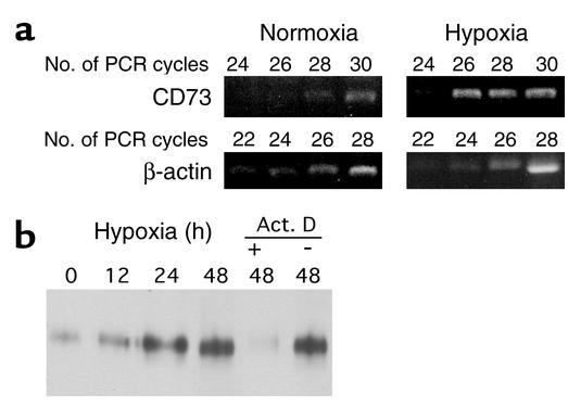
We have previously demonstrated that intestinal epithelial cells are uniquely resistant to changes in barrier function elicited by hypoxia (15). A transcriptional profiling approach similar to that used previously (15, 29) was used to identify potential hypoxia-regulated genes that might influence barrier in model epithelia (T84 cells). Microarray analysis (17) identified a qualitative induction of CD73 following epithelial subjection to hypoxia. Since CD73 has been shown to influence both vascular endothelial (30) and intestinal epithelial barrier function (31), we pursued CD73 as a potential protective mechanism for hypoxia-induced barrier disruption. Semiquantitative RT-PCR analysis (comparison of CD73 and control β-actin with increasing PCR cycle numbers) was employed to verify microarray results (Figure 1a) and revealed vigorous induction of CD73 mRNA expression after 18 hours’ exposure to hypoxia (6.3 ± 0.5–fold increase of integrated band density in cells exposed to hypoxia compared with those exposed to normoxia, P < 0.01).
Figure 1.
Induction of epithelial CD73 by hypoxia. (a) Confluent T84 monolayers were exposed to normoxia (pO2 147 torr, 18 hours) or hypoxia (pO2 20 torr, 18 hours). Total RNA was isolated, and CD73 mRNA levels were determined by RT-PCR using semiquantitative analysis (increasing cycle numbers, as indicated). As shown, β-actin transcript was determined in parallel and used as an internal standard. (b) Confluent T84 monolayers were exposed to indicated periods of hypoxia (with or without actinomycin D [Act. D], as indicated), monolayers were washed, surface proteins were biotinylated, and cells were lysed. CD73 was immunoprecipitated with mAb 1E9 and resolved by SDS-PAGE, and resultant Western blots were probed with avidin-peroxidase. Representative experiments of three are shown in each case.
Previous studies have suggested that intestinal epithelial cells express functional CD73 predominantly on the apical membrane surface, and, as such, membrane CD73 can readily be labeled with biotin and detected by avidin blots of immunoprecipitation with mAb 1E9 (3). This antibody specifically recognizes the ecto-5′-nucleotidase as opposed to intracellular forms of the nucleotidase (32). As shown in Figure 1b, immunoprecipitation of surface biotinylated protein and avidin blot from epithelial cells subjected to hypoxia (range 12–48 hours) revealed a time-dependent increase in expression of an approximately 70-kDa protein consistent with CD73, with maximal protein levels observed by 48 hours (no additional increases at 72 hours; data not shown). Incubation of epithelial cells with the transcriptional inhibitor actinomycin D attenuated such induction, suggesting that this pathway is likely transcriptional in nature.
Induction of functional surface CD73.
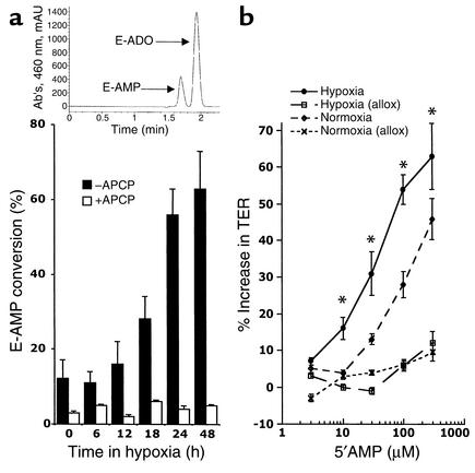
We next assessed whether hypoxia-induced CD73 was functional. As shown in Figure 2a, T84 cell exposure to hypoxia induced a time-dependent increase in functional CD73 (ANOVA, P < 0.01), as determined by HPLC analysis of E-AMP conversion to E-ADO (Figure 2a, inset) (33), with a 5.2 ± 0.6–fold increase at 48 hours’ hypoxia exposure (P < 0.001). Shorter periods of incubation in hypoxia (i.e., <12 hours) revealed no significant change in CD73 activity (P = not significant). Selectivity for CD73 in this assay was demonstrated by parallel incubation with APCP (5 μM) and revealed a significant reduction in CD73 bioactivity at each time point examined (P < 0.01 by ANOVA). Such data indicate that hypoxia induces functional surface CD73.
Figure 2.
Functional increase in CD73 surface activity by hypoxia. (a) Epithelial monolayers were exposed to indicated periods of hypoxia and washed, and surface CD73 activity was determined by HPLC analysis of E-AMP conversion to E-ADO (black bars). To determine specificity, a similar analysis was performed in the presence of the CD73 inhibitor APCP (white bars). Data are derived from five to seven monolayers in each condition, and results are expressed as mean percent E-AMP conversion ± SEM. The inset is a representative HPLC tracing demonstrating peak resolution of E-AMP and E-ADO. mAu, milli-absorbance unit. (b) Confluent T84 monolayers were subjected to hypoxia for 24 hours and treated with 2 mM EDTA for 5 minutes, followed by incubation in cell culture media with normo-calcium in the presence of indicated concentrations of 5′-AMP. TER was monitored over time, and the results shown represent the percent recovery of TER over 2 hours relative to no 5′-AMP. Also shown are plots of monolayers coincubated with the adenosine A2B receptor antagonist alloxazine (allox; 10 μM). Data are mean ± SEM from three separate experiments. *P < 0.025 compared with normoxia.
Our previous studies have indicated that CD73 is important for the maintenance of barrier function during interactions with polymorphonuclear leukocytes (PMN) or soluble PMN mediators (e.g., 5′-AMP) (30). Similarly, it was previously shown that 5′-AMP enhances epithelial barrier recovery via activation of surface adenosine A2B receptors (31). Thus, we examined whether conditions of hypoxia (24 hours’ exposure) that increase CD73 expression (Figure 1a) and function (Figure 2a) also enhance 5′-AMP–induced barrier function recovery following Ca2+ switch. As shown in Figure 2b, the addition of 5′-AMP following Ca2+ switch resulted in a concentration-dependent increase in barrier recovery over a 2-hour period (P < 0.01 for both hypoxia and normoxia). Moreover, at 5′-AMP concentrations of 10–300 μM, epithelial cells subjected to hypoxia demonstrated an enhanced recovery compared with those in normoxia (P < 0.025 by ANOVA), indicating the likelihood that hypoxia-induced CD73 functionally enhances 5′-AMP conversion to adenosine. To determine the relative role of adenosine A2B receptors in this response, parallel series of monolayers were also exposed to the A2B receptor antagonist alloxazine, and they revealed a nearly 80% decrease in 5′-AMP–induced barrier recovery, suggesting a prominent role for the A2B receptor. Taken together, these findings suggest that the increment of CD73 induced by hypoxia is relevant to epithelial functional responses.
CD39 is hypoxia-inducible.
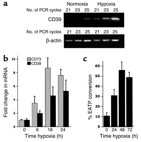
We next determined whether other surface nucleotidases might be similarly hypoxia-responsive. Specifically, we examined whether CD39, an ecto-apyrase that converts ATP/ADP to 5′-AMP (34), might also respond to hypoxia. As shown in Figure 3a, semiquantitative RT-PCR analysis (comparison of CD39 and control β-actin with increasing PCR cycle numbers) was employed to determine the general features of this response. It revealed prominent induction of CD39 mRNA expression after 18 hours’ exposure to hypoxia (5.3 ± 0.6–fold increase of integrated band density in cells exposed to hypoxia compared with those exposed to normoxia, P < 0.01), suggesting that, similar to CD73, CD39 is induced in parallel.
Figure 3.
Induction of functional CD39 by hypoxia. (a) Confluent T84 monolayers were exposed to normoxia (pO2 147 torr, 18 hours) or hypoxia (pO2 20 torr, 18 hours). Total RNA was isolated, and CD39 mRNA levels were determined by RT-PCR using semiquantitative analysis (increasing cycle numbers, as indicated). As shown, β-actin transcript was determined in parallel and used as an internal standard. (b) More quantitative real-time PCR was employed to directly compare hypoxia-inducibility of CD39 and CD73. Data were calculated relative to internal control genes (β-actin) and are expressed as fold increase over normoxia ± SEM at each indicated time. Results are derived from two experiments in each condition. (c) Epithelial monolayers were exposed to indicated periods of hypoxia and washed, and surface CD39 activity was determined by HPLC analysis of E-ATP conversion to E-AMP. Data are derived from five to seven monolayers in each condition, and results are expressed as mean percent E-AMP conversion ± SEM.
As a direct comparison for hypoxia-inducibility between CD39 and CD73, more quantitative real-time PCR was employed. As depicted in Figure 3b, a comparison of CD39 and CD73 revealed a time-dependent induction (P < 0.01 for both CD39 and CD73 by ANOVA) with maximal responses increased five- to tenfold at 18 hours’ exposure. Similar results were observed in other cell types (e.g., endothelia; data not shown), suggesting that this hypoxia-inducible pathway is likely not restricted to epithelia. In addition to these mRNA findings, functional CD39 surface expression was demonstrable through examination of E-ATP conversion to E-AMP (Figure 3c). Indeed, a time course of hypoxia revealed that surface enzyme activity was maximally increased 4.6 ± 0.6–fold in epithelia subjected to 48 hours’ hypoxia (P < 0.01). Such findings indicate a parallel induction of at least two adenine nucleotidases (CD39 and CD73) by hypoxia.
Role of HIF-1 in CD73 induction.
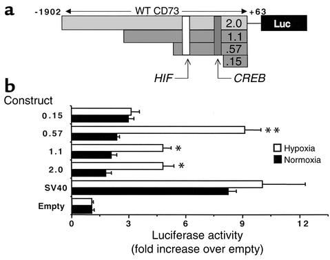
In an attempt to gain specific insight into the mechanisms of CD73 induction, we began examining induction pathways from hypoxia response genes. In the course of our experiments, we identified a previously unappreciated HIF-1 binding site in the CD73 gene promoter (DNA consensus motif 5′-CCGTG-3′ located at positions –367 to –371 relative to the major transcription start site) (11). Thus, luciferase reporter constructs expressing varied lengths of the CD73 promoter (Figure 4a) were used to address hypoxia-inducibility and, specifically, the role of HIF-1. As shown in Figure 4b, cells transiently transfected with the full-length CD73 promoter (pGL22.0NT, including nucleotides –1902 to +63), when exposed to hypoxia (48 hours), showed a 3.1 ± 0.3–fold increase in luciferase activity over normoxia controls (P < 0.01). Similar results were observed with an approximately 1-kb 5′ truncation (pGL21.1NT, encoding bp –993 to +63; Figure 4a). A larger truncation of this promoter sequence, to bp –518 to +63 (pGL20.57NT), resulted in a 4.1 ± 0.5–fold increase in hypoxia-inducibility over normoxia controls (P < 0.01), suggesting the presence of partial repressor activity in the –993 to –518 region. However, as shown in Figure 4b, further truncation to bp –92 to +63 (pGL20.15NT construct), which deletes the putative HIF-1α binding site at positions –367 to –371, resulted in a complete loss of hypoxia-inducibility (P < 0.01 compared with the wild-type promoter), indicating at least the possibility that HIF-1 contributes to CD73 hypoxia-inducibility.
Figure 4.
CD73 luciferase reporter assays. (a) The orientation of the HIF-1 and CREB binding sites in the CD73 luciferase reporter constructs and the location of truncations used for transient transfections (see Methods for details). WT, wild-type. (b) Confluent BAE monolayers were transiently transfected with plasmids expressing sequence corresponding to full-length CD73 (pGL22.0NT, bp –1902 to +63) or to the 5′ truncations pGL21.1NT (bp –993 to +63), pGL20.57NT (bp –518 to +63), or pGL20.15NT (bp –92 to +63), as well as with the SV40 promoter upstream from the luc reporter gene. Twelve hours later, cells were exposed to hypoxia or normoxia for 48 hours and assessed for luciferase activity. All transfections were normalized to cotransfected β-galactosidase activity. Data are mean ± SEM from three separate experiments.*P < 0.01, significantly different from normoxia; **P < 0.025, significantly different than other hypoxia conditions.
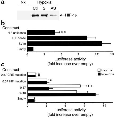
To extend these data, we assessed hypoxia-inducibility in the pGL20.57NT promoter construct in cells depleted of HIF-1α through the use of antisense oligonucleotides (Figure 5a). As shown in Figure 5b, luciferase activity in cells depleted of HIF-1 was diminished compared to control cells (P < 0.025), providing additional evidence for HIF-1 in hypoxia-induced expression of CD73. These findings were not a result of differences in background luciferase, since parallel transfections with promoter-less luciferase vectors showed no differences in activity between sense and antisense oligonucleotides directed against HIF-1α (1.1 ± 0.2–fold and 1.0 ± 0.3–fold increase over mock-transfected for sense and antisense, respectively, P not significant). In subsets of experiments, we examined whether depletion of HIF-1α might influence barrier recovery from Ca2+ switch (see conditions in Figure 2b). To do this, we used Caco2 epithelial cells, since antisense treatment is problematic in T84 cells (15). Using these conditions, we were able to both deplete HIF-1α (72% decrease as determined by densitometry of Western blots, n = 2) and attenuate barrier recovery associated with induced CD73 (50% ± 12% decrease in barrier recovery, P < 0.025, n = 2) with antisense oligonucleotides. Such findings support our hypothesis that adenosine liberated by hypoxia-induced CD73 promotes barrier function in vitro.
Figure 5.
Role of HIF-1 and CREB in CD73 hypoxia-inducibility. (a) Confluent BAE monolayers were exposed to mock treatment (Ctl), HIF-1α sense oligonucleotides (S), or HIF-1α antisense oligonucleotides (AS) for 48 hours. Total protein was solubilized, and HIF-1α expression was examined by Western blot. Nx, normoxia. (b) Confluent BAE monolayers were transiently transfected with plasmids expressing sequence corresponding to truncations at the 5′ end (pGL20.57NT, bp –518 to +63) in the presence or absence of HIF-1α sense or antisense oligonucleotides. Twelve hours later, cells were exposed to hypoxia for 48 hours and assessed for luciferase activity. Data are mean ± SEM from three separate experiments. **P < 0.01. (c) Confluent BAE monolayers were transiently transfected with plasmids expressing sequence corresponding to truncations at the 5′ end (pGL20.57NT, bp –518 to +63) or plasmids encoding HIF-1 or CRE mutations, as indicated. Twelve hours later, cells were exposed to hypoxia or normoxia for 48 hours and assessed for luciferase activity. All transfections were normalized to cotransfected β-galactosidase activity. Data are mean ± SEM from three separate experiments. *P < 0.025 compared with the corresponding nonmutated control; **P < 0.01 compared with normoxia.
To rule out the possibility that truncations at the 5′ end of the CD73 promoter simply reflect the deletion of a large promoter segment, we examined the influence of HIF-1α binding site mutations on hypoxia-inducibility. An HIF-1α binding site mutation was introduced in the pGL20.57NT construct, and as shown in Figure 5c. A two-nucleotide mutation (consensus 5′-CGTGC-3′ mutated to 5′-CATGG-3′ within the HIF-1 site) resulted in a 67% ± 9% decrease in luciferase activity under hypoxic conditions (P < 0.01), with no significant change in basal expression under normoxic conditions (P not significant). Taken together, these reporter construct data provide strong evidence for a functional HRE, mediated by HIF-1, within the CD73 promoter.
The cloned CD73 promoter also bears a classical CRE (TGACGTC at positions –115 to –121 relative to the transcription start site) (11). This DNA motif is the functional binding site for the transcriptional coactivator CREB in many genes (12). Since we and others have previously shown that CREB can mediate hypoxia responses in some genes (14, 35, 36), and truncations eliminating CRE result in a loss of hypoxia-inducibility (Figure 4), we addressed the role of CREB through mutational analysis of this site. Interestingly, as shown in Figure 5c, CRE site mutations (consensus 5′-TGACGTCG-3′ mutated to 5′-TGAATTCG-3′ within the CRE site) resulted in a nearly complete loss of reporter expression in both normoxia and hypoxia, indicating the likelihood that CREB regulates basal expression of CD73.
Induction and function of CD73 in vivo.
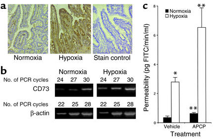
As proof of principle for CD73 function in epithelia, we extended these in vitro observations to an in vivo model. We recently showed that the unique resistance of intestinal epithelial barrier function to changes elicited by hypoxia in vitro and in vivo is, at least in part, mediated by intestinal trefoil peptide (15). Since CD73 has been shown to contribute to barrier properties of both vascular endothelial cells and intestinal epithelial cells in vitro (30, 31), we examined the relative role of CD73 in hypoxia using a murine mouse model (15). As shown in Figure 6a, we monitored in vivo hypoxia using the nitroimidazole compound EF5. Due to the lipophilicity of the molecule, EF5 enters cells readily by diffusion and is reduced in all tissues. In the absence of adequate levels of oxygen, EF5 undergoes further steps of reduction to more reactive products, which then bind to cellular proteins and are thus retained (37). Detection of the retained compound is possible using mAb’s directed against adducts of EF5 (38). Staining for EF5 adducts was more evident in small-intestinal sections of mice subjected to hypoxia than in those maintained in room air. In parallel, and as shown in Figure 6b, CD73 mRNA expression in mucosal scrapings (enriched with epithelial cells) from the small intestine of mice subjected to whole-animal hypoxia (8% O2, 4 hours) was increased compared with those from mice subjected to normoxia (2.7 ± 0.41–fold increase relative to β-actin by densitometry, P < 0.025). Consistent with previous studies (15), and as shown in Figure 6c, hypoxia significantly increased intestinal permeability to 4-kDa FITC-labeled dextran tracer (P < 0.025 compared with normoxia control). Inclusion of the CD73 inhibitor APCP within gavage fluid resulted in moderately increased permeability in normoxic animals (P < 0.05 compared with vehicle control) and increased barrier disruption by hypoxia (P < 0.025 compared with vehicle control), suggesting that CD73 normally maintains intestinal barrier and that hypoxia-induced CD73 in vivo may provide a protective mechanism for barrier during episodes of decreased oxygen delivery.
Figure 6.
Role of hypoxia-induced CD73 in vivo. BL/6/129 mice were gavaged with vehicle (PBS) or APCP (2 mg/100 g body weight, based on previous work in rats [ref. 27]) in combination with permeability tracer (60 mg/100 g body weight of FITC-labeled dextran, mol wt 4,000 daltons, as indicated) and exposed to ambient hypoxia (8% O2, 92% N2) or ambient room air for 4 hours. (a) Small-intestine hypoxia was monitored by localization of EF5 relative to the normoxia control. Also shown is a stain control (primary antibody omitted). (b) Total RNA was isolated from mucosal scrapings (enriched with epithelial cells), and CD73 mRNA levels were determined by RT-PCR using semiquantitative analysis (increasing cycle numbers, as indicated). As shown, β-actin transcript was determined in parallel and used as an internal standard. (c) Serum analysis of FITC concentration was performed as an indicator of intestinal permeability. Data are mean permeability ± SEM pooled from four to six animals per condition. *P < 0.025 compared with normoxia in vehicle controls; **P < 0.05 for APCP treatment compared with animals exposed to vehicle only.
Discussion
Adenosine exerts paracrine and autocrine functions on most cell types. Pathophysiologic conditions of hypoxia/ischemia result in numerous adenine nucleotide metabolic changes, and adenosine has a demonstrated role in organ function under such conditions. In the present studies, we explored the mechanisms and impact of CD73 induction by hypoxia, a primary determinant of localized production of adenosine at tissue interfaces (39). These studies revealed that ambient hypoxia transcriptionally regulates CD73 and that one mechanism of such induction involves HIF-1. Extensions of these studies revealed that CD73 induction may subserve intestinal permeability during hypoxia in vivo.
Adenosine is a critical mediator during ischemia and hypoxia (9). While the source of interstitial adenosine in hypoxic tissue has been the basis of much debate, it is generally accepted that the dephosphorylation of AMP by CD73 represents the major pathway of extracellular adenosine formation during oxygen supply imbalances (9). Extracellular adenosine production in the ischemic myocardium, for example, is attributable to activity of CD73 (40), and both CD73 activity and adenosine metabolism have been demonstrated in cardiac preconditioning by brief periods of ischemia (41, 42). Increased CD73 activity in ischemic preconditioning has been attributed to a variety of acute activation pathways (10), and a recent study provides direct evidence that CD73 is transcriptionally regulated by hypoxia in pheochromocytoma cells in vitro (13). Once liberated in the extracellular space, adenosine either is taken up into the cell (through dipyridamole-sensitive carriers) or interacts with cell surface adenosine receptors (5). Presently, four subtypes of G protein–coupled adenosine receptors exist, designated A1, A2A, A2B, and A3. These receptors are classified according to utilization of pertussis toxin–sensitive pathways (A1 and A3) or adenylate cyclase activation pathways (A2A and A2B) (5). Epithelial cells of many origins constitutively express adenosine receptors (5), primarily of the A2A and A2B subtypes (43–46). As such, the present findings of functional CD39/CD73 induction during hypoxia help to clarify at least some of the issues related to increased enzyme activity during hypoxia. Much still remains unknown about these pathways. For example, we do not know whether intracellular nucleotidases are similarly regulated at the protein level. The antibody used here to determine protein levels (mAb 1E9) specifically recognizes the extracellular nucleotidase as opposed to other forms of the molecule (32), and thus, no general conclusions can be drawn about generation of total cellular adenosine. Similarly, little is known about the regulation of CD39 at the transcriptional/posttranscriptional level, and whether similar HIF-1 pathways may also contribute to hypoxia-inducibility.
Given the temporal and robust hypoxia response observed in the induction of CD73, a candidate regulator was HIF-1, a member of the rapidly growing Per-ARNT-Sim family of basic helix-loop-helix transcription factors (47, 48). HIF-1 exists as an αβ heterodimer, the activation of which is dependent upon stabilization of an O2-dependent degradation domain of the α subunit by the ubiquitin-proteasome pathway (49). A search of the cloned CD73 gene promoter revealed a classic HIF-1–binding DNA consensus motif, 5′-CCGTG-3′, located at positions –367 to –371 relative to the major transcription start site (11). However, the existence of an HIF-1α binding consensus is not evidence for an HIF-1α–mediated response; instead, the HRE is defined as a cis-acting transcriptional regulatory sequence located within 5′-flanking, 3′-flanking, or intervening sequences of target genes (50). Three approaches were used to define a role for HIF-1α in the induction of CD73. First, the use of previously published antisense oligonucleotides (23), but not sense controls, resulted in a nearly complete blockade of CD73 induction. Second, the combination of antisense oligonucleotides and transient reporter construct transfections was used to add further evidence for HIF-1 and revealed a complete blockade of CD73 induction. A third approach using luciferase reporter constructs was used to identify the hypoxia-responsive region of the promoter. Results from these studies narrowed the region to –518 to +63, and mutations of this HIF-1α site resulted in a greater than 85% decrease in hypoxia-inducibility. A two-nucleotide mutation of this site resulted in a loss of hypoxia-inducibility and provided additional evidence for a functional HRE. Of note, studies with reporter constructs also indicated that repressor elements may also regulate CD73 expression in hypoxia. For example, the larger 5′ truncations of the promoter sequence to bp –518 to +63 (pGL20.57NT) indicated increased hypoxia-inducibility compared with the full-length promoter, suggesting the presence of partial repressor activity (bp –993 to –518). Transcription factor binding analysis (e.g., TFSEARCH) (51) of this region indicated consensus sites for CdxA, SRY, GATA-1, GATA-2, and HNF-3b. While we have not directly addressed this issue, at least two of these transcription factors (GATA-1 and GATA-2) have been recently implicated in repression of genes in hypoxia (52, 53). Thus, it is likely that both positive and negative regulatory pathways contribute to overall CD73 promoter activity.
Recent work from a number of laboratories has indicated that CREB may mediate hypoxia-elicited induction of a number of genes (14, 35, 36). Based on these findings, and previous observations that the CRE site of the CD73 promoter is functional (30), mutational analysis was employed to define the function of this site. Surprisingly, these studies revealed that CREB binding is critical for basal expression of CD73. These results may have broader implications. For example, it is not presently known how tightly CD73 expression is regulated and whether such regulation is transcriptionally coupled. In addition, it is not known what physiologically relevant mediators (e.g., hormones, chemokines, nucleotides, cytokines) might influence basal expression of CD73. It is possible that basal, low-level maintenance of CD73 expression occurs as a bystander process of adenosine A2A or A2B receptor activation (i.e., activation of CREB). In this regard, and as we have hypothesized previously (30), adenosine may serve as a feed-forward mechanism to regulate nucleotide metabolic enzymes, such as CD73.
Our previous studies suggested that surface CD73 likely represents a protective pathway for the maintenance of barrier function in epithelia (31) and vascular endothelia (4, 30). We provide in vivo evidence here that CD73, and likely CD39, functions as an overall barrier-protective element in the intestine. In control animals exposed to luminal APCP, significantly increased permeability was observed, suggesting that CD73 provides a physiologic function in this regard, and this influence was enhanced in parallel to induction of CD73 mRNA in hypoxic animals. These findings are in line with previous studies indicating that the intestinal mucosa supports barrier-protective pathways during hypoxia in vivo. Work addressing the role of intestinal trefoil factor (ITF) suggested that hypoxia-induced ITF (via HIF-1 activation) contributes, in part, an endogenous protective mechanism for intestinal epithelia (15). Those studies were noteworthy in that there were likely other important molecules with similar functions. It is possible that CD73 is an additional molecule with a similar function. Of note, it was recently reported that the MDR1 gene product P-glycoprotein could also contribute a similar function during hypoxia (54), particularly since MDR has been associated with barrier abnormalities in intestinal disease (55). Taken together, a number of molecules likely contribute to this interesting pathway, and the redundancy likely indicates the biologic importance of this protective mechanism.
While the present studies are the first, to our knowledge, to define a physiologic role for CD73 in barrier function in vivo, significant work has implicated CD73 in barrier regulation in vitro. During modeled inflammation, neutrophils release a number of biologically active mediators, including 5′-AMP (7), and metabolism of 5′-AMP to adenosine requires CD73 (1, 56). Inhibitor-based studies have implicated CD73 in regulation of both epithelial and endothelial permeability during PMN transmigration and have suggested that the metabolism of 5′-AMP to adenosine by CD73 may be rate-limiting (i.e., increased CD73 results in parallel increases in 5′-AMP–mediated bioactivity) (4, 31). The competitive inhibitor APCP abolishes the influence of 5′-AMP, whereas the less potent, noncompetitive inhibitor mAb 1E9 substantially diminishes the influence of 5′-AMP (4). Our findings that the CD73 antagonist APCP increases intestinal permeability are consistent with previous studies in rats showing that APCP inhibits ATP-dependent (i.e., cAMP-dependent) peripheral vasodilation. Importantly, in this regard, it will be necessary to define the exact role of CD39 in barrier function, particularly since a number of cell types, especially activated platelets, are able to release large quantities of CD39 substrates (i.e., ATP and ADP) at sites of inflammation or hypoxia (57). Recent studies with stroke models in CD39–/– mice suggest that CD39 provides a protective thromboregulatory function during ischemia and stroke (58). Such observations suggest that the hypoxic microenvironment may liberate large amounts of both CD39 and CD73 substrates, and, as a result, large quantities of extracellular adenosine. While we do not know the exact mechanism(s) of barrier promotion by adenosine, recent studies suggest that phosphorylation of the tight junction–associated protein vasodilator-stimulated phosphoprotein (VASP) may be critical in both epithelial and endothelial permeability (20, 59).
In these studies, we assessed mucosal hypoxia using the nitroimidazole EF5. In the absence of adequate levels of oxygen, EF5 undergoes reduction to more reactive products, which then bind to cellular proteins and are thus retained and detected with antibodies directed against the adducts (37). Noteworthy in these experiments was the nearly complete lack of staining in connective tissue under all experimental conditions, but evident staining in the epithelium of normoxic animals. This basal staining in normoxic animals was not evident in other mucosal tissues examined (e.g., liver and kidney; data not shown) and may be related to previous observations that the pO2 of the healthy intestine is relatively low (∼30–35 mmHg, depending on the specific region examined) (60). Given these features, it stands to reason that the relative hypoxia of the healthy intestine could be an endogenous mechanism to maintain expression of hypoxia-responsive genes, such as CD39 and CD73. This hypothesis has not been directly tested.
In summary, these results define hypoxia-regulated CD39 and CD73 expression in the intestinal mucosa and identify a previously unappreciated HIF-1 regulatory binding site in the CD73 promoter. This regulatory pathway extends to the transcriptional level in vitro and in vivo and identifies an important role for CD39/CD73 in the regulation of intestinal permeability during hypoxia.
Acknowledgments
This work was supported by NIH grants DK-50189, DE-13499, and HL-60569, and by a grant from the Crohn’s and Colitis Foundation of America.
Footnotes
Conflict of interest: No conflict of interest has been declared.
Nonstandard abbreviations used: cAMP response element (CRE); cAMP response element–binding protein (CREB); bovine aortic endothelium (BAE); α,β-methylene ADP (APCP); etheno-AMP (E-AMP); ethenoadenosine (E-ADO); etheno-ATP (E-ATP); transepithelial electrical resistance (TER); hypoxia-inducible factor-1 (HIF-1); hypoxia response element (HRE); polymorphonuclear leukocytes (PMN).
References
- 1.Pearson JD, Gordon JL. Nucleotide metabolism by endothelium. Annu Rev Physiol. 1985;47:617–627. doi: 10.1146/annurev.ph.47.030185.003153. [DOI] [PubMed] [Google Scholar]
- 2.Zimmermann H, Braun N. Ecto-nucleotidases: molecular structures, catalytic properties, and functional roles in the nervous system. Prog Brain Res. 1999;120:371–385. [PubMed] [Google Scholar]
- 3.Strohmeier GR, et al. Surface expression, polarization, and functional significance of CD73 in human intestinal epithelia. J Clin Invest. 1997;99:2588–2601. doi: 10.1172/JCI119447. [DOI] [PMC free article] [PubMed] [Google Scholar]
- 4.Lennon PF, Taylor CT, Stahl GL, Colgan SP. Neutrophil-derived 5′-adenosine monophosphate promotes endothelial barrier function via CD73-mediated conversion to adenosine and endothelial A2Breceptor activation. J Exp Med. 1998;188:1433–1443. doi: 10.1084/jem.188.8.1433. [DOI] [PMC free article] [PubMed] [Google Scholar]
- 5.Stiles GL. Adenosine receptors. J Biol Chem. 1992;267:6451–6454. [PubMed] [Google Scholar]
- 6.Mertens S, Noll T, Spahr R, Krutzfeldt A, Piper HM. Energetic response of coronary endothelial cells to hypoxia. Am J Physiol. 1990;258:H689–H694. doi: 10.1152/ajpheart.1990.258.3.H689. [DOI] [PubMed] [Google Scholar]
- 7.Madara JL, et al. 5′-Adenosine monophosphate is the neutrophil-derived paracrine factor that elicits chloride secretion from T84 intestinal epithelial cell monolayers. J Clin Invest. 1993;91:2320–2325. doi: 10.1172/JCI116462. [DOI] [PMC free article] [PubMed] [Google Scholar]
- 8.Airas L, et al. CD73 is involved in lymphocyte binding to the endothelium: characterization of lymphocyte-vascular adhesion protein 2 identifies it as CD73. J Exp Med. 1995;182:1603–1608. doi: 10.1084/jem.182.5.1603. [DOI] [PMC free article] [PubMed] [Google Scholar]
- 9.Linden J. Molecular approach to adenosine receptors: receptor-mediated mechanisms of tissue protection. Annu Rev Pharmacol Toxicol. 2001;41:775–787. doi: 10.1146/annurev.pharmtox.41.1.775. [DOI] [PubMed] [Google Scholar]
- 10.Kitakaze M, et al. Adenosine and cardioprotection in the diseased heart. Jpn Circ J. 1999;63:231–243. doi: 10.1253/jcj.63.231. [DOI] [PubMed] [Google Scholar]
- 11.Hansen KR, Resta R, Webb CF, Thompson LF. Isolation and characterization of the promoter of the human 5′-nucleotidase (CD73)-encoding gene. Gene. 1995;167:307–312. doi: 10.1016/0378-1119(95)00574-9. [DOI] [PubMed] [Google Scholar]
- 12.Montminy M. Transcriptional regulation by cyclic AMP. Annu Rev Biochem. 1997;66:807–822. doi: 10.1146/annurev.biochem.66.1.807. [DOI] [PubMed] [Google Scholar]
- 13.Kobayashi S, Zimmermann H, Millhorn DE. Chronic hypoxia enhances adenosine release in rat PC12 cells by altering adenosine metabolism and membrane transport. J Neurochem. 2000;74:621–632. doi: 10.1046/j.1471-4159.2000.740621.x. [DOI] [PubMed] [Google Scholar]
- 14.Taylor CT, Furuta GT, Synnestvedt K, Colgan SP. Phosphorylation-dependent targeting of cAMP response element binding protein to the ubiquitin/proteasome pathway in hypoxia. Proc Natl Acad Sci USA. 2000;97:12091–12096. doi: 10.1073/pnas.220211797. [DOI] [PMC free article] [PubMed] [Google Scholar]
- 15.Furuta GT, et al. Hypoxia-inducible factor 1-dependent induction of intestinal trefoil factor protects barrier function during hypoxia. J Exp Med. 2001;193:1027–1034. doi: 10.1084/jem.193.9.1027. [DOI] [PMC free article] [PubMed] [Google Scholar]
- 16.Zünd G, et al. Hypoxia enhances stimulus-dependent induction of E-selectin on aortic endothelial cells. Proc Natl Acad Sci USA. 1996;93:7075–7080. doi: 10.1073/pnas.93.14.7075. [DOI] [PMC free article] [PubMed] [Google Scholar]
- 17.Lockhart DJ, et al. Expression monitoring by hybridization to high-density oligonucleotide arrays. Nat Biotechnol. 1996;14:1675–1680. doi: 10.1038/nbt1296-1675. [DOI] [PubMed] [Google Scholar]
- 18.Higuchi R, Fockler C, Dollinger G, Watson R. Kinetic PCR analysis: real-time monitoring of DNA amplification reactions. Biotechnology. 1993;11:1026–1030. doi: 10.1038/nbt0993-1026. [DOI] [PubMed] [Google Scholar]
- 19.Pfaffl MW. A new mathematical model for relative quantification in real-time RT-PCR. Nucleic Acids Res. 2001;29:2002–2007. doi: 10.1093/nar/29.9.e45. [DOI] [PMC free article] [PubMed] [Google Scholar]
- 20.Lawrence DW, Comerford KM, Colgan SP. Role of VASP in reestablishment of epithelial tight junction assembly after Ca2+ switch. Am J Physiol Cell Physiol. 2002;282:C1235–C1245. doi: 10.1152/ajpcell.00288.2001. [DOI] [PubMed] [Google Scholar]
- 21.Parkos CA, Colgan SP, Delp C, Arnaout MA, Madara JL. Neutrophil migration across a cultured epithelial monolayer elicits a biphasic resistance response representing sequential effects on transcellular and paracellular pathways. J Cell Biol. 1992;117:757–764. doi: 10.1083/jcb.117.4.757. [DOI] [PMC free article] [PubMed] [Google Scholar]
- 22.Dharmsathaphorn K, Madara JL. Established intestinal cell lines as model systems for electrolyte transport studies. Methods Enzymol. 1990;192:354–389. doi: 10.1016/0076-6879(90)92082-o. [DOI] [PubMed] [Google Scholar]
- 23.Caniggia I, et al. Hypoxia-inducible factor-1 mediates the biological effects of oxygen on human trophoblast differentiation through TGFβ3. J Clin Invest. 2000;105:577–587. doi: 10.1172/JCI8316. [DOI] [PMC free article] [PubMed] [Google Scholar]
- 24.Ueda K, Pastan I, Gottesman MM. Isolation and sequence of the promoter region of the human multidrug-resistance (P-glycoprotein) gene. J Biol Chem. 1987;262:17432–17436. [PubMed] [Google Scholar]
- 25.Chen CJ, et al. Genomic organization of the human multidrug resistance (MDR1) gene and origin of P-glycoproteins. J Biol Chem. 1990;265:506–514. [PubMed] [Google Scholar]
- 26.Napolitano LM, Koruda MJ, Meyer AA, Baker CC. The impact of femur fracture with associated soft tissue injury on immune function and intestinal permeability. Shock. 1996;5:202–207. doi: 10.1097/00024382-199603000-00006. [DOI] [PubMed] [Google Scholar]
- 27.Skinner MR, Marshall JM. Studies on the roles of ATP, adenosine and nitric oxide in mediating muscle vasodilatation induced in the rat by acute systemic hypoxia. J Physiol. 1996;495:553–560. doi: 10.1113/jphysiol.1996.sp021615. [DOI] [PMC free article] [PubMed] [Google Scholar]
- 28.Evans SM, et al. Detection of hypoxia in human squamous cell carcinoma by EF5 binding. Cancer Res. 2000;60:2018–2024. [PubMed] [Google Scholar]
- 29.Narravula S, Colgan SP. Hypoxia-inducible factor 1-mediated inhibition of peroxisome proliferator-activated receptor alpha expression during hypoxia. J Immunol. 2001;166:7543–7548. doi: 10.4049/jimmunol.166.12.7543. [DOI] [PubMed] [Google Scholar]
- 30.Narravula S, Lennon PF, Mueller BU, Colgan SP. Regulation of endothelial CD73 by adenosine: paracrine pathway for enhanced endothelial barrier function. J Immunol. 2000;165:5262–5268. doi: 10.4049/jimmunol.165.9.5262. [DOI] [PubMed] [Google Scholar]
- 31.Friedman GB, Taylor CT, Parkos CA, Colgan SP. Epithelial permeability induced by neutrophil transmigration is potentiated by hypoxia: role of intracellular cAMP. J Cell Physiol. 1998;176:76–84. doi: 10.1002/(SICI)1097-4652(199807)176:1<76::AID-JCP9>3.0.CO;2-5. [DOI] [PubMed] [Google Scholar]
- 32.Thompson LF, et al. Production and characterization of monoclonal antibodies to the glycosyl phosphatydylinositol-anchored lymphocyte differentiation antigen ecto-5′-nucleotidase (CD73) Tissue Antigens. 1990;35:9–19. doi: 10.1111/j.1399-0039.1990.tb01750.x. [DOI] [PubMed] [Google Scholar]
- 33.Jamal Z, Afkham-Ebrahimi A, Saggerson ED. A novel assay for 5′ectonucleotidase using 1,N6-etheno-AMP as substrate, and comments on the properties of the reaction product, ethenoadenosine. Biochem J. 1988;250:369–373. doi: 10.1042/bj2500369. [DOI] [PMC free article] [PubMed] [Google Scholar]
- 34.Wang TF, Guidotti G. CD39 is an ecto-(Ca2+,Mg2+)-apyrase. J Biol Chem. 1996;271:9898–9901. [PubMed] [Google Scholar]
- 35.Beitner-Johnson D, Millhorn DE. Hypoxia induces phosphorylation of the cyclic AMP response element-binding protein by a novel signaling mechanism. J Biol Chem. 1998;273:19834–19839. doi: 10.1074/jbc.273.31.19834. [DOI] [PubMed] [Google Scholar]
- 36.Taylor CT, Fueki N, Agah A, Hershberg RM, Colgan SP. Critical role of cAMP response element binding protein expression in hypoxia-elicited induction of epithelial TNFα. J Biol Chem. 1999;274:19447–19450. doi: 10.1074/jbc.274.27.19447. [DOI] [PubMed] [Google Scholar]
- 37.Li Z, et al. Roles of PLC-beta2 and -beta3 and PI3Kgamma in chemoattractant-mediated signal transduction. Science. 2000;287:1046–1049. doi: 10.1126/science.287.5455.1046. [DOI] [PubMed] [Google Scholar]
- 38.Hirsch E, et al. Central role for G protein-coupled phosphoinositide 3-kinase gamma in inflammation. Science. 2000;287:1049–1053. doi: 10.1126/science.287.5455.1049. [DOI] [PubMed] [Google Scholar]
- 39.Zimmerman H. 5′-Nucleotidase: molecular structure and functional aspects. Biochem J. 1992;285:345–365. doi: 10.1042/bj2850345. [DOI] [PMC free article] [PubMed] [Google Scholar]
- 40.Imai, S., Nakazawa, M., Imai, H., and Jin, H. 1987. 5′-Nucleotidase inhibitors and the myocardial reactive hyperemia and adenosine content. In Topics and perspectives in adenosine research. E. Gerlach and B.F. Becker, editors. Springer-Verlag. Berlin, Germany. 416–426.
- 41.Minamino T, et al. Cardioprotection due to preconditioning correlates with increased ecto-5′-nucleotidase activity. Am J Physiol. 1996;270:H238–H244. doi: 10.1152/ajpheart.1996.270.1.H238. [DOI] [PubMed] [Google Scholar]
- 42.de Jonge R, Bradamante S, de Jong JW. Cardioprotection by ischemic preconditioning. Role of adenosine and glycogen. Adv Exp Med Biol. 1998;431:279–282. [PubMed] [Google Scholar]
- 43.Schiele JO, Scwabe U. Characterization of the adenosine receptor in microvascular coronary endothelial cells. Eur J Pharmacol. 1994;269:51–58. doi: 10.1016/0922-4106(94)90025-6. [DOI] [PubMed] [Google Scholar]
- 44.Sexl V, et al. Stimulation of the mitogen-activated protein-kinase via the A2A-adenosine receptor on primary human endothelial cells. J Biol Chem. 1997;272:5792–5799. doi: 10.1074/jbc.272.9.5792. [DOI] [PubMed] [Google Scholar]
- 45.Iwamoto T, et al. Identification of the adenosine A2 receptor-cAMP system in human aortic endothelial cells. Biochem Biophys Res Commun. 1994;199:905–910. doi: 10.1006/bbrc.1994.1314. [DOI] [PubMed] [Google Scholar]
- 46.Watanabe H, Kuhne W, Schwartz P, Piper HM. A2-adenosine receptor stimulation increases macromolecular permeability of coronary endothelial cells. Am J Physiol. 1992;262:H1174–H1181. doi: 10.1152/ajpheart.1992.262.4.H1174. [DOI] [PubMed] [Google Scholar]
- 47.Wang GL, Jiang BH, Rue EA, Semenza GL. Hypoxia-inducible factor 1 is a basic-helix-loop-helix-PAS heterodimer regulated by cellular oxygen tension. Proc Natl Acad Sci USA. 1995;92:5510–5514. doi: 10.1073/pnas.92.12.5510. [DOI] [PMC free article] [PubMed] [Google Scholar]
- 48.Wang GL, Semenza GL. Purification and characterization of hypoxia-inducible factor 1. J Biol Chem. 1995;270:1230–1237. doi: 10.1074/jbc.270.3.1230. [DOI] [PubMed] [Google Scholar]
- 49.Semenza GL. HIF-1 and human disease: one highly involved factor. Genes Dev. 2000;14:1983–1991. [PubMed] [Google Scholar]
- 50.Semenza GL. Hypoxia-inducible factor 1: master regulator of O2 homeostasis. Curr Opin Genet Dev. 1998;8:588–594. doi: 10.1016/s0959-437x(98)80016-6. [DOI] [PubMed] [Google Scholar]
- 51.Heinemeyer T, et al. Databases on transcriptional regulation: TRANSFAC, TRRD and COMPEL. Nucleic Acids Res. 1998;26:362–367. doi: 10.1093/nar/26.1.362. [DOI] [PMC free article] [PubMed] [Google Scholar]
- 52.Tarumoto T, et al. N(G)-monomethyl-L-arginine inhibits erythropoietin gene expression by stimulating GATA-2. Blood. 2000;96:1716–1722. [PubMed] [Google Scholar]
- 53.De Maria R, et al. Negative regulation of erythropoiesis by caspase-mediated cleavage of GATA-1. Nature. 1999;401:489–493. doi: 10.1038/46809. [DOI] [PubMed] [Google Scholar]
- 54.Comerford KM, et al. Hypoxia-inducible factor-1-dependent regulation of the multidrug resistance (MDR1) gene. Cancer Res. 2002;62:3387–3394. [PubMed] [Google Scholar]
- 55.Panwala CM, Jones JC, Viney JL. A novel model of inflammatory bowel disease: mice deficient for the multiple drug resistance gene, mdr1a, spontaneously develop colitis. J Immunol. 1998;161:5733–5744. [PubMed] [Google Scholar]
- 56.Plesner L. Ecto-ATPases: identities and functions. Int Rev Cytol. 1995;158:141–214. doi: 10.1016/s0074-7696(08)62487-0. [DOI] [PubMed] [Google Scholar]
- 57.Marcus AJ, et al. Thrombosis and inflammation as multicellular processes: significance of cell-cell interactions. Thromb Haemost. 1995;74:213–217. [PubMed] [Google Scholar]
- 58.Pinsky DJ, et al. Elucidation of the thromboregulatory role of CD39/ectoapyrase in the ischemic brain. J Clin Invest. 2002;109:1031–1040. doi:10.1172/JCI200210649. doi: 10.1172/JCI10649. [DOI] [PMC free article] [PubMed] [Google Scholar]
- 59.Comerford KM, Lawrence DW, Synnestvedt K, Levi BP, Colgan SP. Role of vasodilator-stimulated phosphoprotein in PKA-induced changes in endothelial junctional permeability. FASEB J. 2002;16:583–585. doi: 10.1096/fj.01-0739fje. [DOI] [PubMed] [Google Scholar]
- 60.Sheridan WG, Lowndes RH, Young HL. Intraoperative tissue oximetry in the human gastrointestinal tract. Am J Surg. 1990;159:314–319. doi: 10.1016/s0002-9610(05)81226-7. [DOI] [PubMed] [Google Scholar]


