Abstract
Antagonists of growth hormone-releasing hormone (GH-RH) inhibit the growth of various tumors through mechanisms that involve the suppression of the insulin-like growth factor I and/or insulin-like growth factor II levels or secretion. In the present study, we tested the hypothesis that the tumor inhibition is associated with a decrease in telomerase activity because telomerase is considered obligatory for continued tumor growth. Nude mice bearing xenografts of U-87MG human glioblastomas were treated with GH-RH antagonist MZ-5-156. Telomerase activity was assessed by the telomerase repeat amplification protocol. Treatment with MZ-5-156 reduced levels of telomerase activity as compared with controls. When U-87 glioblastomas, H-69 small cell lung carcinomas, H-23 non-small cell lung carcinomas, and MDA-MB-468 breast carcinoma cells were cultured in vitro, addition of 3 μM MZ-5-156 also inhibited telomerase activity. Reverse transcription–PCR analysis revealed that in U-87MG glioblastomas, the expression of the hTRT gene encoding for the telomerase catalytic subunit was significantly decreased by MZ-5-156, whereas the levels of mRNA for hTR and TP1, which encode for the telomerase RNA and telomerase-associated protein, respectively, were unaffected. The repression of the telomerase activity was not accompanied by a significant decrease of mRNA level for the c-myc protooncogene that regulates telomerase. Our findings suggest that tumor inhibition induced by the GH-RH antagonists in U-87MG glioblastomas is associated with the down-regulation of the hTRT gene, resulting in a decrease in telomerase activity. Further studies are needed to establish whether GH-RH antagonists produce telomerase inhibition in other tumors.
Keywords: telomerase repeat amplification assay, telomerase catalytic subunit regulation, tumor regression, down-regulation of telomerase gene
Growth hormone-releasing hormone (GH-RH) is secreted by the hypothalamus and through specific GH-RH receptors in the anterior pituitary stimulates the secretion of the GH (1). In addition to its physiological role in regulating GH release, GH-RH may play a role in the development of some neoplasms (2, 3). It has been demonstrated that GH-RH antagonists MZ-4-71 and MZ-5-156, synthesized in our laboratory, inhibit the proliferation of various experimental human and animal tumors in vivo and in vitro (4–10). Thus, GH-RH antagonists suppress growth of androgen-independent human DU-145 and PC-3 and rat Dunning R-3327-AT-1 prostate cancers (5, 7), H-69 small cell lung carcinomas (SCLC) and H-157 non-SCLC (6), CAKI-1 renal adenocarcinomas (8), SK-ES-1 and MNNG-HOS osteosarcomas (9), and other cancers (4). GH-RH antagonists appear to inhibit the growth of cancers through indirect or direct pathways. The indirect mechanism operate through a suppression of the GH release from the pituitary and the resulting inhibition of the hepatic production of insulin-like growth factor (IGF) I (4, 7). In addition, a significant reduction in concentrations of IGF-I and/or IGF-II produced in osteosarcomas, renal cancers, and prostatic tumors as well as in non-SCLCs after treatment of nude mice with MZ-4-71 or MZ-5-156, points to a likely direct effect of GH-RH antagonists on tumors (5–9). A strong suppression of IGF-II mRNA expression in DU-145 tumors after treatment with MZ-5-156 supports this concept (7). In vitro studies also demonstrate that GH-RH antagonists cause a direct inhibition of growth, IGF-II production, and expression of IGF-II mRNA in human cancer cell lines (V. Csernus and A.V.S., unpublished work). IGF-I and IGF-II are involved in the regulation of normal and malignant growth through endocrine, paracrine, or autocrine mechanisms (4, 11–14).
Telomerase is a ribonucleoprotein that functions as a specific DNA polymerase that is involved in the maintenance of telomeres, specialized structures at the ends of the eukaryotic chromosomes, by replacing the loss of telomeric DNA that occurs at each cell division (15–18). Telomerase maintains telomere length and chromosome stability, which are required for cellular immortality and subsequent malignant transformations (19–21). A striking association exists between telomerase activity and malignancy: 85–90% of the primary human tumors express this activity, as compared with approximately 24% of benign tumors, whereas it is absent in most normal somatic tissues (22–24). Three components of the telomerase ribonucleoprotein complex have been identified: the telomerase catalytic subunit (hTRT) (24–27), the telomerase RNA (hTR) (16), and telomerase-associated protein (TP1) (28, 29). Among them, hTRT is considered the rate-limiting determinant of the telomerase activity because a correlation was found between telomerase activity and hTRT mRNA, but not hTR and TP1 mRNA levels (25, 30). In addition, the introduction of the hTRT gene in telomerase-negative normal human fibroblasts restores telomerase activity and extends their life span (20). The mechanism for the reactivation of telomerase in tumor cells, which is a necessary event for the acquisition of cellular immortality, remains poorly understood, but an important role for the c-myc oncogene has been recognized (31, 32).
In an attempt to investigate further the mechanism of antitumor action of MZ-5-156, we evaluated whether this analog affects the levels of telomerase activity in human U-87MG glioblastomas in vitro and in vivo. GH-RH antagonists such as MZ-5-156 are highly effective in inhibiting growth of U-87MG glioblastomas xenografted into nude mice through mechanisms that involve the down-regulation of IGF-II and K-ras genes (unpublished work). Telomerase activity is detectable in most primary human brain tumors and is associated with the progression of the disease (33–35). In this study we investigated whether growth inhibition of U-87MG glioblastomas by the GH-RH antagonist MZ-5-156 is accompanied by a decrease in the levels of telomerase activity.
MATERIALS AND METHODS
Peptides.
GH-RH antagonist MZ-5-156 ([PhAc0, d-Arg2, Phe(4-Cl)6, Abu15, Nle27]hGH-RH(1–28)Agm) and agonistic analog of GH-RH JI-38 [Dat1, Gln8, Orn12,21, Abu15, Nle27, Asp28, Agm29]hGH-RH(1–29) [where PhAc is phenylacetyl, Phe(p-Cl) is parachlorophenylalanyl, Abu is α-aminoisobutyryl, Nle is norleucyl, Agm is agmatine, Dat is desaminotyrosine, and Orn is ornithine] were synthesized by solid-phase methods (10, 36). GH-RH(1–29)NH2 also was synthesized and characterized in our laboratory as described (10, 36).
Animals.
Five- to 6-week-old male athymic (Ncr nu/nu) nude mice were obtained from the National Cancer Institute (Bethesda, MD), housed in sterile cages under laminar flow hoods in a temperature-controlled room with a 12-hr light/12-hr dark schedule, and fed autoclaved chow and water ad libitum. Their care was in accord with institutional guidelines.
Cell Lines.
The human glioblastoma cell line U-87MG, the SCLC cell line NCI-H-69, the non-SCLC cell line NCI-H-23, and the breast carcinoma cell line MDA-MB-468 were obtained from American Type Culture Collection. The medium for U-87MG was MEM with 1 mM pyruvate, the lung carcinoma cell lines were cultured in RPMI medium 1640 (GIBCO), and the medium for MDA-MB-468 was Eagle’s improved MEM (IMEM), all supplemented with 2 mM l-glutamine, 100 units/ml of penicillin G sodium, 100 μg/ml of streptomycin sulfate, 0.25 μg/ml of amphotericin B, and 10% fetal bovine serum. The cells were grown at 37°C in a humidified 95% air/5% carbon dioxide atmosphere, passaged weekly, and routinely monitored for mycoplasma contamination by using a detection kit (Boehringer Mannheim). All culture media components were purchased from GIBCO except IMEM (Biofluids, Rockville, MD). Cells growing exponentially were harvested by a brief incubation with 0.25% trypsin-EDTA solution.
Experimental Protocol.
U-87MG cells growing exponentially were implanted into one male nude mouse by s.c. injection of 1 × 10⁷ cells in the right flank. The tumor xenograft resulting after 1 week was aseptically dissected, mechanically minced, and 3-mm3 pieces of tumor tissue were transplanted s.c. by trocar needle into six mice under methoxyflurane (Metofane, Pittman–Moore, Mundelein, IL) anesthesia. Three weeks after transplantation, when tumors reached a volume of approximately 75 mm3, mice were randomized and divided into two experimental groups of three animals each, which received the following treatments for 4 weeks: (i) injections of saline containing 0.1% dimethyl sulfoxide (control); and (ii) MZ-5-156 injected s.c. at a dose of 20 μg/animal per day.
Extract Preparation and Quantitation of Telomerase Activity.
Telomerase activity was essentially assessed as described (22) with some modifications. Briefly, 5–10 mg of pulverized tissue or 2 × 10⁶ cells was washed in a Ca2+- and Mg2+-free PBS and homogenized in 50 μl of ice-cold lysis buffer containing 10 mM Tris⋅HCl (pH 7.5), 1 mM MgCl2, 1 mM EGTA, 0.1 mM phenylmethylsulfonyl fluoride, 5 mM β-mercaptoethanol, 0.5% (3-[(3-cholamidopropyl)dimethylammonio]-1-propane-sulfonate), and 10% glycerol. The suspension was incubated on ice for 30 min and consequently centrifuged at 12,000 g for 30 min at 4°C. The supernatant was removed and quick-frozen, and the protein concentration was measured by the Bradford method (37) using a Bio-Rad protein assay kit. Subsequently, telomerase activity was assessed as follows: 1 μg of protein or, alternatively, the equivalent of 103 cells was added to an Eppendorf tube containing 20 mM Tris⋅HCl (pH 8.3), 1.5 mM MgCl2, 50 mM KCl, 0.005% Tween-20, 1 mM EGTA, 0.1 μg/μl BSA, 50 μM deoxynucleoside triphosphates, 1 unit of Taq DNA polymerase (Perkin–Elmer), 50 ng TS primer (5′-AATCCGTCGAGCAGAGTT-3′), 50 ng of nontelomerase internal control primer (5′-GATGATGATAAGTCTGTGA-3′), 10−2 attomole (10−18) telomerase substrate–nontelomerase internal control (5′-AATCCGTCGAGCAGAGTTTCACAGACTTATCATCATC-3′), and diluted with diethylpyrocarbonate-treated H2O to a final volume up to 25 μl. The mixture was incubated at room temperature for 30 min, and the reaction was terminated with heating at 95°C for 5 min. Subsequently, PCR amplification of the telomerase products was performed with the supplementation of 50 ng of the CX primer (5-[CCCTTA]3CCCTAA-3′) at 95°C (hot start). The PCR program consisted of 26 cycles at 95°C for 30 sec, 52°C for 30 sec, and 72°C for 30 sec. The number of cycles was decided in preliminary experiments to remain within the dose-responsive phase of the PCR amplification and thus, quantitative results could be obtained. Five microliters of the PCR product were electrophoresed on a 10% polyacrylamide gel, stained with silver, and quantified densitometrically by using a scanning densitometer (model GS-700, Bio-Rad) coupled with the Bio-Rad personal computer analysis software. The telomerase activity for each specimen was expressed as the ratio of the total telomerase product versus the internal control. Each experiment was repeated at least twice. RNase pretreated (50 ng/μl, 15 min at 37°C) samples for each extract also were included in the analysis as negative controls for the specificity of the telomerase repeat amplification protocol (TRAP) assay. All chemicals were purchased from Sigma unless otherwise indicated.
RNA Extraction.
Cells were washed with 1× PBS and total RNA was isolated by using the RNAzol B reagent (Tel-Test, Friendswood, TX) following the manufacturer’s instructions. The quantity and the quality of the RNA was assessed by spectrophotometry at 260 nm and 280 nm.
Reverse Transcription (RT).
One microgram of total RNA was added in a test tube containing 10 mM Tris⋅HCl (pH 8.3), 50 mM KCl, 5 mM MgCl2, 1 mM of each deoxyribonucleoside triphosphate, 2.5 μM random hexamers, 1 unit of RNase inhibitor, and double-distilled H2O in a final volume up to 19 μl. After heating for 10 min at 65°C and quenching on ice, 2.5 units of Moloney murine leukemia virus reverse transcriptase (Perkin–Elmer) was added, and the reaction mixture was incubated for 10 min at room temperature after incubation at 42°C for 1 hr. The reaction was terminated by heating at 95°C for 5 min and quenching on ice.
PCR Amplification.
One microliter of the cDNA was amplified in a 50-μl solution containing 10 mM Tris⋅HCl (pH 8.3), 50 mM KCl, 1.5 mM MgCl2, 200 nM of each dNTP, 2.5 units of Taq, and 0.4 μM of each primer. The primers used were 5′-TCCTCTGACTTCAACAGCGACACC-3′ and 5′-TCTCTCTTCCTCTTGTGCTCTTGG-3′ for human glyceraldehyde-3-phosphate dehydrogenase (hGAPDH) (38), 5′-CGGAAGAGTGTCTGGAGCAA-3′ and 5′-GGATGAAGCGGAGTCTGGA-3′ for hTRT (25), 5′-TCAAGCCAAACCTGAATCTGAG-3′ and 5′-CCCCGAGTGAATCTTTCTACGC-3′ for TP1 (25), 5′-TCTAACCCTAACTGAGAAGGGCGTAG-3′ and 5′-GTTTGCTCTAGAATGAACGGTGGAAG-3′ for hTR (25), and 5′-CCAGCAGCGACTCTGAGG-3′ and 5′-CCAAGACGTTGTGTGTTC-3′ for c-myc (39). PCR consisted of one cycle at 95°C for 3 min, 62°C for 1 min, and 72°C for 1 min and subsequently 26 cycles of 95°C for 35 sec, 62°C for 40 sec, and 72°C for 40 sec by using a Stratagene Robocycler 40 system. The number of cycles was determined in preliminary experiments to be within the exponential range of PCR amplification. Five microliters of each PCR product was electrophoresed on an 8% polyacrylamide gel and visualized with silver staining. The intensity of the bands was analyzed as described for the assessment of telomerase activity, and the relative mRNA levels of each gene were normalized versus the corresponding levels of hGAPDH.
Statistical Analyses.
The data are expressed as the mean ± SEM. Statistical analyses of the data were performed with the use of Student’s t test (two-tailed) and Duncan’s multiple range test. Differences were considered statistically significant when P < 0.05. All P values listed were based on Student’s t test.
RESULTS
The Effect of MZ-5-156 on the Levels of Telomerase Activity.
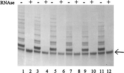
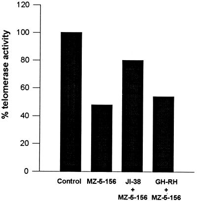
Telomerase activity in U-87MG glioblastomas xenografted into nude mice is shown in Table 1. Electrophoretic analyses of PCR-amplified telomerase extension products are illustrated in Fig. 1. Telomerase activity was assessed by using the TRAP assay and internal control (telomerase substrate–nontelomerase) for the estimation of the efficiency of the PCR was included in the analysis of each sample (see Materials and Methods). The results presented in Table 1 indicate that treatment of nude mice bearing xenografted U-87MG glioblastomas with the GH-RH antagonist MZ-5-156 significantly decreased telomerase activity after 4 weeks. The levels of telomerase activity, expressed in arbitrary units, were 5.1 ± 0.8 in the treated group as compared with 7.5 ± 0.3 in the control group (P < 0.05), which represents a decrease of 32% (Fig. 1, Table 1). When U-87MG glioblastomas were treated in vitro for 4 hr with 3 μM MZ-5-156, densitometric quantification of TRAP assay products showed that telomerase activity decreased by 52% (Fig. 2). Incubation of H-69 SCLC, H-23 non-SCLC, and MDA-MB-468 breast carcinoma cells with 3 μM MZ-5-156 for 4 hr produced 17%, 35%, and 45% inhibition, respectively.
Table 1.
Effect of the treatment with the GH-RH antagonist MZ-5-156 on the levels of telomerase activity and mRNA levels of hTRT, hTR, TP1, and c-myc genes in U-87MG glioblastomas xenografted into nude mice
| Treatment | Telomerase activity | mRNA levels
|
|||
|---|---|---|---|---|---|
| hTRT | TP1 | hTR | c-myc | ||
| Control | 7.5 ± 0.3 | 1.18 ± 0.008 | 4.5 ± 0.3 | 1.5 ± 0.1 | 1.5 ± 0.2 |
| MZ-5-156 | 5.1 ± 0.8* | 0.54 ± 0.08* | 3.9 ± 0.3 | 1.5 ± 0.1 | 1.2 ± 0.3 |
| % decrease | 32 | 54 | 12 | 0 | 18 |
Telomerase activity was assessed by the TRAP assay and expressed as the ratio of the total telomerase product versus the internal control telomerase substrate–nontelomerase. mRNA levels of hTRT, hTR, TP1 and c-myc were assessed by RT-PCR and normalized versus hGAPDH. All values are expressed in arbitrary units. The % decrease in the treated group vs. the controls also is indicated. The results were obtained by using three tumors.
*P < 0.05 vs. control.
Figure 1.
Effect of the GH-RH antagonist MZ-5-156 on the levels of telomerase activity of U-87MG glioblastomas xenografted into nude mice. PCR-amplified telomerase extension products (TRAP assay) are shown after electrophoresis on a 10% polyacrylamide gel. Lanes 1, 3, and 5, telomerase activity in untreated tumors; lanes 7, 9, and 11, telomerase activity of tumor samples from animals treated with MZ-5-156; lanes 2, 4, 6, 8, 10, and 12, telomerase activity in RNase-pretreated extracts from control and MZ-5-156-treated tumors, respectively. The arrow indicates the position of the internal control telomerase substrate–nontelomerase. Telomerase activity decreased significantly (P < 0.05) in the MZ-5-156-treated group (Table 1).
Figure 2.
Telomerase activity of U-87MG glioblastomas cultured in vitro without any treatment (control), treated with the MZ-5-156 (MZ-5-156), and pretreated with JI-38 (JI-38 + MZ-5-156) or GH-RH(1–29) (GH-RH + MZ-5-156) before the treatment with MZ-5-156. Telomerase activity was assessed by quantification of TRAP assay products after electrophoresis on a 10% polyacrylamide gel. Treatment with MZ-5-156 decreased telomerase activity to levels corresponding to 48% of the untreated cells whereas pretreatment with JI-38 restored telomerase activity to levels corresponding to 80% of the controls. Pretreatment with GH-RH(1–29) had no significant effect.
Effects of GH-RH Agonists on the Levels of Telomerase Activity.
Treatment of U-87MG glioblastomas, H-23 non-SCLC, and MDA-MB-468 breast carcinoma cells in vitro for 4 hr with 3 μM GH-RH(1–29)NH2, or a more potent GH-RH agonist JI-38, did not produce an increase in the levels of telomerase activity (data not shown). This finding could be the result of telomerase activity being expressed at very high levels, and further stimulation could not be achieved. However, when U-87MG glioblastomas were pretreated in vitro for 1 hr with 3 μM GH-RH agonist JI-38 before the incubation with equimolar concentration of MZ-5-156 for 4 hr, the inhibitory effect of the GH-RH antagonist on the telomerase activity was partially blocked (Fig. 2). While the treatment of U-87MG glioblastomas with MZ-5-156 resulted in 52% inhibition of telomerase activity, the pretreatment of these cells with agonist JI-38 decreased the inhibition to 20%. When the same experiment was repeated by using GH-RH(1–29)NH2 instead of agonist JI-38, the effect of MZ-5-156 was not blocked (Fig. 2).
Effects of MZ-5-156 on the mRNA Levels of the Genes Encoding for the Telomerase Subunits and the c-myc Protooncogene.
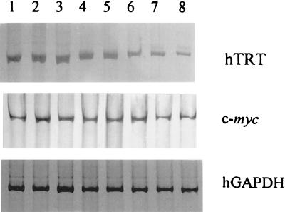
The levels of mRNA for hTRT, hTR, and TP1, which encode for the telomerase catalytic subunit, the telomerase RNA, and the telomerase-associated protein, respectively, as well as the levels of mRNA for c-myc, which regulates telomerase, also were analyzed in U-87MG glioblastomas xenografted into nude mice and chronically treated with MZ-5-156 (Table 1, Fig. 3). Densitometric analysis of RT-PCR products revealed that the hTRT mRNA was significantly decreased by 54% in the treated group as compared with levels in the control group (P < 0.05). A slight, but insignificant, decrease also was found in the mRNA levels of TP1 and c-myc mRNA (12% and 18%, respectively) (Fig. 3, Table 1). MZ-5-156 had no effect on the mRNA levels of hTR. In vitro treatment of U-87MG cells with 3 μM MZ-5-156 for 4 hr resulted in 21%, 5%, 11%, and 2% decreases in the levels of hTRT, TP1, hTR, and c-myc genes, respectively.
Figure 3.
Expression of hTRT, c-myc, and hGAPDH genes in U-87MG glioblastomas xenografted into nude mice or cultured in vitro and treated with MZ-5-156. Total RNA was extracted from the cells or the tumors, amplified by RT-PCR, and electrophoresed on 8% polyacrylamide gel. Lanes 1–3, untreated tumors; lanes 4–6, tumors treated in vivo with MZ-5-156; lane 7, U-87MG cells untreated; and lane 8, U-87MG cells treated in vitro with MZ-5-156. Densitometric quantification of the PCR products showed a 54% decrease (P < 0.05) in the mRNA levels of the hTRT gene in the group treated with MZ-5-156 in vivo as compared with those in the control group. Treatment in vitro of U-87MG glioblastomas with MZ-5-156 decreased the levels of hTRT mRNA by 21% as compared with the controls. MZ-5-156 had no significant effect on the mRNA levels of TP1, hTR, and c-myc after in vivo or in vitro treatment.
DISCUSSION
We previously have demonstrated that GH-RH antagonists, such as MZ-4-71 and MZ-5-156, inhibit growth of human prostatic and renal cancers, SCLC and non-SCLC, osteosarcomas, and other tumors (4–9). In the present study, we showed that human U-87MG glioblastomas xenografted into nude mice display lower levels of telomerase activity after in vivo treatment with the GH-RH antagonist MZ-5-156. A down-regulation of the hTRT gene, which encodes for the catalytic subunit of telomerase, also was found, whereas the levels of the telomerase RNA (hTR) and the mRNA for the telomerase-associated protein (TP1) remained essentially unaffected. Collectively, these findings suggest that the decrease in the levels of telomerase activity reported here can be attributed, at least partially, to the decreased mRNA levels of hTRT gene. This finding is in agreement with previous observations in normal and tumoral cells that established the hTRT gene as the limiting factor for telomerase activity (25, 30). However, a regulatory role for TP1 and hTR at the posttranscriptional level is still possible (28, 29, 40).
Chronic treatment of U-87MG glioblastomas with MZ-5-156 resulted in a significant tumor inhibition (unpublished work). It thus could be argued that the decrease in the levels of telomerase activity is a secondary consequence of tumor inhibition and not the result of a direct (causative) action of the GH-RH antagonist MZ-5-156. Although this possibility cannot be ruled out by the present study, the observation that a decrease in telomerase activity in U-87MG glioblastomas was found not only in vivo after a chronic treatment, but also in vitro after 4-hr exposure to MZ-5-156 argues in favor of the specificity of these findings. A decrease at the levels of telomerase activity after treatment with MZ-5-156 likewise was found in H-69 SCLC, H-23 non-SCLC, and MDA-MB-468 breast carcinoma cells cultured in vitro, suggesting that the involvement of telomerase in the mechanism of action of the GH-RH antagonists is not restricted to the U-87MG glioblastomas, but also occurs in other tumor types.
Treatment of U-87MG glioblastomas cultured in vitro, with GH-RH(1–29)NH2 or the more potent GH-RH agonist JI-38 failed to further increase the levels of telomerase activity, perhaps because the endogenous levels of telomerase activity in U-87MG cells were very high and could not be further stimulated. However, pretreatment with agonist JI-38 partially inhibited the decrease in the levels of telomerase activity by MZ-5-156, providing further evidence for the specificity of the effects of the GH-RH antagonists on telomerase activity (Fig. 2). The observation that agonist JI-38 failed to block completely the effect of the MZ-5-156 could be attributed to the fact that the affinity of the antagonist for the GH-RH binding sites was three times higher (10, 36).
The inhibitory effect of GH-RH antagonists on tumor growth involves the suppression of IGF-I and/or IGF-II levels or secretion (4–9). IGF-I and IGF-II may act as autocrine, paracrine, or endocrine growth factors in the development of various tumors. Among the effects of IGF-I at the subcellular level is the stimulation of the expression of the c-myc oncogene (41, 42). Thus, we also investigated whether the decrease in the levels of telomerase activity produced by the GH-RH antagonist MZ-5-156 involves the reduction in the expression of c-myc protooncogene. RT-PCR revealed that MZ-5-156 did not decrease significantly the levels of c-myc mRNA in U-87MG glioblastomas, either in vivo or in vitro. Considering that c-myc regulates telomerase in some tissues, the present observations indicate that the repression of hTRT, and consequently of the telomerase activity by MZ-5-156, involves a c-myc-independent mechanism, at least in U-87MG glioblastomas. However, these findings should be interpreted with caution because the posttranscriptional regulation of c-myc is also possible, as suggested by Wang et al. (32), who found that E6-induced alterations in Myc protein did not reflect changes in the abundance of the myc mRNA. In addition, the suppression of IGF-II by GH-RH antagonists, which is probably independent of the c-myc levels of expression, could be considered for the regulation of telomerase.
Telomerase is necessary for the continuous proliferation of the cancer cells and although a causative relationship between telomerase activity and neoplastic transformation has not been proven, the reactivation of telomerase appears to be an essential event in somatic cell immortalization. A decrease in the levels of telomerase activity would result in telomere instability and, after a certain number of cell divisions, in cell death. Although alternative mechanisms involving genetic recombination for the maintenance of telomeres in the cancer cells have been suggested (43–46), the inhibition of telomerase activity has been proposed as a target of cancer therapy (47).
The effect of MZ-5-156 at the levels of telomerase activity in U-87MG glioblastomas was higher in vitro than in vivo. Several reasons may account for this difference. The effects of the chronic treatment in vivo may be the result of decrease in the expression of genes downstream of the GH-RH antagonists, including hTRT, whereas the reduction in telomerase activity after in vitro treatment of the U-87MG cells with MZ-5-156 may reflect a rapid response of these cells to the GH-RH antagonist. Under in vivo conditions, a selection process in favor of the GH-RH antagonist-resistant cells may occur and cells with higher telomerase activity may have a proliferative advantage in the presence of a GH-RH antagonist. The doses used for the in vivo and in vitro treatment of the U-87MG glioblastomas were also different, and thus the results obtained may not be comparable.
Surprisingly, the stronger effect of MZ-5-156 on the levels of telomerase activity in vitro was not accompanied by a greater inhibition of the hTRT gene expression, as compared with the results obtained in vivo: hTRT mRNA levels decreased by only 21% after in vitro treatment with MZ-5-156 as compared with the 54% decrease in vivo. We may postulate that although hTRT is important for the regulation of telomerase activity, additional mechanism(s) also may be implicated, particularly in cases that involve a rapid response to exogenous signals, such as the growth inhibition signal mediated by MZ-5-156 and that may bypass the regulatory role of hTRT, at least during an initial period of treatment.
The present evaluation of telomerase activity was based on the effects of the GH-RH antagonist MZ-5-156 on tumor cells, particularly the U-87MG glioblastomas. However, the present findings could have implications for other pathological and physiological conditions as well. Although we failed to observe an induction in the levels of telomerase activity in cancer cell lines after treatment with GH-RH or its agonistic analog JI-38, probably for aforementioned reasons, it is possible that under certain physiological conditions and developmental stages, GH-RH may induce the expression of hTRT and consequently telomerase activity in normal cells. It already has been shown that an ectopic expression of the c-myc protooncogene in normal human cells is capable of inducing telomerase activity and extending the life span of these cells in vitro (32). Thus, the participation of telomerase in various processes controlled by GH-RH/GH axis also could be considered.
In conclusion, we have demonstrated that the antitumor effects of the GH-RH antagonist MZ-5-156 in U-87MG glioblastomas and probably in other tumor models such as H-69 SCLC, H-23 non-SCLC, and MDA-MB-468 breast carcinoma are accompanied by a decrease at the levels of telomerase activity in vivo and in vitro. We also have shown that the decrease in telomerase activity was associated with a decrease in the expression of hTRT gene, which encodes for the telomerase catalytic subunit. Nevertheless, the regulatory pathways of telomerase are complex and our observations must be considered as preliminary. However, our work suggests the merit of further investigations with GH-RH antagonists aimed at restoration of cellular senescence and designing new strategies for cancer therapy involving telomerase inhibition. A possible link between GH-RH, GH, and telomerase activity also should attract some attention.
Acknowledgments
We thank P. Armatis and H. Valerio for their technical assistance. We are grateful to Professor J. K. Field, Professor D. A. Spandidos, Dr. A. J. Lustig, and Dr. T. Reissmann for critical reading of the manuscript. This work was supported by the Medical Research Service of the Veterans Affairs Department and a grant from ASTA Medica (Frankfurt on Main, Germany) to Tulane University (to A.V.S.).
ABBREVIATIONS
- GH
growth hormone
- GH-RH
GH-releasing hormone
- hGAPDH
human glyceraldehyde-3-phosphate dehydrogenase
- IGF
insulin-like growth factor
- SCLC
small cell lung carcinoma
- TRAP
telomerase repeat amplification protocol
- hTRT
telomerase catalytic subunit
- hTR
telomerase RNA
- TP1
telomerase-associated protein
- RT
reverse transcription
References
- 1.Schally A V, Comaru-Schally A M. In: Growth Hormone Secretagogues in Clinical Practice. Bercu B B, Walker R F, editors. New York: Dekker; 1998. pp. 131–144. [Google Scholar]
- 2.Benlot C, Levy L, Fontanaud P, Roche A, Rouannet P, Joubert D. J Clin Endocrinol Metab. 1997;82:690–696. doi: 10.1210/jcem.82.2.3754. [DOI] [PubMed] [Google Scholar]
- 3.Kahan, Z., Arencibia, J. M., Csernus, V. J., Groot, K., Kineman, R. D., Robinson, W. R. & Schally, A. V. (1999) J. Clin. Endocrinol. Metab., in press. [DOI] [PubMed]
- 4.Schally A V, Kovács M, Toth K, Comaru-Schally A M. In: Growth Hormone Secretagogues in Clinical Practice. Bercu B B, Walker R F, editors. New York: Dekker; 1998. pp. 145–162. [Google Scholar]
- 5.Jungwirth A, Schally A V, Pinski J, Halmos G, Groot K, Armatis P, Vadillo-Buenfil M. Br J Cancer. 1997;75:1585–1592. doi: 10.1038/bjc.1997.271. [DOI] [PMC free article] [PubMed] [Google Scholar]
- 6.Pinski J, Schally A V, Jungwirth A, Groot K, Halmos G, Szepeshazi K, Zarandi M, Armatis P. Int J Oncol. 1996;9:1099–1105. doi: 10.3892/ijo.9.6.1099. [DOI] [PubMed] [Google Scholar]
- 7.Lamharzi N, Schally A V, Koppán M, Groot K. Proc Natl Acad Sci USA. 1998;95:8864–8869. doi: 10.1073/pnas.95.15.8864. [DOI] [PMC free article] [PubMed] [Google Scholar]
- 8.Jungwirth A, Schally A V, Pinski J, Groot K, Armatis P, Halmos G. Proc Natl Acad Sci USA. 1997;94:5810–5813. doi: 10.1073/pnas.94.11.5810. [DOI] [PMC free article] [PubMed] [Google Scholar]
- 9.Pinski J, Schally A V, Groot K, Halmos G, Szepeshazi K, Zarandi M, Armatis P. J Natl Cancer Inst. 1995;87:1787–1794. doi: 10.1093/jnci/87.23.1787. [DOI] [PubMed] [Google Scholar]
- 10.Zarandi M, Kovács M, Horváth J E, Toth K, Halmos G, Groot K, Nagy A, Kele Z, Schally A V. Peptides. 1997;18:423–430. doi: 10.1016/s0196-9781(96)00344-0. [DOI] [PubMed] [Google Scholar]
- 11.Toretsky J A, Helman L J. J Endocrinol. 1996;149:367–372. doi: 10.1677/joe.0.1490367. [DOI] [PubMed] [Google Scholar]
- 12.Westley B R, May F E B. Br J Cancer. 1995;72:1065–1066. doi: 10.1038/bjc.1995.465. [DOI] [PMC free article] [PubMed] [Google Scholar]
- 13.Daughaday W H. Endocrinology. 1990;127:1–4. doi: 10.1210/endo-127-1-1. [DOI] [PubMed] [Google Scholar]
- 14.Schally A V, Comaru-Schally A M. In: Cancer Medicine. 4th Ed. Holland J F, Frei E III, Bast R C Jr, Kufe D E, Morton D L, Weichselbaum R R, editors. Baltimore: Williams & Wilkins; 1997. pp. 1067–1085. [Google Scholar]
- 15.Watson J D. Nature (London) 1972;239:197–201. doi: 10.1038/newbio239197a0. [DOI] [PubMed] [Google Scholar]
- 16.Blackburn E H. Nature (London) 1990;350:569–573. doi: 10.1038/350569a0. [DOI] [PubMed] [Google Scholar]
- 17.Feng J, Funk W D, Wang S S, Weinrich A A A, Chiu C P, Adams R R, Chang E, Allsopp R C, Yu J, Le S, et al. Science. 1995;269:1236–1241. doi: 10.1126/science.7544491. [DOI] [PubMed] [Google Scholar]
- 18.Greider C W, Blackburn E H. Cell. 1985;43:405–413. doi: 10.1016/0092-8674(85)90170-9. [DOI] [PubMed] [Google Scholar]
- 19.Rowley P T. Cancer Invest. 1998;16:170–174. doi: 10.3109/07357909809050032. [DOI] [PubMed] [Google Scholar]
- 20.Bodnar A G, Ouellette M, Frolkis M, Holt S E, Chiu C P, Morin G B, Harley C B, Shay J W, Lichtsteiner S, Wright W E. Science. 1998;279:349–352. doi: 10.1126/science.279.5349.349. [DOI] [PubMed] [Google Scholar]
- 21.Sedivy J M. Proc Natl Acad Sci USA. 1998;95:9078–9081. doi: 10.1073/pnas.95.16.9078. [DOI] [PMC free article] [PubMed] [Google Scholar]
- 22.Kim N W, Piatyszek M A, Prowse K R, Harley C B, West M D, Ho P L C, Coviello G M, Wright W E, Weinrich S L, Shay J W. Science. 1994;266:2011–2015. doi: 10.1126/science.7605428. [DOI] [PubMed] [Google Scholar]
- 23.Shay J W, Bacchetti S. Eur J Cancer. 1997;33:787–791. doi: 10.1016/S0959-8049(97)00062-2. [DOI] [PubMed] [Google Scholar]
- 24.Shay J W, Wright W E. Curr Opin Oncol. 1996;8:66–71. doi: 10.1097/00001622-199601000-00012. [DOI] [PubMed] [Google Scholar]
- 25.Nakamura T M, Morin G B, Chapman K B, Weinrich S L, Andrews W H, Lingner J, Harley C B, Cech T R. Science. 1997;277:955–959. doi: 10.1126/science.277.5328.955. [DOI] [PubMed] [Google Scholar]
- 26.Meyerson M, Counter C M, Eaton E N, Ellisen L W, Steiner P, Caddle S D, Ziaugra L, Beijersbergen R L, Davidoff M J, Liu Q, et al. Cell. 1997;90:785–795. doi: 10.1016/s0092-8674(00)80538-3. [DOI] [PubMed] [Google Scholar]
- 27.Killian A, Bowtell D D, Abud H E, Hime G R, Venter D J, Keese P K, Duncan E L, Reddel R R, Jefferson R A. Hum Mol Genet. 1997;6:2011–2019. doi: 10.1093/hmg/6.12.2011. [DOI] [PubMed] [Google Scholar]
- 28.Harrington L, McPhail T, Mar V, Zhou W, Oulton R, Bass M B, Arruda I, Robinson M O Amgen EST Program. Science. 1997;275:973–977. doi: 10.1126/science.275.5302.973. [DOI] [PubMed] [Google Scholar]
- 29.Nakayama J, Saito M, Nakamura H, Matsuura A, Ishikawa F. Cell. 1997;88:875–884. doi: 10.1016/s0092-8674(00)81933-9. [DOI] [PubMed] [Google Scholar]
- 30.Takakura M, Kyo S, Kanaya T, Tanaka M, Inoue M. Cancer Res. 1998;58:1558–1561. [PubMed] [Google Scholar]
- 31.Fujimoto K, Takahashi M. Biochem Biophys Res Commun. 1997;241:775–781. doi: 10.1006/bbrc.1997.7806. [DOI] [PubMed] [Google Scholar]
- 32.Wang J, Xie L Y, Allan S, Beach D, Hannon G J. Genes Dev. 1998;12:1769–1774. doi: 10.1101/gad.12.12.1769. [DOI] [PMC free article] [PubMed] [Google Scholar]
- 33.Hiyama E, Hiyama K, Yokoyama T, Matsuura Y, Piatyszek M A, Shay J W. Nat Med. 1995;1:249–255. doi: 10.1038/nm0395-249. [DOI] [PubMed] [Google Scholar]
- 34.Sano T, Asai A, Mishima K, Fujimaki T, Kirino T. Br J Cancer. 1998;77:1633–1637. doi: 10.1038/bjc.1998.267. [DOI] [PMC free article] [PubMed] [Google Scholar]
- 35.Hiraga S, Ohnishi T, Izumoto S, Miyahara E, Kanemura Y, Matsumura H, Arita N. Cancer Res. 1998;58:2117–2125. [PubMed] [Google Scholar]
- 36.Izdebski J, Pinski J, Horvath J E, Halmos G, Groot K, Schally A V. Proc Natl Acad Sci USA. 1995;92:4872–4876. doi: 10.1073/pnas.92.11.4872. [DOI] [PMC free article] [PubMed] [Google Scholar]
- 37.Bradford M M. Anal Biochem. 1976;72:248–254. doi: 10.1016/0003-2697(76)90527-3. [DOI] [PubMed] [Google Scholar]
- 38.Gordon A W, Joyce C P, Gibbes R J, Bhanu K, Nelly A, Stromberg K. Cancer Lett. 1995;89:63–71. doi: 10.1016/0304-3835(95)90159-0. [DOI] [PubMed] [Google Scholar]
- 39.Bernard O, Cory S, Gerondakis S, Webb E, Adams J M. EMBO J. 1983;2:2375–2383. doi: 10.1002/j.1460-2075.1983.tb01749.x. [DOI] [PMC free article] [PubMed] [Google Scholar]
- 40.Soder A I, Hoare S F, Muir S, Going J J, Parkinson E K, Keith W N. Oncogene. 1997;14:1013–1021. doi: 10.1038/sj.onc.1201066. [DOI] [PubMed] [Google Scholar]
- 41.Banskota N K, Taub R, Zellner K, King G L. Mol Endocrinol. 1989;3:1183–1190. doi: 10.1210/mend-3-8-1183. [DOI] [PubMed] [Google Scholar]
- 42.Conover C A, Bale L K. Exp Cell Res. 1998;238:122–127. doi: 10.1006/excr.1997.3815. [DOI] [PubMed] [Google Scholar]
- 43.Wang S-S, Zakian V A. Nature (London) 1990;345:356–358. doi: 10.1038/345456a0. [DOI] [PubMed] [Google Scholar]
- 44.Bryan T M, Englezou A, Gupta J, Bacchetti S, Reddel R R. EMBO J. 1995;14:4240–4248. doi: 10.1002/j.1460-2075.1995.tb00098.x. [DOI] [PMC free article] [PubMed] [Google Scholar]
- 45.Bryan T M, Reddel R R. Eur J Cancer. 1997;33:767–773. doi: 10.1016/S0959-8049(97)00065-8. [DOI] [PubMed] [Google Scholar]
- 46.Li B, Lustig A J. Genes Dev. 1996;10:1310–1326. doi: 10.1101/gad.10.11.1310. [DOI] [PubMed] [Google Scholar]
- 47.Shay J W, Werbin H, Wright W E. Leukemia. 1996;10:1255–1261. [PubMed] [Google Scholar]


