

Abstract
One hundred twenty-four cases of external endometriosis and 95 cases of adenomyosis were analyzed. The two are clinically different diseases which have one feature in common—a reactive fibrosis to aberrant endometrial tissue. They are coexistent in about the same frequency as would result from a noncausal relationship.
The origin of external endometriosis from the epithelial “inclusion” cyst is considered proven histologically. This is the source of origin of most external endometriosis, although occasional involvement from regurgitated endometrium probably occurs. Both the endometrial and the serous cysts have a common parentage in this anlage.
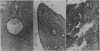
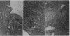
Certain histological features that are considered pathognomonic of endometriosis are: (1) the minimal lesion, (2) the characteristic cuboidal lined cyst, (3) the siderophagic cyst without lining, and (4) the siderophagic nest.
Recognition of the siderophagic nest will permit identification of extinct endometriosis and thus aid in studies to determine the spontaneous or therapeutically induced regression of the disease.
The coexistence of endometriosis with other pelvic pathological changes, notably carcinoma, indicates the need for further studies to search the possibility of relationship.
The ability of ectopic deposits of endometrium to become malignant on rare occasions would appear to be proven, but it is a rare occurrence and there is no justification for regarding endometriosis as a premalignant disease.
Full text
PDF


Images in this article
Selected References
These references are in PubMed. This may not be the complete list of references from this article.
- Meigs J. V. Endometriosis. Ann Surg. 1948 May;127(5):795–808. [PMC free article] [PubMed] [Google Scholar]