Abstract
In this study we investigated the role of the bacterial flagellum in Burkholderia pseudomallei entry to Acanthamoeba astronyxis trophozoites. B. pseudomallei cells were tethered to the external amoebic surface via their flagella. MM35, the flagellum-lacking fliC knockout derivative of B. pseudomallei NCTC 1026b did not demonstrate flagellum-mediated endocytosis in timed coculture, confirming that an intact flagellar apparatus assists B. pseudomallei entry into A. astronyxis.
Burkholderia pseudomallei is the etiologic agent of melioidosis, a potentially fatal infection endemic in tropical Australia and southeast Asia (1). B. pseudomallei is a facultative intracellular bacterial pathogen with the capacity to invade a variety of cell types including macrophages, in which the bacteria can survive for prolonged periods (7). Escape from sequestered cellular locations is thought to explain recrudescent and delayed-onset B. pseudomallei infection (12). In a recent study it was shown that B. pseudomallei entry into macrophages results in cellular effects similar to those caused by other intracellular bacteria (8).
In common with other intracellular bacterial pathogens such as Legionella pneumophila and Listeria monocytogenes (9, 11), B. pseudomallei has been shown to enter and survive within free-living amoebae belonging to the genus Acanthamoeba (6). We noted previously that an unusual form of tethered bacterial motility occurred shortly after B. pseudomallei cells attached to the amoebic trophozoite (6), prompting the present investigation into whether or not adherence by B. pseudomallei flagella was required to initiate bacterial entry into the amoebic trophozoite.
Bacterial strains and growth conditions.
The B. pseudomallei strains used in this investigation were NCTC 13177 (also known as WKo97, the cause of a small outbreak of melioidosis in Western Australia during 1997 [4, 5]), NCTC 1026b, and the flagellin-lacking mutant MM35, derived from NCTC 1026b (2). MM35 contains Tn5-OT182 integrated within the flagellin structural gene, fliC. The mutation is nonpolar and is complemented by cloned fliC in trans (2). These bacteria were stored in 20% glycerol at −70°C until required and were then recovered by culture onto fresh 5% horse blood agar at 37°C in air for 18 h. Five colonies were picked and used to inoculate 15 ml of fresh Trypticase soy broth (Excel Laboratory Products, Bentley, Western Australia) and incubated at 37°C in air for 18 h. A 1:10 dilution of this 18-h culture was then made in fresh Trypticase soy broth and incubated for a further 1 h 30 min in air at 37°C to obtain mid-lag-phase bacteria, as previously described (6).
Amoeba culture.
Acanthamoeba astronyxis (CCAP 1534/1) was maintained in axenic form in PYG broth (Excel Laboratory Products) in tissue culture flasks at 28°C. An aliquot of supernatant was harvested from the culture flask immediately before coculture procedures and concentrated by centrifugation at 1,000 × g for 5 min, and the supernatant was replaced with sterile 0.8% NaCl solution. The cell count was obtained by examining an aliquot of this suspension in a counting chamber (Fuchs-Rosenthal). This was adjusted by dilution to 105 cells/ml.
Coculture conditions.
Coculture of B. pseudomallei with A. astronyxis was performed by adding the mid-lag-phase bacterial suspension to the amoebic suspension either in a small volume (1.0 ml) for light microscopy or in larger volumes (10 ml) in capped tubes for electron microscope preparations. In the timed-coculture experiment, NCTC 1026b and MM35 were set up in parallel as coverslip preparations. These preparations were examined alternately, 20 consecutive cells were viewed on each occasion, and features previously described (6) were noted. In view of the negative results obtained with MM35, a coculture preparation of this strain was centrifuged onto amoebic trophozoites (3,000 × g for 15 min) and examined after 60 min at 20°C. All aerosol-generating procedures were conducted in a class II biological safety cabinet.
Electron microscopy.
One-, 5-, and 30-min coculture preparations were used for gold sputter coating. Ten-millimeter-diameter glass coverslips containing the organism were fixed in 2.5% glutaraldehyde in 0.05 M cacodylate buffer at pH 7.4 overnight, washed well in 0.05 M cacodylate buffer, postfixed in 1% aqueous osmium tetroxide, dehydrated through graded alcohol, and dried in a Polaron critical-point drying apparatus using superdry alcohol as the chamber liquid and liquid CO2 as the exchange gas. They were mounted on aluminum stubs with carbon conductive adhesive tape and coated with a 15-nm layer of gold and palladium in a Polaron E5100 sputter coating unit. An E5500 quartz crystal monitor was used to control the thickness of the metal coat. The studs were stored in a desiccating chamber containing silica gel until viewed in a XL30-TM scanning electron microscope (Philips, Eindhoven, The Netherlands).
Scanning electron microscopy of cocultures.
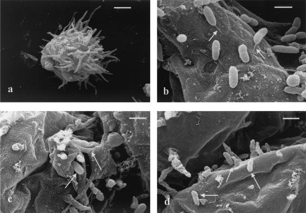
Scanning electron microscopy of A. astronyxis trophozoites demonstrated an external surface covered with tufts of filamentous pseudopodia or filopodia (Fig. 1a). In coculture preparations NCTC 13177 bacilli were found on the external trophozoite surface. Many bacilli were observed to demonstrate end-on adherence (Fig. 1b), and in several a thickened flagellum tethering the bacillus to the trophozoite external surface could be seen (Fig. 1c). Individual bacilli were also seen in clefts between extensions of the trophozoite surface (Fig. 1d). NCTC 1026b exhibited the same series of interactions with A. astronyxis (results not shown).
FIG. 1.
Scanning electron microscope views of B. pseudomallei and A. astronyxis coculture. (a) A. astronyxis trophozoite prior to inoculation with B. pseudomallei NCTC 13177. Scale bar = 10 μm. (b) Trophozoite surface covered with adherent B. pseudomallei NCTC 13177 cells, some of whose flagella (arrows) are visible. Scale bar = 2 μm. (c) B. pseudomallei NCTC 13177 cells (arrows) tucked in folds of the trophozoite surface during phagocytosis. Scale bar = 2 μm. (d) B. pseudomallei NCTC 13177 cells (arrows) remaining on the external trophozoite surface showing end-on adherence. Scale bar = 2 μm.
Phase-contrast microscopy of coculture.
In a timed coculture, NCTC 1026b followed the sequence of cellular events noted previously for other strains of B. pseudomallei (6), including flagellar adherence, tethered motility, incorporation into single-bacillus vacuoles, and formation of tufts of bacilli on the trophozoite surface (Table 1). Tethered motility was inferred from the high-frequency rotation of a bacillus around a fixed point on the external amoebic surface in a helicopter blade-like motion. Bacillary tufts were noted when a cluster of five or more bacilli were observed in parallel alignment with each other, all adhering end-on to the amoebic surface. The flagellin-negative derivative MM35 did not demonstrate any of these phenomena during the period of observation in the series of five repeat experiments (Table 1). During the first 60-min observation, MM35 was observed in single-bacillus vacuoles in 5 of the 100 trophozoites examined. No multibacillary vacuoles were seen. When the MM35 preparation was examined after a 24-h incubation, intracellular bacilli were noted in unibacillary vacuoles in only 7 of the total of 100 trophozoites examined (five preparations, each of 20 cells). Only two multibacillary vacuoles were seen at 24 h, and each of these contained only two bacilli. Repetition of the coculture with MM35 precipitated onto optimally receptive A. astronyxis trophozoites by centrifugation did not increase uptake over a 60-min observation period.
TABLE 1.
Cellular outcomes of B. pseudomallei cocultured with A. astronyxis
| B. pseudomallei strain | Time (min) | In amoebic trophozoitesa no. of:
|
||||||
|---|---|---|---|---|---|---|---|---|
| Adherent bacillib | Rotating bacillic | Single-bacillus vacuolesd | Multibacillary vacuolese | Bacillary tuftsf | Bacilli in chainsg | Bacillary tanglesh | ||
| NCTC 1026b | 0 | 9.4 ± 4.83 | 3.8 ± 3.6 | 0.8 ± 0.8 | 0.00 ± 0.00 | 0.00 ± 0.00 | 0.00 ± 0.00 | 0.00 ± 0.00 |
| 20 | 11.8 ± 3.35 | 5.2 ± 1.3 | 7.2 ± 2.6 | 10.4 ± 4.4 | 0.6 ± 0.9 | 0.00 ± 0.00 | 0.00 ± 0.00 | |
| 40 | 10.4 ± 2.9 | 5.0 ± 1.4 | 4.8 ± 1.3 | 12.8 ± 2.8 | 0.8 ± 0.8 | 0.00 ± 0.00 | 0.00 ± 0.00 | |
| 60 | 6.4 ± 3.0 | 1.8 ± 1.6 | 4.0 ± 0.7 | 13.6 ± 2.3 | 0.6 ± 0.9 | 0.00 ± 0.00 | 0.00 ± 0.00 | |
| 1,440 | 17.4 ± 3.1 | 15.4 ± 5.1 | 2.4 ± 2.2 | 6.2 ± 2.6 | 5.2 ± 2.7 | 2.2 ± 2.0 | 1.6 ± 3.0 | |
| MM35 | 0 | 0.00 ± 0.00 | 0.00 ± 0.00 | 0.00 ± 0.00 | 0.00 ± 0.00 | 0.00 ± 0.00 | 0.00 ± 0.00 | 0.00 ± 0.00 |
| 20 | 0.00 ± 0.00 | 0.00 ± 0.00 | 0.20 ± 0.45 | 0.00 ± 0.00 | 0.00 ± 0.00 | 0.00 ± 0.00 | 0.00 ± 0.00 | |
| 40 | 0.00 ± 0.00 | 0.00 ± 0.00 | 0.80 ± 0.45 | 0.00 ± 0.00 | 0.00 ± 0.00 | 0.00 ± 0.00 | 0.00 ± 0.00 | |
| 60 | 0.00 ± 0.00 | 0.00 ± 0.00 | 0.00 ± 0.00 | 0.00 ± 0.00 | 0.00 ± 0.00 | 0.00 ± 0.00 | 0.00 ± 0.00 | |
| 1,440 | 0.20 ± 0.45 | 0.00 ± 0.00 | 1.40 ± 1.52 | 0.40 ± 0.90 | 0.00 ± 0.00 | 0.00 ± 0.00 | 0.00 ± 0.00 | |
At each time interval, 20 amoebic trophozoites were examined by phase-contrast microscopy for the presence of each feature indicated. Each series of observations was repeated with a total of five complete cocultures for both B. pseudomallei strains. Values shown are the means ± standard deviations of the numbers of trophozoites exhibiting each feature from five consecutive replicate experiments.
Bacilli adhering to the trophozoite surface via the bacillus end.
One or more bacilli in tethered high-frequency rotation around a point on the external trophozoite surface.
One or more trophozoite vacuoles containing single bacilli.
One or more trophozoite vacuoles containing more than one distinct bacillus.
Five or more bacilli in a single tuft of parallel bacilli adhering to the external trophozoite surface.
Five or more bacilli in one or more chains of bacilli adherent to the external trophozoite surface.
Bacilli in a tangled web of chains external to the trophozoite or cyst surface.
The role of the B. pseudomallei flagellum in adherence to phagocytic eukaryotic cells was inferred from a previous subjective observation (6). These preliminary observations were insufficient to establish that the flagellum of B. pseudomallei was necessary for incorporation in an amoebic endocytic vacuole and did not establish a causal relationship between bacterial adherence and eukaryotic cellular invasion.
The results of the present study provide more-direct evidence for the involvement of the B. pseudomallei flagellum in eukaryotic cellular invasion. The striking difference in amoeba-invasive capacity between B. pseudomallei strain NCTC 1026b and the corresponding flagellin-negative motility mutant, MM35, supports a critical role for the flagellum in the early stages of cellular invasion. The lack of motility in MM35 reduces the normally observed frequency of encounter between rapidly motile, mid-lag-phase B. pseudomallei and receptive A. astronyxis trophozoites, but centrifugation of bacteria onto the amoebae did not improve bacterial uptake. The comprehensive lack of other cellular events such as formation of vacuoles containing bacilli, bacterial escape, and tuft formation with strain MM35 indicates that tethered motility is the first in a series of interactions between B. pseudomallei and A. astronyxis. Further study of flagellum-mediated adherence by B. pseudomallei may therefore assist in the selection of suitable targets for subsequent development of antiadhesion vaccine candidates.
We note with interest a recent report that Aeromonas caviae requires motility and flagellar function to maximize adherence to human epithelial cells (10). Other bacterial pathogens appear to employ a flagellar-adhesion strategy. In a recently published report, a nonflagellate Legionella pneumophila flaA mutant was used to demonstrate that the Legionella flagellum was required for invasion of HL-40 cells and Acanthamoeba castellanii trophozoites but not for adherence (3). That study centrifuged bacteria onto the amoebic cells, a procedure that may have obscured bacterial adherence that depends on delicate structures such as flagellar tips. Nevertheless, their results imply a cellular interaction different from that between B. pseudomallei and A. astronyxis, in which flagellar adherence is a necessary precursor to invasion. Detailed comparison of these two bacterium-eukaryote pairs may provide useful insights into the cellular mechanisms involved.
In conclusion, our investigation of the interaction between B. pseudomallei and A. astronyxis trophozoites supports a critical role for the bacterial flagellum in the early stages of cellular invasion. Further work is needed to characterize the nature of the eukaryotic cell surface receptor, the signaling events that trigger cytoskeletal rearrangements in the amoeba, the molecular basis of this interaction, and the extent of parallels with B. pseudomallei-mammalian phagocytic cell systems. The B. pseudomallei-A. astronyxis system provides a useful model with which to explore the cellular pathogenesis of melioidosis.
Editor: J. D. Clements
REFERENCES
- 1.Dance, D. A. 2000. Melioidosis as an emerging global problem. Acta Trop. 74:115-119. [DOI] [PubMed] [Google Scholar]
- 2.DeShazer, D., P. J. Brett, R. Carlyon, and D. E. Woods. 1997. Mutagenesis of Burkholderia pseudomallei with Tn5-OT182: isolation of motility mutants and molecular characterization of the flagellin structural gene. J. Bacteriol. 179:2116-2125. [DOI] [PMC free article] [PubMed] [Google Scholar]
- 3.Dietrich, C., K. Heuner, B. C. Brand, J. Hacker, and M. Steinert. 2001. Flagellum of Legionella pneumophila positively affects the early phase of infection of eukaryotic host cells. Infect. Immun. 69:2116-2122. [DOI] [PMC free article] [PubMed] [Google Scholar]
- 4.Inglis, T. J. J., S. C. Garrow, C. Adams, M. Henderson, M. Mayo, and B. J. Currie. 1999. Acute melioidosis outbreak in Western Australia. Epidemiol. Infect. 123:437-443. [DOI] [PMC free article] [PubMed] [Google Scholar]
- 5.Inglis, T. J. J., S. C. Garrow, M. Henderson, A. Clair, J. Sampson, L. O'Reilly, and B. Cameron. 2000. Burkholderia pseudomallei traced to water treatment plant in Australia. Emerg. Infect. Dis. 6:56-59. [DOI] [PMC free article] [PubMed] [Google Scholar]
- 6.Inglis, T. J. J., P. Rigby, T. A. Robertson, N. S. Dutton, M. Henderson, and B. J. Chang. 2000. Interaction between Burkholderia pseudomallei and Acanthamoeba species results in coiling phagocytosis, endamoebic bacterial survival, and escape. Infect. Immun. 68:1681-1686. [DOI] [PMC free article] [PubMed] [Google Scholar]
- 7.Jones, A. L., T. J. Beveridge, and D. E. Woods. 1996. Intracellular survival of Burkholderia pseudomallei. Infect. Immun. 64:782-790. [DOI] [PMC free article] [PubMed] [Google Scholar]
- 8.Kespichayawattana, W., S. Rattanachetkul, T. Wanun, P. Utaisincharoen, and S. Sirisinha. 2000. Burkholderia pseudomallei induces cell fusion and action-associated membrane protrusion: a possible mechanism for cell-to-cell spreading. Infect. Immun. 68:5377-5384. [DOI] [PMC free article] [PubMed] [Google Scholar]
- 9.Ly, T. M., and H. E. Muller. 1990. Ingested Listeria monocytogenes survive and multiply in protozoa. J. Med. Microbiol. 33:51-54. [DOI] [PubMed] [Google Scholar]
- 10.Rabaan, A. A., I. Gryllos, J. M. Tomas, and J. G. Shaw. 2001. Motility and the polar flagellum are required for Aeromonas caviae adherence to Hep-2 cells. Infect. Immun. 69:4257-4267. [DOI] [PMC free article] [PubMed] [Google Scholar]
- 11.Rowbotham, T. 1980. Preliminary report on the pathogenicity of Legionella pneumophila for freshwater and soil amoebae. J. Clin. Pathol. 33:1179-1183. [DOI] [PMC free article] [PubMed] [Google Scholar]
- 12.Wong, K. T., S. D. Puthucheary, and J. Vadivelu. 1995. The histopathology of human melioidosis. Histopathology 26:51-55. [DOI] [PubMed] [Google Scholar]


