

Abstract
Genotoxic agents induce chromosomal alterations, such as aberrations, micronuclei, and sister chromatid exchanges as well as mutations both in vivo and in vitro. Ionizing radiation and typical radiomimmetic agents such as bleomycin are very efficient inducers of chromosomal aberrations. The type of aberrations induced by these agents are cell-cycle dependent, i.e., chromosome type in pre-replication stages and chromatid type in post-replication stages of the cell cycle. Under optimal DNA repair conditions, DNA double-strand breaks (DSBs) appear to be the most important lesion responsible for the production of aberrations. In human lymphocytes, fast-repairing DSBs lead to exchange-type aberrations. The fact that the dose-response curves for induction of exchange aberrations induced by ionizing radiation are similar in vitro and in vivo allows one to use the yield of induced aberrations to estimate absorbed radiation dose in the case of accidents. In this respect, frequencies of translocations detected by the chromosome painting technique appear to be more sensitive. Mutations do not express immediately after exposure and require an expression time before they can be detected. In humans, it is estimated that for the mutations induced in bone marrow, it takes about 2 months for them to express and to be detected in peripheral blood lymphocytes. Hence, frequency of mutations is of limited value for estimating radiation doses immediately after an accident. This holds true for chemical exposure as well.(ABSTRACT TRUNCATED AT 250 WORDS)
Full text
PDF
Selected References
These references are in PubMed. This may not be the complete list of references from this article.
- Albertini R. J., Nicklas J. A., O'Neill J. P., Robison S. H. In vivo somatic mutations in humans: measurement and analysis. Annu Rev Genet. 1990;24:305–326. doi: 10.1146/annurev.ge.24.120190.001513. [DOI] [PubMed] [Google Scholar]
- Bryant P. E. Enzymatic restriction of mammalian cell DNA using Pvu II and Bam H1: evidence for the double-strand break origin of chromosomal aberrations. Int J Radiat Biol Relat Stud Phys Chem Med. 1984 Jul;46(1):57–65. doi: 10.1080/09553008414551061. [DOI] [PubMed] [Google Scholar]
- Carrano A. V., Natarajan A. T. International Commission for Protection Against Environmental Mutagens and Carcinogens. ICPEMC publication no. 14. Considerations for population monitoring using cytogenetic techniques. Mutat Res. 1988 Mar;204(3):379–406. doi: 10.1016/0165-1218(88)90036-5. [DOI] [PubMed] [Google Scholar]
- Cremer T., Popp S., Emmerich P., Lichter P., Cremer C. Rapid metaphase and interphase detection of radiation-induced chromosome aberrations in human lymphocytes by chromosomal suppression in situ hybridization. Cytometry. 1990;11(1):110–118. doi: 10.1002/cyto.990110113. [DOI] [PubMed] [Google Scholar]
- Fenech M., Morley A. A. Measurement of micronuclei in lymphocytes. Mutat Res. 1985 Feb-Apr;147(1-2):29–36. doi: 10.1016/0165-1161(85)90015-9. [DOI] [PubMed] [Google Scholar]
- Langlois R. G., Bigbee W. L., Kyoizumi S., Nakamura N., Bean M. A., Akiyama M., Jensen R. H. Evidence for increased somatic cell mutations at the glycophorin A locus in atomic bomb survivors. Science. 1987 Apr 24;236(4800):445–448. doi: 10.1126/science.3563520. [DOI] [PubMed] [Google Scholar]
- Lucas J. N., Tenjin T., Straume T., Pinkel D., Moore D., 2nd, Litt M., Gray J. W. Rapid human chromosome aberration analysis using fluorescence in situ hybridization. Int J Radiat Biol. 1989 Jul;56(1):35–44. doi: 10.1080/09553008914551161. [DOI] [PubMed] [Google Scholar]
- Natarajan A. T., Darroudi F., Mullenders L. H., Meijers M. The nature and repair of DNA lesions that lead to chromosomal aberrations induced by ionizing radiations. Mutat Res. 1986 May;160(3):231–236. doi: 10.1016/0027-5107(86)90132-6. [DOI] [PubMed] [Google Scholar]
- Natarajan A. T., Obe G. Molecular mechanisms involved in the production of chromosomal aberrations. I. Utilization of Neurospora endonuclease for the study of aberration production in G2 stage of the cell cycle. Mutat Res. 1978 Oct;52(1):137–149. doi: 10.1016/0027-5107(78)90102-1. [DOI] [PubMed] [Google Scholar]
- Natarajan A. T., Obe G. Molecular mechanisms involved in the production of chromosomal aberrations. III. Restriction endonucleases. Chromosoma. 1984;90(2):120–127. doi: 10.1007/BF00292448. [DOI] [PubMed] [Google Scholar]
- Natarajan A. T., Obe G., van Zeeland A. A., Palitti F., Meijers M., Verdegaal-Immerzeel E. A. Molecular mechanisms involved in the production of chromosomal aberrations. II. Utilization of Neurospora endonuclease for the study of aberration production by X-rays in G1 and G2 stages of the cell cycle. Mutat Res. 1980 Feb;69(2):293–305. doi: 10.1016/0027-5107(80)90094-9. [DOI] [PubMed] [Google Scholar]
- Natarajan A. T., Ramalho A. T., Vyas R. C., Bernini L. F., Tates A. D., Ploem J. S., Nascimento A. C., Curado M. P. Goiania radiation accident: results of initial dose estimation and follow up studies. Prog Clin Biol Res. 1991;372:145–153. [PubMed] [Google Scholar]
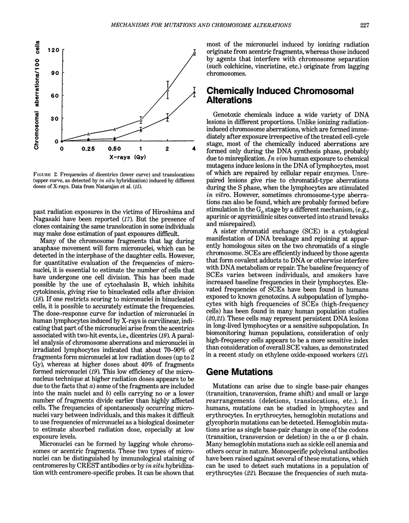
- Natarajan A. T., Vyas R. C., Darroudi F., Vermeulen S. Frequencies of X-ray-induced chromosome translocations in human peripheral lymphocytes as detected by in situ hybridization using chromosome-specific DNA libraries. Int J Radiat Biol. 1992 Feb;61(2):199–203. doi: 10.1080/09553009214550821. [DOI] [PubMed] [Google Scholar]
- Natarajan A. T., Vyas R. C., Wiegant J., Curado M. P. A cytogenetic follow-up study of the victims of a radiation accident in Goiania (Brazil). Mutat Res. 1991 Mar;247(1):103–111. doi: 10.1016/0027-5107(91)90038-p. [DOI] [PubMed] [Google Scholar]
- Ramalho A., Sunjevaric I., Natarajan A. T. Use of the frequencies of micronuclei as quantitative indicators of X-ray-induced chromosomal aberrations in human peripheral blood lymphocytes: comparison of two methods. Mutat Res. 1988 Mar-Apr;207(3-4):141–146. doi: 10.1016/0165-7992(88)90078-4. [DOI] [PubMed] [Google Scholar]
- Rossi A. M., Thijssen J. C., Tates A. D., Vrieling H., Natarajan A. T., Lohman P. H., van Zeeland A. A. Mutations affecting RNA splicing in man are detected more frequently in somatic than in germ cells. Mutat Res. 1990 Aug;244(4):353–357. doi: 10.1016/0165-7992(90)90084-w. [DOI] [PubMed] [Google Scholar]
- Tates A. D., Grummt T., Törnqvist M., Farmer P. B., van Dam F. J., van Mossel H., Schoemaker H. M., Osterman-Golkar S., Uebel C., Tang Y. S. Biological and chemical monitoring of occupational exposure to ethylene oxide. Mutat Res. 1991 Sep-Oct;250(1-2):483–497. doi: 10.1016/0027-5107(91)90205-3. [DOI] [PubMed] [Google Scholar]
- Tates A. D., van Dam F. J., van Mossel H., Schoemaker H., Thijssen J. C., Woldring V. M., Zwinderman A. H., Natarajan A. T. Use of the clonal assay for the measurement of frequencies of HPRT mutants in T-lymphocytes from five control populations. Mutat Res. 1991 Oct;253(2):199–213. doi: 10.1016/0165-1161(91)90133-s. [DOI] [PubMed] [Google Scholar]
- Vrieling H., Thijssen J. C., Rossi A. M., van Dam F. J., Natarajan A. T., Tates A. D., van Zeeland A. A. Enhanced hprt mutant frequency but no significant difference in mutation spectrum between a smoking and a non-smoking human population. Carcinogenesis. 1992 Sep;13(9):1625–1631. doi: 10.1093/carcin/13.9.1625. [DOI] [PubMed] [Google Scholar]
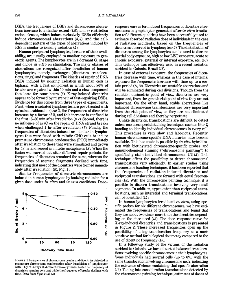
- Vyas R. C., Darroudi F., Natarajan A. T. Radiation-induced chromosomal breakage and rejoining in interphase-metaphase chromosomes of human lymphocytes. Mutat Res. 1991 Jul;249(1):29–35. doi: 10.1016/0027-5107(91)90130-g. [DOI] [PubMed] [Google Scholar]