Most bacteria can, at least to some degree, hunker down during periods of stress and wait for good times to return. No cells, however, do this as effectively as those Bacilli and Clostridia that form spores. Dormant cells produced by these bacteria can survive most environmental challenges found on earth and even a few in outer space and can remain dormant in excess of millions of years (1, 2). Nonetheless, when suitable conditions are present once again, spores rapidly germinate and resume vegetative growth.
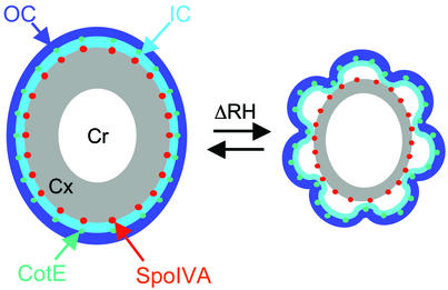
The spore is composed of a set of protective structures arranged in a series of concentric shells (Fig. 1); each component contributes in some essential way to spore durability. Key functions of these structures include locking the DNA into a stable, relatively desiccated crystalline state and excluding toxic molecules via an armored external shell (3). The conception of the spore as a dormant, dry, and hardened vehicle designed to preserve DNA has strongly suggested the notion of a cell that is largely static. In this view, the spore has no moving parts; dormancy depends on being inert. What a surprise, then, to learn from Westphal et al. (4) that Bacillus thuringiensis spore dimensions can change significantly depending on relative humidity, a parameter that is likely to vary in natural environments where spores are found, such as the soil. The authors used a novel automated technique, formerly applied to nonbiological problems such as relativistic heavy ion physics, to nondestructively measure spore dimensions. Their technique gives extremely accurate relative size information and, importantly, allows them to collect multiple measurements on the same spore as environmental conditions are varied. They show that swelling occurs in two phases, the first taking place in less than 1 min, and the second requiring about 8 min. These two phases may reflect different rates of entry of water into different compartments within the spore. To better appreciate the novelty of this finding and the manner in which it obligates us to revise our understanding of the spore, I will review the assembly and function of the spore structures that allow it to resist environmental stress.
Figure 1.
Morphological consequences of changes in relative humidity. The layers comprising the spore are color-coded: white, core (Cr); gray, cortex (Cx); light blue, inner coat (IC); dark blue, outer coat (OC). RH, relative humidity. The exosporium is not illustrated, for simplicity. Thin-section electron microscopic analysis shows significant variation in the degree of folds in the coat, reflected in the two cartoons (the folds in the right spore coat are somewhat exaggerated for clarity). Possibly, the ability of the coat to fold and unfold permits changes in spore size as relative humidity varies.
Resistance depends on three spore substructures: the core, cortex, and coat. The interior compartment of the spore, the core, houses the DNA, which is complexed with small acid-soluble proteins (5). The core is surrounded by a membrane and then a thick layer of specialized, loosely cross linked peptidoglycan called the cortex (6). Finally, the cortex is encased in a multilayered protein shell called the coat (7–10).
In some species, including Bacillus anthracis and its close relatives Bacillus cereus and B. thuringiensis, the spore is enclosed in an additional relatively poorly studied external structure known as the exosporium (11–13). The exosporium does not directly abut the coat. Rather, a gap separates the two structures. The nature of the material in the gap, if any, is unknown. No role in resistance for the exosporium has been documented. Because it is absent in species, such as Bacillus subtilis, which tolerate a wide range of environmental stresses, the exosporium may have more to do with accommodation to specific niches or lifestyles than with general protection or germination.
The tight girdle formed by the cortex acts to keep the DNA in the core relatively dry. This desiccation, in turn, helps preserve the DNA during long periods of spore dormancy. Because the cortex has a structure resembling a woven fabric, small molecules, such as water, can pass through it. Despite this, the core remains dry, in large part due to the squeezing action of the cortex on the core. Although the view that the cortex does nothing other than constrain the core volume is appealing in its simplicity, it is probably incomplete. For example, the degree of crosslinking of the cortex peptidoglycan strands varies along the spore radius. This variation could alter the flexibility of the cortex as well as its ability to expand and contract in response to ionic changes. Although it was initially proposed that this variation contributed to core dehydration (14), later experiments using peptidoglycan biosynthesis mutants with altered crosslinking revealed that when the gradient of crosslinking is absent, dehydration is normal (15) (see discussion in ref. 6). This result can be taken as a clue that the role of the cortex is more complex than has been appreciated so far.
Surrounding the cortex is the coat. The coat has been deeply studied only in the model organism B. subtilis. By thin-section electron microscopy, the B. subtilis coat can be seen to possess two major layers (10, 16): a lightly staining, finely striated inner coat and a more darkly staining coarsely layered outer coat. Frequently, the outer circumference of the spore appears scalloped, due to folds in the coat (exaggerated in Fig. 1). The coat's morphological complexity is mirrored by the complexity of the coat's polypeptide composition. Genetic analysis has revealed over 25 coat proteins, some of which have been subject to detailed analysis (8, 9). More recently, two proteomic studies have identified over 20 strong candidate coat proteins (17, 18).
The coat apparently has a variety of functions. It has long been known to serve as a barrier against entry of large toxic molecules, such as lysozyme, and to have a role in germination (16). The more recent identification of a germination-specific protein (19) in the coat should help to clarify its role in germination, and the discovery of coat protein oxidases (18, 20, 21) points to additional active roles that have yet to be elucidated.
Most coat proteins appear to be dispensable for the coat's barrier functions, as measured in the laboratory, because the deletion of any one of the corresponding genes has no detectable consequence. Two important exceptions are SpoIVA and CotE. SpoIVA resides between the coat and cortex (22, 23). As this location suggests, SpoIVA connects the coat to the spore surface; without it, the coat is built but is not attached to the spore (24–26). CotE is positioned at the inner coat/outer coat interface (22). It is responsible for the assembly of the entire outer coat as well as several inner coat proteins (27–30). Several other proteins have more intermediate roles. The best studied of these proteins are SpoVID and SafA, which are likely to participate in early stages of coat assembly (31–35).
Both the cortex and coat possess a degree of complexity that is surprising if these structures have only the simple protective roles assigned to them historically. As discussed above, the cortex has variations in crosslinking density that are dispensable for maintaining dehydration, at least in the laboratory. Similarly, the coat possesses proteins that seem to be dispensable for resistance to known environmental assaults. These observations strongly suggest that both structures have as-yet-undiscovered activities. It is in this context that the results of Westphal et al. (4) are particularly illuminating. They reveal that the spore is capable of relatively rapid expansion and contraction without breaking dormancy. Therefore, the exosporium, the coat, and/or the cortex are sufficiently flexible to accommodate these volume changes without impairing spore integrity. The details of how this is accomplished are not obvious and will be fascinating to discover. The exosporium is not readily detected by light microscopy, and it is plausible that it has little impact on the measurements of spore dimensions reported by Westphal et al. (4). Among the questions raised, therefore, is whether the gradient of crosslinking in the cortex somehow permits it to swell in conditions of high humidity without compromising core dryness. Likewise, the process by which the tough coat tolerates volume changes represents an intriguing problem.
The spore is capable of relatively rapid expansion and contraction without breaking dormancy.
An important clue to a possible solution comes from classical studies of germination in B. subtilis. The hallmark of germination is rehydration of the core, resulting in an increase in spore volume (36). During this process, the folds in the dormant spore coat disappear. It is reasonable to suppose that the mechanical properties of the coat that permit this apparent unfolding are the same as those permitting expansion of spore dimensions in conditions of high relative humidity. This idea is illustrated by Fig. 1. The two cartoons represent the extremes of folding of the coat seen in electron micrographs. In the speculative view presented here, a decrease in cortex volume allows the coat to adopt a more folded state. Possibly, the two phases of swelling detected by the authors reflect a mechanical property of coat unfolding. As Westphal et al. note (4), they now have the opportunity to identify mutants defective in dynamic size changes and, therefore, to begin to identify molecular determinants of coat and cortex flexibility.
Efforts to discover determinants of coat plasticity may be aided by recent collaborative work between my own laboratory and that of R. Wang (Illinois Institute of Technology). We used atomic force microscopy (AFM) to show that the formation of ridges in the coat surface is altered (but not entirely eliminated) by mutations in any of three coat protein genes. We infer that the ridges correspond to the folds in the coat seen by thin-section electron microscopy. Most likely, building a ridge involves several coat proteins acting together. Our AFM analysis dovetails nicely with the work of Westphal et al. (4), suggesting that changes in spore size in response to varying relative humidity require properly formed ridges.
The discovery that spore dimensions change under conditions that likely fluctuate in natural environments suggests that coat flexibility is probably widespread among spore-formers. Genome sequence data on Bacilli and Clostridia are still too sparse to estimate how similar the coat protein compositions of these organisms are, in general. Nonetheless, the currently available data suggest that coat composition as well as structure can vary considerably among these diverse species. Taken together with our AFM analysis, one might speculate that coat flexibility is not the result of just one or a few specific coat protein species and is selected at the level of the entire coat.
Westphal et al. (4) have developed a particularly sophisticated and ecologically realistic system in which to study spore responses to the environment. Their striking findings should encourage others in the field to continue development of novel assays to measure other dynamic parameters of the natural environments in which spores are found.
Acknowledgments
I gratefully acknowledge very helpful comments from David Popham, David Keating, and members of my department. My work is supported by Grants GM53989 and AI53365 from the National Institutes of Health.
Footnotes
See companion article on page 3461.
References
- 1.Nicholson W L. Cell Mol Life Sci. 2002;59:410–416. doi: 10.1007/s00018-002-8433-7. [DOI] [PMC free article] [PubMed] [Google Scholar]
- 2.Nicholson W L, Munakata N, Horneck G, Melosh H J, Setlow P. Microbiol Mol Biol Rev. 2000;64:548–572. doi: 10.1128/mmbr.64.3.548-572.2000. [DOI] [PMC free article] [PubMed] [Google Scholar]
- 3.Driks A, Setlow P. In: Prokaryotic Development. Brun Y V, Shimkets L J, editors. Washington, DC: Am. Soc. Microbiol.; 2000. pp. 191–218. [Google Scholar]
- 4.Westphal A J, Price P B, Leighton T J, Wheeler K E. Proc Natl Acad Sci USA. 2003;100:3461–3466. doi: 10.1073/pnas.232710999. [DOI] [PMC free article] [PubMed] [Google Scholar]
- 5.Setlow P. Annu Rev Microbiol. 1995;49:29–54. doi: 10.1146/annurev.mi.49.100195.000333. [DOI] [PubMed] [Google Scholar]
- 6.Popham D L. Cell Mol Life Sci. 2002;59:426–433. doi: 10.1007/s00018-002-8435-5. [DOI] [PMC free article] [PubMed] [Google Scholar]
- 7.Takamatsu H, Watabe K. Cell Mol Life Sci. 2002;59:434–444. doi: 10.1007/s00018-002-8436-4. [DOI] [PMC free article] [PubMed] [Google Scholar]
- 8.Henriques A O, Moran C P., Jr Methods. 2000;20:95–110. doi: 10.1006/meth.1999.0909. [DOI] [PubMed] [Google Scholar]
- 9.Driks A. In: Bacillus subtilis and Its Closest Relatives. Sonenshein A L, Hoch J A, Losick R, editors. Washington, DC: Am. Soc. Microbiol.; 2002. pp. 527–536. [Google Scholar]
- 10.Driks A. Microbiol Mol Biol Rev. 1999;63:1–20. doi: 10.1128/mmbr.63.1.1-20.1999. [DOI] [PMC free article] [PubMed] [Google Scholar]
- 11.Driks A. Trends Microbiol. 2002;10:251–254. doi: 10.1016/s0966-842x(02)02373-9. [DOI] [PubMed] [Google Scholar]
- 12.Matz L L, Beaman T C, Gerhardt P. J Bacteriol. 1970;101:196–201. doi: 10.1128/jb.101.1.196-201.1970. [DOI] [PMC free article] [PubMed] [Google Scholar]
- 13.Sylvestre P, Couture-Tosi E, Mock M. Mol Microbiol. 2002;45:169–178. doi: 10.1046/j.1365-2958.2000.03000.x. [DOI] [PubMed] [Google Scholar]
- 14.Popham D L, Gilmore M E, Setlow P. J Bacteriol. 1999;181:126–132. doi: 10.1128/jb.181.1.126-132.1999. [DOI] [PMC free article] [PubMed] [Google Scholar]
- 15.Meador-Parton J, Popham D L. J Bacteriol. 2000;182:4491–4499. doi: 10.1128/jb.182.16.4491-4499.2000. [DOI] [PMC free article] [PubMed] [Google Scholar]
- 16.Aronson A I, Fitz-James P. Bacteriol Rev. 1976;40:360–402. doi: 10.1128/br.40.2.360-402.1976. [DOI] [PMC free article] [PubMed] [Google Scholar]
- 17.Kuwana R, Kasahara Y, Fujibayashi M, Takamatsu H, Ogasawara N, Watabe K. Microbiology. 2002;148:3971–3982. doi: 10.1099/00221287-148-12-3971. [DOI] [PubMed] [Google Scholar]
- 18.Lai E-M, Phadke N D, Kachman M T, Giorno R, Vazquez S, Vazquez J A, Maddock J R, Driks A. J Bacteriol. 2003;185:1443–1454. doi: 10.1128/JB.185.4.1443-1454.2003. [DOI] [PMC free article] [PubMed] [Google Scholar]
- 19.Bagyan I, Setlow P. J Bacteriol. 2002;184:1219–1224. doi: 10.1128/jb.184.4.1219-1224.2002. [DOI] [PMC free article] [PubMed] [Google Scholar]
- 20.Martins L O, Soares C M, Pereira M M, Teixeira M, Costa T, Jones G H, Henriques A O. J Biol Chem. 2002;277:18849–18859. doi: 10.1074/jbc.M200827200. [DOI] [PubMed] [Google Scholar]
- 21.Hullo M F, Moszer I, Danchin A, Martin-Verstraete I. J Bacteriol. 2001;183:5426–5430. doi: 10.1128/JB.183.18.5426-5430.2001. [DOI] [PMC free article] [PubMed] [Google Scholar]
- 22.Driks A, Roels S, Beall B, Moran C P J, Losick R. Genes Dev. 1994;8:234–244. doi: 10.1101/gad.8.2.234. [DOI] [PubMed] [Google Scholar]
- 23.Pogliano K, Harry E, Losick R. Mol Microbiol. 1995;18:459–470. doi: 10.1111/j.1365-2958.1995.mmi_18030459.x. [DOI] [PubMed] [Google Scholar]
- 24.Piggot P J, Coote J G. Bacteriol Rev. 1976;40:908–962. doi: 10.1128/br.40.4.908-962.1976. [DOI] [PMC free article] [PubMed] [Google Scholar]
- 25.Roels S, Driks A, Losick R. J Bacteriol. 1992;174:575–585. doi: 10.1128/jb.174.2.575-585.1992. [DOI] [PMC free article] [PubMed] [Google Scholar]
- 26.Stevens C M, Daniel R, Illing N, Errington J. J Bacteriol. 1992;174:586–594. doi: 10.1128/jb.174.2.586-594.1992. [DOI] [PMC free article] [PubMed] [Google Scholar]
- 27.Little S, Driks A. Mol Microbiol. 2001;42:1107–1120. doi: 10.1046/j.1365-2958.2001.02708.x. [DOI] [PubMed] [Google Scholar]
- 28.Bauer T, Little S, Stöver A G, Driks A. J Bacteriol. 1999;181:7043–7051. doi: 10.1128/jb.181.22.7043-7051.1999. [DOI] [PMC free article] [PubMed] [Google Scholar]
- 29.Takamatsu H, Chikahiro Y, Kodama T, Koide H, Kozuka S, Tochikubo K, Watabe K. J Bacteriol. 1998;180:2968–2974. doi: 10.1128/jb.180.11.2968-2974.1998. [DOI] [PMC free article] [PubMed] [Google Scholar]
- 30.Zilhao R, Naclerio G, Henriques A O, Baccigalupi L, Moran C P, Jr, Ricca E. J Bacteriol. 1999;181:2631–2633. doi: 10.1128/jb.181.8.2631-2633.1999. [DOI] [PMC free article] [PubMed] [Google Scholar]
- 31.Ozin A J, Henriques A O, Yi H, Moran C P., Jr J Bacteriol. 2000;182:1828–1833. doi: 10.1128/jb.182.7.1828-1833.2000. [DOI] [PMC free article] [PubMed] [Google Scholar]
- 32.Ozin A J, Costa T, Henriques A O, Moran C P., Jr J Bacteriol. 2001;183:2032–2040. doi: 10.1128/JB.183.6.2032-2040.2001. [DOI] [PMC free article] [PubMed] [Google Scholar]
- 33.Ozin A J, Samford C S, Henriques A O, Moran C P., Jr J Bacteriol. 2001;183:3041–3049. doi: 10.1128/JB.183.10.3041-3049.2001. [DOI] [PMC free article] [PubMed] [Google Scholar]
- 34.Takamatsu H, Kodama T, Nakayama T, Watabe K. J Bacteriol. 1999;181:4986–4994. doi: 10.1128/jb.181.16.4986-4994.1999. [DOI] [PMC free article] [PubMed] [Google Scholar]
- 35.Beall B, Driks A, Losick R, Moran C P., Jr J Bacteriol. 1993;175:1705–1716. doi: 10.1128/jb.175.6.1705-1716.1993. [DOI] [PMC free article] [PubMed] [Google Scholar]
- 36.Santo L Y, Doi R H. J Bacteriol. 1974;120:475–481. doi: 10.1128/jb.120.1.475-481.1974. [DOI] [PMC free article] [PubMed] [Google Scholar]


