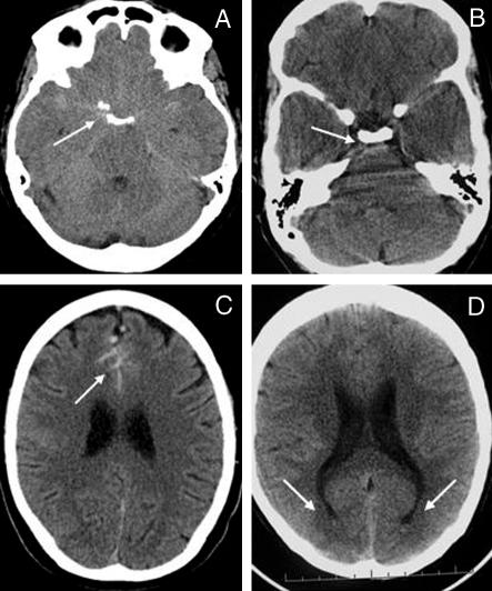
Fig 1.
(A) Subarachnoid haemorrhage from right posterior communicating artery aneurysm with hyperdense or isodense blood in anterior and posterior basal cisterns (arrow). (B) Blood in prepontine cistern (arrow). (C) Blood in anterior interhemispheric fissure (arrow). (D) Blood sedimenting in occipital horns of lateral ventricles (arrows)

