Figure 4.
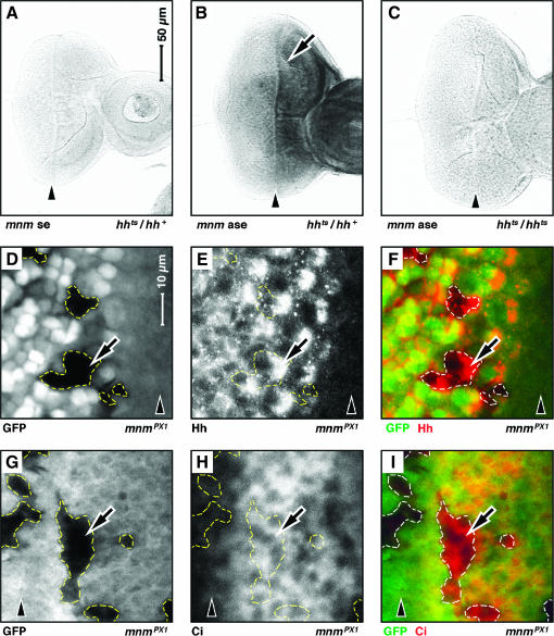
mnm is genetically downstream of hedgehog signaling. Third instar eye-imaginal discs: anterior, right. A–C and D–I are to the same scale; see bars in A and D. Arrowheads indicate the position of the morphogenetic furrow. (A–C) RNA in situ hybridization experiments: (A) hhts2/+, mnm sense (se) strand control; (B) hhts2 heterozygote, mnm anti-sense (ase) strand [note elevated level of mnm mRNA anterior to the morphogenetic furrow (arrow)]; (C) hhts2/hhts2, mnm anti-sense strand (mnm signal is lost in hhts2/hhts2 animals raised at the nonpermissive temperature). (D–I) Mosaic clones of mnmPX1 cells. Clones are negatively marked with GFP and are outlined (white in D and G and green in F and I). E (in white) and F (in red) show the expression of Hedgehog antigen. Note that Hedgehog is not lost from mnmPX1 mutant cells (arrow). H (in white) and I (in red) show the expression of activated Ci antigen. Note that activated Ci is not lost from mnmPX1 mutant cells (arrow).

