Figure 6.
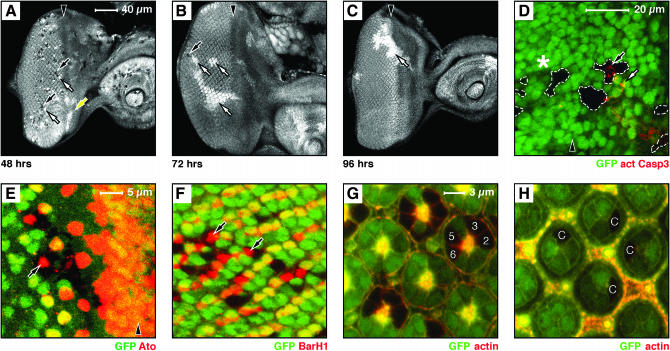
mnm is required for the survival of proliferating cells in the developing eye. (A–H) Eye-imaginal discs containing mnmPX1 mosaic clones, negatively marked by GFP; anterior, right. (A–F) Late third instar eye discs; (G–H) 48-hr pupal eye discs. A–C are to the same scale (bar in A), D is at higher magnification (see bar), E and F are to the same scale (bar in E), and G and H are to the same scale (bar in G). Arrowheads indicate the position of the morphogenetic furrow in A–E. (A–C) hs:FLP-induced clones, with the induction time before dissection indicated below: (A) 48 hr, (B) 72 hr, and (C) 96 hr. (A–C) GFP is shown as white. Note that 48 hr after induction (A) many small mnmPX1 homozygous clones are seen (black clones and black arrows) together with their homozygous wild-type twin spots (white twin spots and white arrow) posterior to the furrow. Note that the twin spots immediately anterior to the furrow lack clones (white twin spots and yellow arrow). At 72 hr (B) only very rare and small mutant clones are seen (black arrow) and the twin spots are far larger than the clones (white arrows). By 96 hr (C) only twin spots are seen (white arrow). (D) Activated Caspase3 antigen within (black arrow) or near (white arrow) mnmPX1 clones, anterior to the furrow. Note that there is no activated Caspase3 staining associated with mnm clones posterior to the furrow (asterisk). (E) Atonal antigen (red) and (F) BarH1 antigen (red) expressed within mnmPX1 clones (arrows). G and H show that mnmPX1 mutant cells can persist into pupal life and can differentiate normally [numbers indicate examples of R cell types (G) and cone cells (H)].

