Abstract
We report a case of a large, B-cell lymphoma of the atria in a 65-year-old man who presented with obstructive right-heart failure, shortness of breath, cirrhosis, and ascites. A computed tomographic scan revealed a large cardiac tumor occupying both atria. The patient underwent debulking of the tumor and postoperative chemotherapy. Six months postoperatively, he was alive and his symptoms of obstructive right-heart failure had improved; however, he had developed brain metastasis. (Tex Heart Inst J 2003;30:74–5)
Key words: Chemotherapy, adjuvant; heart neoplasms/diagnosis/surgery; lymphoma, B-cell/diagnosis/pathology/surgery
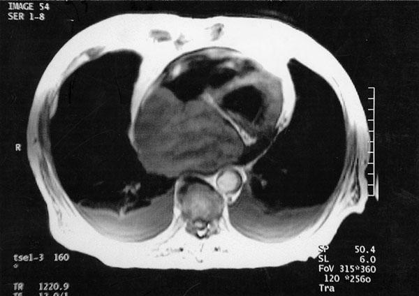
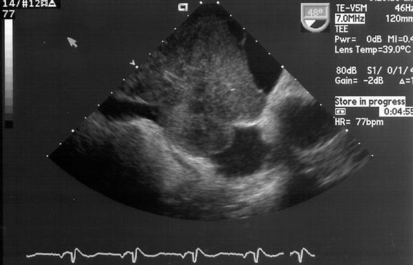
On 14 November 2001, a 65-year-old man with a history of chronic obstructive pulmonary disease and alcohol abuse presented at our institution with worsening shortness of breath and fatigue. He had clinical evidence of cirrhosis, ascites, and lower-extremity edema. A computed tomographic (CT) scan of the abdomen and chest revealed the presence of a large intracardiac tumor occupying both atria (Fig. 1). Two-dimensional echocardiography and transesophageal echocardiography, which were performed to better delineate the mass, showed a large globular tumor (Fig. 2).

Fig. 1 Computed tomography of the chest shows a large atrial tumor occupying both atria.

Fig. 2 Transesophageal echocardiogram shows a tumor occupying the atrium.
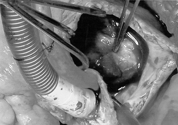
At surgery (27 November), the patient was found to have a very large, unresectable tumor that occupied the right atrium and invaded the inferior vena cava, with involvement of the septum and the left atrium (Fig. 3). Partial excision of the right atrial mass was performed to relieve the symptoms caused by the obstruction. 1 Postoperatively, the patient underwent chemotherapy, with subsequent improvement of his symptoms of obstructive right-heart failure. However, 6 months postoperatively, he developed brain metastasis.

Fig. 3 Intraoperative view. Forceps point at the tumor in the right atrium.
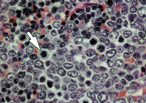
Pathology. A cross-section of the excised specimen revealed a whitish-to-tan, solid surface with irregular, extensive necrosis. Histologically, there were large lymphocytes with moderate pleomorphism. The cells had open-type nuclei with an irregular outline, and granular chromatin with chromocenters. Patchy areas throughout the tumor had irregular zones with smaller lymphoid cells (Fig. 4). The morphologic appearance and immunophenotypic features were those of a diffuse, large, B-cell, malignant lymphoma. 1–4

Fig. 4 Histologic photomicrograph of the atrial tumor shows closely packed lymphoid cells with more than 4 mitoses (arrow) (H&E, orig. ×40).
Footnotes
Address for reprints: Dr. Masoud AlZeerah,1215 Coulter, Suite 301, Amarillo, TX 79106
E-mail: malzeerah@hotmail.com
References
- 1.Takagi M, Kugimiya T, Fujii T, Yamauchi H, Shibata R, Narimatsu M, Tsuda N. Extensive surgery for primary malignant lymphoma of the heart. J Cardiovasc Surg (Torino) 1992;33:570–2. [PubMed]
- 2.Burke A, Virmani R, editors. Tumors of the heart and great vessels. Atlas of tumor pathology. 3rd series; fasc. 16. Bethesda (MD): Armed Forces Institute of Pathology; 1996. p. 21–46.
- 3.McAllister HA, Fenoglio JJ, editors. Tumors of the cardiovascular system. Atlas of tumor pathology. 2nd series; fasc. 15. Bethesda (MD): Armed Forces Institute of Pathology; 1978.
- 4.Smith C. Tumors of the heart. Arch Pathol Lab Med 1986; 110:371–4. [PubMed]


