Abstract
We report a case of Carney's complex in a 12-year-old boy who had the characteristic features of multiple cutaneous tumors, pigmentation, and biatrial myxoma. His large right atrial myxoma almost occluded the tricuspid valve and presented a life-threatening emergency. Surgery saved his life, but recurrence of myxoma was noted on follow-up. The familial nature of the condition is highlighted by the case of the patient's 44-year-old mother, who also presented with features of Carney's complex: multiple cutaneous tumors and a tiny, asymptomatic, left atrial myxoma, which was detected during routine echocardiographic screening. (Tex Heart Inst J 2003;30:80–2)
Key words: Carney's complex; endocrine diseases/complications; heart atrium/ultrasonography; heart neoplasms/ultrasonography; melanosis; myxoma/diagnosis; neoplasm recurrence/local; neoplasms, multiple primary; pigmentation disorders; myxoma/genetics; skin neoplasms; syndrome
Cardiac myxoma is the most common primary tumor of the heart. 1 Familial cardiac myxomas constitute 7% of cardiac myxomas. 2 Singh and Lansing 3 have published a comprehensive review of 26 familial cardiac myxomas in 68 family members. Familial cardiac myxomas are characterized by involvement in sites other than the left atrium, including biatrial involvement 4 and ventricular involvement, and by a high recurrence rate. 5,6 Familial cardiac myxomas may present as part of a syndrome (Carney's complex, LAMB, or NAME syndrome*) 7–10 or as nonsyndromic familial cardiac myxomas. 11 Carney's complex, as described by J.A. Carney, 7 is characterized by the association of cutaneous pigmentation, fibromyxoid tumors of the skin, myxomas of the heart, endocrine overactivity, and autosomal dominant inheritance. We report cardiac myxoma in a mother and son, both of whom had cutaneous pigmentation and multiple fibromyxoid tumors of the skin over the face, extremities, and back, but no evidence of endocrine involvement.
Case Reports
Patient 1
In July 1999, a 12-year-old boy presented with a history of fatigability, swelling of the legs, facial puffiness, abdominal distention of 3 months' duration, and progressive breathlessness of 1 month's duration. He had a history of multiple swellings over the face, extremities, and back since birth and had undergone surgical removal of some of the swellings. The biopsy report confirmed the pathologic features of fibromyxoid tumors. On examination, the patient was orthopneic and had gross pedal edema, facial puffiness, and elevated jugular venous pressure. His blood pressure was 110/80 mmHg, and the pulse was 130/min and regular. He had large, black, pigmented patches and soft-tissue swellings over the trunk, face (Fig. 1), and extremities. Auscultation revealed a grade 2/6 pansystolic murmur in the tricuspid area with an early diastolic sound. He had hepatomegaly with systolic pulsations and ascites. Two-dimensional color-flow Doppler transthoracic echocardiography (Fig. 2) revealed an 8- × 4-cm myxoma arising from the right atrium, with attachment to the lower part of the atrial septum and extension through the tricuspid valve into the right ventricular cavity. The mass was seen to almost occlude the tricuspid orifice; there was very little flow across the valve. Another pedunculated myxoma, 3 × 1.9 cm, was seen in the left atrium, attached to the interatrial septum 12 at a site opposite the attachment point of the right atrial myxoma. There was moderate pericardial effusion. The patient underwent surgical removal of the tumor on an emergency basis. At surgery, we used a biatrial approach. 13,14 A large myxoma occupied the entire right atrium, with its stalk attached to the fossa ovalis region of the atrial septum. The stalk was excised and an incision was made in the region of the fossa ovalis. In the left atrium, the stalk of that myxoma was attached between the mitral annulus and the atrial septum. We excised part of the atrial septum adjacent to the myxoma's stalk, and closed the defect with a pericardial patch.
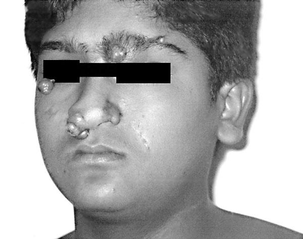
Fig. 1 Patient 1: The son shows multiple cutaneous tumors and surgical scars.
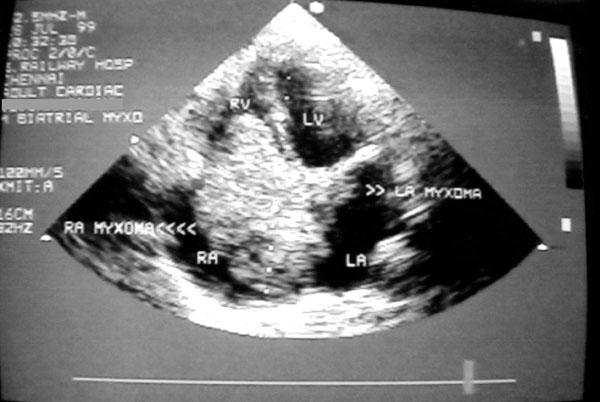
Fig. 2 Patient 1: Transthoracic echocardiogram in apical 4-chamber view shows a large right atrial myxoma extending into the right ventricle, and a small left atrial myxoma.
LA = left atrium; LV = left ventricle; RA = right atrium; RV = right ventricle
The patient was asymptomatic on follow-up; however, transthoracic and transesophageal echocardiographic examinations in January 2001 revealed a recurrent, pedunculated, left atrial myxoma, 1.5 × 1 cm, attached to the atrial septum at the base of the anterior mitral leaflet (Fig. 3). We advised surgery for the recurrent myxoma, but the patient did not return for follow-up until October 2001, when transthoracic echocardiography showed that the tumor had increased in size to 4 × 2 cm and had prolapsed across the mitral valve, which produced moderate obstruction with a mean gradient of 7 mmHg. We recommended early surgery, but the patient developed severe emotional stress at the prospect of another surgical procedure. Currently, he is under psychiatric care, after which surgery is planned.
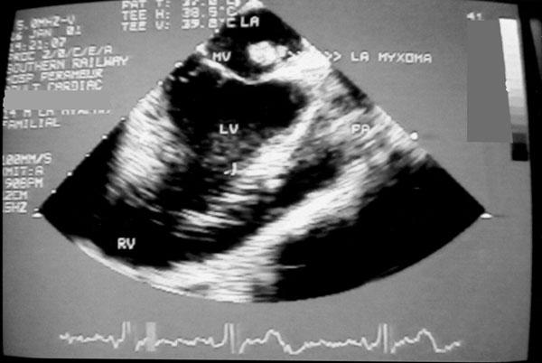
Fig. 3 Patient 1: Transesophageal echocardiogram shows the recurrent, pedunculated, left atrial myxoma attached to the atrial septum at the base of the anterior mitral leaflet.
LA = left atrium; LV = left ventricle; MV = mitral valve; PA = pulmonary artery; RV = right ventricle
Patient 2
The mother of Patient 1, aged 44 years, was asymptomatic for cardiac disease, but gave a history of multiple cutaneous swellings over her face (Fig. 4), back, and extremities. She had undergone surgical excision of some of the swellings. Two-dimensional color-flow Doppler transesophageal echocardiography performed in January 2001, during the follow-up examination of her son, revealed a tiny, pedunculated, left atrial myxoma, 0.6 × 0.56 cm, attached to the interatrial septum just below the anterior mitral leaflet. The echocardiogram delineated the tumor well (Fig. 5). In view of the very small size of the tumor, we advised close periodic follow-up in lieu of surgery. However, upon follow-up in October 2001, her tumor had increased in size to 1.5 × 1 cm. On that occasion, she also complained of mild weakness of the right hand and left leg, of 6 months' duration. A neurologic examination suggested mononeuritis multiplex. A computed tomographic brain scan revealed a left parietal infarct. Magnetic resonance imaging showed that the cervical spine was normal. She had a 10-year history of diabetes. The neurologist hypothesized that the parietal infarct was silent and that the mononeuritis multiplex might be due to the diabetes. In view of the embolic episode to the brain, we advised early surgery. This patient has 2 other sons, neither of whom has cutaneous involvement nor cardiac tumors. She also has 8 siblings. No member of her family has cutaneous involvement or a medical history that would suggest cardiac tumor.
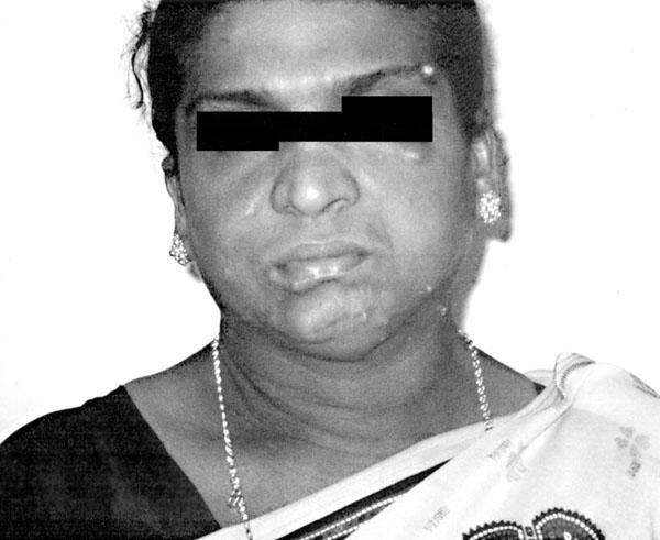
Fig. 4 Patient 2: The mother also shows multiple cutaneous tumors and surgical scars.
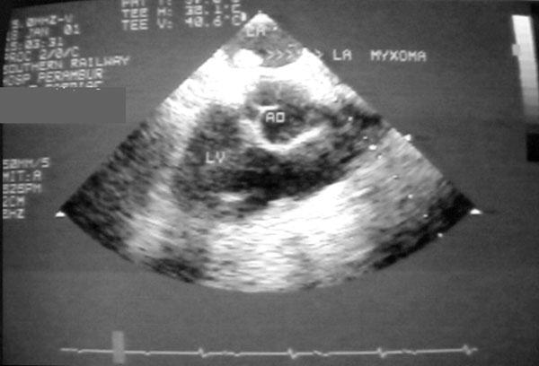
Fig. 5 Patient 2: Transesophageal echocardiogram shows a small, pedunculated, left atrial myxoma attached to the atrial septum at the base of the anterior mitral leaflet.
AO = aorta; LA = left atrium; LV = left ventricle
Discussion
Familial syndromic myxomas are rare. Characteristic features include involvement of multiple sites (often biatrial tumors), young age of occurrence, and high recurrence rate—10% to 21%, compared with 4% to 7% in sporadic myxomas. 15,16 These features are reflected in our case reports. An interesting feature was the delay in the development of symptomatic myxoma in the mother until the age of 44 years, although she had experienced cutaneous manifestations since birth. This emphasizes the need for periodic echocardiographic screening of family members for the occurrence of cardiac tumors, even when they have had normal results on previous examinations. The growth rate of recurrent myxomas in previous reports is 1.8 cm/year, although there are 2 reports of initial left atrial myxomas that grew at a rate of 5 cm/year. 17 In our 2 patients, the size of the tumors doubled over a period of 9 months, and growth of the initial and recurrent atrial myxomas was equally rapid.
Footnotes
* LAMB = Lentigines, Atrial Myxoma, and Blue nevi; NAME = Nevi, Atrial Myxoma, and neurofibroma Ephelides
Address for reprints: Dr. K.A. Abraham, Chief Cardiologist, Railway Hospital, Ayanavaram, Chennai 600023, India
E-mail: abibaby@md3.vsnl.net.in
References
- 1.Bulkley BH, Hutchins GM. Atrial myxomas: a fifty year review. Am Heart J 1979;97:639–43. [DOI] [PubMed]
- 2.Farah MG. Familial cardiac myxoma. A study of relatives of patients with myxoma. Chest 1994;105:65–8. [DOI] [PubMed]
- 3.Singh SD, Lansing AM. Familial cardiac myxoma—a comprehensive review of reported cases. J Ky Med Assoc 1996;94(3):96–104. [PubMed]
- 4.Amir O, Merdler A, Shiran A. Bilateral atrial myxomas associated with hyperpigmented skin lesions. N Engl J Med 2001;344:938–9. [DOI] [PubMed]
- 5.Vidaillet HJ Jr, Seward JB, Fyke FE 3rd, Su WP, Tajik AJ. “Syndrome myxoma”: a subset of patients with cardiac myxoma associated with pigmented skin lesions and peripheral and endocrine neoplasms. Br Heart J 1987;57:247–55. [DOI] [PMC free article] [PubMed]
- 6.Powers JC, Falkoff M, Heinle RA, Nanda NC, Ong LS, Weiner RS, Barold SS. Familial cardiac myxoma: emphasis on unusual clinical manifestations. J Thorac Cardiovasc Surg 1979;77:782–8. [PubMed]
- 7.Carney JA, Gordon H, Carpenter PC, Shenoy BV, Go VL. The complex of myxomas, spotty pigmentation, and endocrine overactivity. Medicine (Baltimore) 1985;64:270–83. [DOI] [PubMed]
- 8.Carney JA. Psammomatous melanotic schwannoma. A distinctive, heritable tumor with special associations, including cardiac myxoma and the Cushing syndrome. Am J Surg Pathol 1990;14:206–22. [PubMed]
- 9.Yen RS, Allen B, Ott R, Brodsky M. The syndrome of right atrial myxoma, spotty skin pigmentation, and acromegaly. Am Heart J 1992;123:243–4. [DOI] [PubMed]
- 10.Krause S, Adler LN, Reddy PS, Magovern GJ. Intracardiac myxoma in siblings. Chest 1971;60:404–6. [DOI] [PubMed]
- 11.Dandolu BR, Iyer KS, Das B, Venugopal P. Nonsyndrome familial atrial myxoma in two generations. J Thorac Cardiovasc Surg 1995;110:872–4. [DOI] [PubMed]
- 12.Tway KP, Shah AA, Rahimtoola SH. Multiple biatrial myxomas demonstrated by two-dimensional echocardiography. Am J Med 1981;71:896–9. [DOI] [PubMed]
- 13.Imperio J, Summers D, Krasnow N, Piccone VA Jr. The distribution patterns of biatrial myxomas. Ann Thorac Surg 1980;129:467–73. [DOI] [PubMed]
- 14.Bhan A, Mehrotra R, Choudhary SK, Sharma R, Prabhakar D, Airan B, et al. Surgical experience with intracardiac myxomas: long-term follow-up. Ann Thoracic Surg 1998;66: 810–3. [DOI] [PubMed]
- 15.Murphy MC, Sweeney MS, Putnam JB Jr, Walker WE, Frazier OH, Ott DA, Cooley DA. Surgical treatment of cardiac tumors: a 25-year experience. Ann Thorac Surg 1990;49: 612–8. [DOI] [PubMed]
- 16.van Gelder HM, O'Brien DJ, Staples ED, Alexander JA. Familial cardiac myxoma. Ann Thorac Surg 1992;53:419–24. [DOI] [PubMed]
- 17.Malekzadeh S, Roberts WC. Growth rate of left atrial myxoma. Am J Cardiol 1989;64:1075–6. [DOI] [PubMed]


