The advancement of our knowledge of biochemical phenomena depends on our ability to implement increasingly more powerful analytical techniques. Traditional means to characterize the chemical constituents of a solution include spectroscopic approaches and separation techniques to identify, isolate and quantify analytes. Unfortunately, these techniques are not perfect. Often, spectral properties of similar species are difficult to resolve in a complex mixture. Alternatively, separation methods for analysis of biological samples generally demand times of at least several seconds or longer, and unstable products can often degrade in solution before ever reaching the detection window. The article by Plenert and Shear (1) in this issue of PNAS represents a significant advance in ultrafast separation and detection of biologically relevant compounds.
In their study, Plenert and Shear (1) adapted an unusual capillary geometry, the hourglass, to locally enhance the electric field strength at the separation region, and added ultrafast optical-based sample injection to enable separations on the microsecond time frame. Although voltage is directly proportional to separation efficiency and to electrophoretic flow (and hence inversely proportional to total analysis time), there is a practical limit to the highest separation voltage that can be used because of Joule heating effects. Under ordinary capillary zone electrophoresis (CZE) conditions, with an unmodified cylindrical capillary, the 100 kV/cm field strengths used by Plenert and Shear would produce significant Joule heating, resulting in broadened peaks and a concomitant limiting of separation efficiency. However, their narrow capillary dimensions and hourglass geometry allows more efficient dissipation of heat not normally available in traditional CZE, allowing for the use of greater field strengths. Although the hourglass shape itself is not unique (2), the application of such a geometry for local field enhancement, without resorting to submicron capillaries with impractically small capillary lengths, is novel.
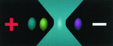
Plenert and Shear also demonstrate a methodology for improving sampling by using 1-μs injections via an approach similar to the optical gating injection method demonstrated by Jorgenson and coworkers (3); in the current implementation, hydroxyindole photoproducts are generated by the “injection” laser pulse, which are then electrophoretically separated (Fig. 1). Instead of being limited by voltage switching times, as is the case for electrophoretic injection, plug sizes are instead fashioned by properties of the laser and speed of the flow in the capillary.
Figure 1.
Schematic of a tightly focused laser beam for detection of electrophoretically resolved components.
Their application is exciting: the probing of short-lived reaction intermediates with similar spectroscopic properties but contrasting electrophoretic mobilities. In this initial demonstration, Plenert and Shear demonstrate the ability to separate two compounds using an ultrafast separation so that each compound's fluorescence intensity can be probed individually.
Although using electrophoresis to separate analytes from each other is not new, separations completed, from start to finish, in <25 μs represents a large decrease in analysis time. From Tiselius' first demonstration of an electrophoretic separation that lasted hours (4) to Jacobson and coworker's millisecond separations (5), there has been a dramatic decrease in the time required to perform a separation, and Plenert and Shear take this to the next level. How much faster a separation is possible with this approach? In the work of Plenert and Shear, they are limited by their ability to perform injections to ≈1 μs, which are accomplished by a Pockel's cell optical gating method. With improved switching technology, they expect their separation time to move into the nanosecond regime, fast enough to follow even shorter-lived chemical species.
The application of the hourglass geometry for local field enhancement is novel.
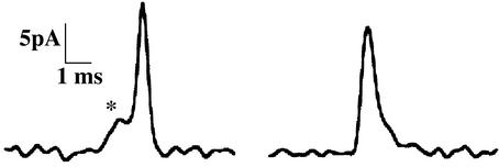
What are such fast separations good for? Obviously, studying reaction intermediates is an important area. Besides looking for reaction intermediates, this new technology has potential when looking at small volume biological systems that have millisecond changes in chemical composition. The obvious application involves the process of exocytosis, or the release of neurotransmitters from neurons (6, 7). Traditionally, one employs various microscopy techniques to obtain spatially resolved information, a separation to gain snapshots of the chemical microenvironment around the cell, or electrochemistry to acquire dynamic information. Shear's current approach may allow rapid, simultaneous acquisition of chemical and temporal information. Dynamic release events have been observed through assorted electrochemical techniques such as amperometry thanks to its fast time scale and high sensitivity (7, 8); as shown in Fig. 2, this has recently been accomplished with spontaneous dopamine release in retinal neurons (9) and adrenal chromaffin cells (10). However, compounds of interest are sometimes not electroactive, and related substances could interfere with or otherwise complicate detection.
Figure 2.
Two amperometric spikes caused by dopamine release of a single neuron at a carbon fiber microelectrode; at the start of the rising phase, a small “foot” signal is observed (asterisk); it will be interesting to see whether the ultrafast separation approach can be adapted to such biological measurements. [Figure reproduced with permission from ref. 9 (Copyright 2001, Elsevier).]
Plenert and Shear used serotonin as their model compound; obviously the system has application for following exocytosis from serotonergic neurons. Indeed, serotonin is an important neurotransmitter in both the central and peripheral nervous systems of mammals. It is implicated in a variety of physiological tasks, including learning and memory in the central nervous system and emesis and peristaltic reflex in the enteric nervous system (11, 12). The improvements made by Shear and coworkers (1) may allow more rapid separations of subcellular components, such as indoleamine-containing single vesicles, and should contribute to studies aimed at characterizing diseases of the nervous system. In particular, dynamic release events, such as those that occur from neurons and endocrine cells, can be monitored on a rapid time scale as is required for adequate time resolution of release. It is no coincidence that the authors employ a neurotransmitter, serotonin, for demonstrating the power of their method. Of course, for their approach to be successful with cellular samples, a number of refinements are required and a number of questions need to be addressed. For example, can they detect and quantify the small amount of serotonin released from a neuron? Unlike the applications demonstrated where a number of separations are coadded to obtain adequate signal, this becomes difficult when probing the response of a cell to an electrical or chemical stimulation, and so sensitivity may be an issue. In addition, the chemical complexity of the released materials may require a higher resolution than they obtain in these microsecond separations, though they may be able to trade speed for separation efficiency. Of course, few compounds become fluorescent with multiphoton excitation, and so the separation efficiency need not be as great as with other detection schemes. A last question involves the negative effect of the high field strengths on the cell. Often, cells lyse undesirably in the presence of large electric fields, preventing the study of cellular release. On the other hand, with an appropriately shaped capillary, whether hourglass or other, it may be possible to maintain the cell in a relatively field free region and still employ the 100 kV/cm fields required for the fast separation.
Their work opens new possibilities in the separation of chemicals in volume-limited samples where high throughput and enhanced separation efficiencies are required. We certainly expect a number of exciting applications to present themselves for what is now the ultimate in fast separation.
Footnotes
See companion article on page 3853.
References
- 1.Plenert M L, Shear J B. Proc Natl Acad Sci USA. 2003;100:3853–3857. doi: 10.1073/pnas.0637211100. [DOI] [PMC free article] [PubMed] [Google Scholar]
- 2.Lundqvist A, Pihl J, Orwar O. Anal Chem. 2000;72:5740–5743. doi: 10.1021/ac000600g. [DOI] [PubMed] [Google Scholar]
- 3.Moore A W, Jr, Jorgenson J W. Anal Chem. 1993;65:3550–3560. doi: 10.1021/ac00072a004. [DOI] [PubMed] [Google Scholar]
- 4.Tiselius A. Trans Faraday Soc. 1937;33:524–531. [Google Scholar]
- 5.Jacobson S C, Hergenroder R, Koutny L B, Ramsey J M. Anal Chem. 1994;66:1114–1118. [Google Scholar]
- 6.Murthy V N. Curr Opin Neurobiol. 1999;9:314–320. doi: 10.1016/s0959-4388(99)80046-4. [DOI] [PubMed] [Google Scholar]
- 7.Chow R H, von Ruden L. In: Single Channel Recording. Sakmann B, Neher E, editors. New York: Plenum; 1995. pp. 245–275. [Google Scholar]
- 8.Travis E R, Wightman R M. Annu Rev Biophys Biomol Struct. 1998;27:77–103. doi: 10.1146/annurev.biophys.27.1.77. [DOI] [PubMed] [Google Scholar]
- 9.Puopolo M, Hochstetler S E, Gustincich S, Wightman R M, Raviola E. Neuron. 2001;30:211–225. doi: 10.1016/s0896-6273(01)00274-4. [DOI] [PubMed] [Google Scholar]
- 10.Chen P, Xu B, Tokranova N, Feng X, Castracane J, Gillis K D. Anal Chem. 2003;75:518–524. doi: 10.1021/ac025802m. [DOI] [PubMed] [Google Scholar]
- 11.Buhot H C, Martin S, Segu L. Ann Med. 2000;32:210–221. doi: 10.3109/07853890008998828. [DOI] [PubMed] [Google Scholar]
- 12.Endo T, Minami M, Hirafujo M, Ogawa T, Akita K, Nemoto M, Saito H, Yoshioka M, Parvez S H. Toxicology. 2000;153:189–201. doi: 10.1016/s0300-483x(00)00314-0. [DOI] [PubMed] [Google Scholar]


