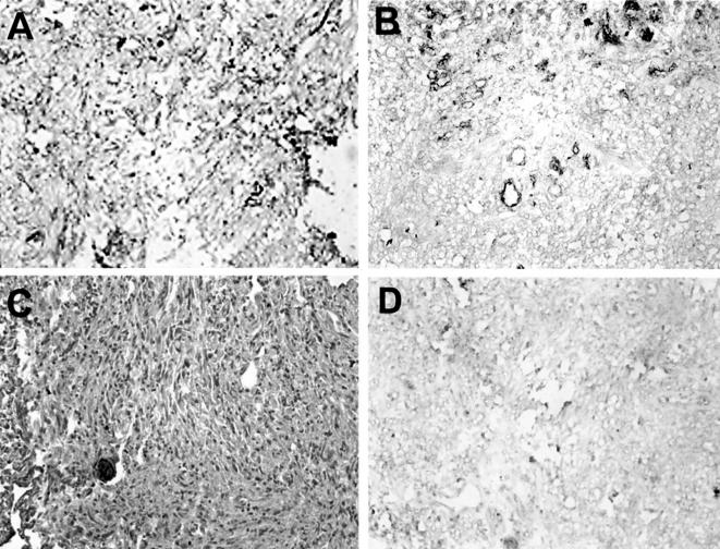
Figure 1.
Photomicrograph (original magnification, x200) illustrating immunohistochemical staining of Gb3 in human meningiomas. (A) A malignant meningioma representing very strong (+ + + +) Gb3 immunoreactivity (brown) where staining is both vascular and extravascular. (B) A MM semiquantitated as strong staining (+ + +) primarily due to the predominantly vascular localization of Gb3. (C) A benign meningioma showing no evidence of Gb3 positivity. (D) A MM (same as in B) showing no Gb3 immunoreactivity when VT1 is omitted.

