Abstract
Two proteins representing the heavy-chain subunits of botulinum neurotoxin types C and D were expressed in Escherichia coli, and their vaccine potential was evaluated. Mice were vaccinated with doses ranging from 0.5 to 10 μg and were challenged with 10 to 105 50% lethal doses of toxin. For the type C subunit protein, C50, two doses of 2 μg were required for full protection, while, for type D subunit protein, D50, two 1-μg doses were required. A bivalent vaccine consisting of a mixture of these two proteins also provided protection against both botulinum neurotoxin type C and type D challenge. Antibody levels in serum were determined by both enzyme-linked immunosorbent assays and serum neutralization assays
Botulism is an intoxication caused by neurotoxins produced by Clostridium botulinum. Seven antigenically distinct botulinum neurotoxins (BoNTs), designated A through G, are produced with C. botulinum strains identified according to the major neurotoxin type produced. The different toxin types are pharmacologically and structurally similar, consisting of a single polypeptide chain that is posttranslationally nicked to yield a dichain molecule with a light chain (50 kDa), joined by a disulfide bond to a 100-kDa heavy chain (HC) (4, 5). The 50-kDa carboxy-terminal end of the HC contains the major determinants responsible for specific toxin binding at the neuromuscular junction (1, 11, 13). BoNT types C and D have both been isolated from cattle suffering paralytic disease, and only these strains are associated with botulism in cattle in Australia (3, 14). To combat the disease, a bivalent vaccine consisting of formalin-inactivated type C and D toxins is presently available, and although it is efficacious, various concerns regarding reliable vaccine availability have been expressed by members of the Australian cattle industry. Previous studies have shown that the HC subunits of the tetanus toxin and various BoNTs are capable of evoking a protective immune response in mice (2, 6, 7, 9, 12). Such a subunit vaccine for BoNT types C and D would offer several advantages over the present vaccine, potentially eliminating or minimizing production problems that can sometimes be experienced.
Primers were designed to amplify the HC-encoding region of the BoNT type C and type D genes. For ease of subsequent manipulations, appropriate restriction enzyme sites were incorporated into the primers. The templates for the PCRs were DNA fragments CH3, derived from the BoNT type C gene (8) (GenBank accession no. D90210), and H1, derived from BoNT type D (15) (GenBank accession no. S49407), inserted into the cloning plasmid pUC19 (New England BioLabs, Beverly, Mass.). Table 1 shows the primers, sequences, and enzymes.
TABLE 1.
Primers and sequencesa
| Primer | Sequence | Restriction enzyme |
|---|---|---|
| C5′ | 5′-AATACCATGGAAAATACAATACCCTTTAAT-3′ | NcoI |
| C3′ | 5′-TCTTCTGAATTCACTCTGGAAGATTATAGACAG-3′ | EcoRI |
| D5′ | 5′-GAAACCATGGTCCCTTTTAATATTTTTTCA-3′ | NcoI |
| FUP (D3′) | 5′-GTTTTCCCAGTCACGAC-3′ |
Incorporated restriction enzyme sites are underlined. PCRs contained 1× PCR buffer; dATP, dCTP, dGTP, and dTTP (200 μM each); 5′ and 3′ primers (0.5 μM each); DNA template (1 or 0.5 ng); MgCl2 (1.25 mM); and Taq DNA polymerase (2.5 U) (Applied Biosystems, Foster City, Calif.). Typical PCR protocol was as follows: initial heating to 94°C for 1 min and then cycles of 94°C, 45 s; 54°C, 30 s; and 72°C, 45 s, for a total of 30 cycles. Products were initially cloned into pGEM-T (Promega Corp., Madison, Wis.) prior to subcloning into ProEXHT-A (Life Technologies, Mt. Waverley, Australia) expression plasmid. All constructs were confirmed by DNA sequencing.
Expression plasmids were used to transform Escherichia coli DH5α cells (Life Technologies), which were then grown overnight in Luria-Bertani broth supplemented with ampicillin and were then subcultured into Terrific broth containing ampicillin. Expression was induced by the addition of 1 mM isopropyl-β-d-thiogalactopyranoside (final concentration) when cultures reached an optical density (OD) at 600 nm of approximately 0.8 to 1.5, and the cultures were transferred to a 25°C shaking incubator for growth overnight. Protein purifications were carried out by using a 50% nickel-nitrilotriacetic acid agarose (Ni-NTA) slurry (Qiagen, Valencia, Calif.) following the manufacturer's protocol for insoluble proteins. Expressed proteins were eluted from the Ni-NTA column with buffer E (100 mM NaH2PO4, 10 mM Tris-HCl, 8 M urea, pH 4.5). Proteins were analyzed by sodium dodecyl sulfate-polyacrylamide gel electrophoresis (SDS-PAGE) and Western blotting and were probed with antihistidine antibody or anti-BoNT antisera. Protein visualization for Western blot analysis incorporated an enhanced chemiluminescence method (Amersham Pharmacia Biotech AB, Uppsala, Sweden). Eluted fractions containing expressed proteins were pooled and dialyzed overnight, at 4°C, against phosphate-buffered saline (PBS). Protein concentrations were estimated by using a Bio-Rad DC Protein Assay (Bio-Rad Laboratories, Hercules, Calif.) according to the manufacturer's protocol.
Vaccine antigens were diluted in sterile PBS and were emulsified with an equal volume of complete Freund's adjuvant (Sigma Chemical Co., St. Louis, Mo.). An equal volume of sterile 2% Tween 20-140 mM NaCl was added and was further emulsified. Six-week-old female ddY mice from Shimizu Laboratory Supplies, Kyoto, Japan, were injected via the intraperitoneal (i.p.) route with 200 μl of each vaccine antigen, covering a dose range of 0.5 to 10 μg. Booster injections were prepared in the same manner, but with incomplete Freund's adjuvant, and were given 2 weeks following initial vaccination. Bivalent vaccines were prepared by combining 10 μg each of C50 and D50 subunit proteins, and adjuvant was incorporated as described above. In some cases vaccines were prepared without adjuvant; the protein antigens (10 μg) were diluted in sterile PBS and 200 μl was injected i.p. Five days prior to toxin challenge, blood was obtained from test animals and the serum was stored at −20°C. To establish the 50% lethal dose (LD50), toxin was diluted in sterile 0.01 M PBS (pH 6.0)-0.2% gelatin and each mouse was injected with 500 μl i.p. Two mice were challenged with each dilution of toxin and were observed for 4 days. In vaccine trials, mice were challenged with toxin doses ranging from 10 to 105 LD50s 2 weeks after the final booster dose and were observed for a further 4 days. All animal work was carried out in accordance with the Australian Code of Practice for the Care and Use of Animals for Scientific Purposes (National Health & Medical Research Council).
Antibody levels in serum were determined by enzyme-linked immunosorbent assay (ELISA). Flat-bottomed, 96-well microtiter plates (Costar, Laguna Niguel, Calif.) were coated with 50 μl of either purified C50 or D50 recombinant protein (1 μg/ml in 70 mM Na2HPO4) or BoNT type C or type D 16S toxin (20 μg/ml in PBS, pH 6.0). Plates were washed with 0.5% Tween-PBS, and nonspecific binding sites were blocked with 10% skim milk-PBS prior to the addition of 50 μl per well of mouse serum, serially diluted in skim milk-PBS. Plate contents were incubated for 1 h at room temperature before horseradish peroxidase-conjugated rabbit anti-mouse antibody (Dako Japan Co. Ltd., Kyoto, Japan), diluted 1:1,000 in skim milk-PBS, was added. After a further hour's incubation, the chromogen, o-phenylenediamine, was added. The color reaction was terminated by the addition of 2 M sulfuric acid, and the OD was measured at 490 nm on a Bio-Rad Novapath Microplate Reader (Bio-Rad Laboratories). The ELISA titer was taken as the reciprocal value of the last dilution giving an OD reading greater than the average plus 3 standard deviations of the OD obtained for eight replicates of pooled sera from 20 unvaccinated mice, run in parallel on each plate.
For some animals, antibody levels in serum were also measured by a serum neutralization assay. Mouse serum, diluted 1 in 10 in PBS, was mixed with an equal volume of toxin containing 10 LD50s of BoNT type C or D diluted in 0.01 M PBS (pH 6.0)-0.2% gelatin. Samples were incubated at 37°C for 1 h, and 200 μl was injected i.p. into each of two mice per sample. Mice were observed for 4 days, and all deaths were recorded.
ELISA titers were log transformed to normalize the data. A one-way analysis of variance (P = 0.05) was used to compare the effects of different vaccination regimes. Spearman's correlation was performed to identify significant relationships between different data sets. Correlation coefficient (r), P, and level of significance (α) are reported.
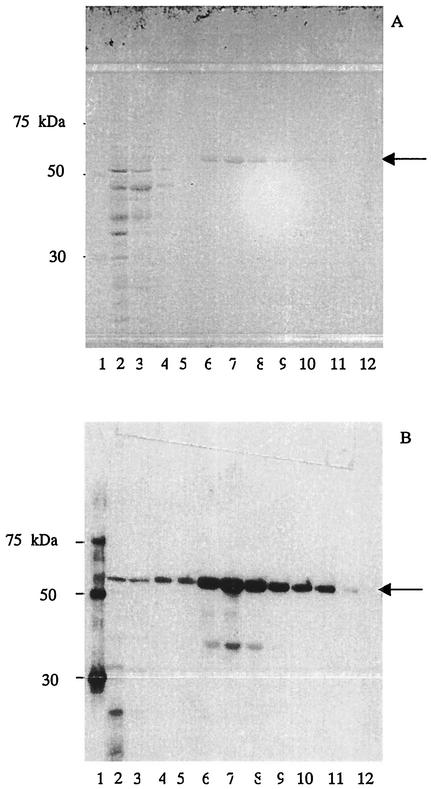
Optimization of expression conditions, specifically the reduction of expression temperature to 25°C, resulted in a significant reduction in the presence of lower-molecular-weight proteins observed following expression at 37°C. Products of approximately 90% purity, as assessed by SDS-PAGE and Western blot analysis, were obtained following purification with Ni-NTA agarose (Fig. 1). Occasional batches contained a minor amount of lower-molecular-weight proteins, presumably degradation products of the full-length protein. These bands reacted with both Penta-His and anti-BoNT antibodies. Initial experiments resulted in yields of purified proteins of approximately 0.5 mg per liter of culture. When the pH of the elution buffer was reduced from 5.9 to 4.5, significantly higher yields in the region of 2.0 to 3.0 mg per liter of culture were obtained. Under these conditions, proteins precipitated upon dialysis against PBS but were still recognized by Penta-His antibodies, while type C derivatives were recognized by anti-BoNT type C serum. The precipitated proteins were stable, with storage for 1 month at −20°C, 4°C, room temperature, and up to 37°C resulting in very little degradation of product as assessed by SDS-PAGE and Western blot analysis.
FIG. 1.
SDS-PAGE (A) and a Western blot, stained with Penta-His antibody (B), of purification of D50 protein (arrowed). Proteins eluted with buffer E, pH 4.8. Lanes: 1, Qiagen His6 protein ladder; 2, broth culture; 3, column flowthrough; 4, column wash 1; 5, wash 2; 6, column elution 1; 7, elution 2; 8, elution 3; 9, elution 4; 10, elution 5; 11, elution 6; and 12, elution 7. Lower-molecular-weight proteins were present in occasional batches for both C50 and D50 expression. These proteins reacted with both anti-His and anti-BoNT antisera and appear to represent degradation products of the full-length products.
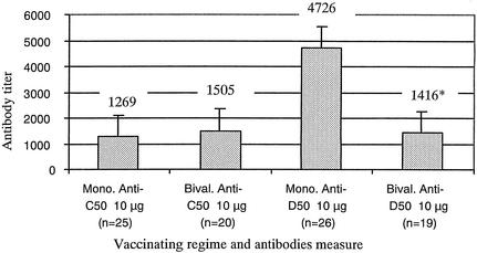
Table 2 summarizes the results of the vaccine efficacy studies for the toxins. None of the control animals that received only adjuvant survived toxin challenge. For full protection against a toxin challenge of 10 LD50s of BoNT type C, two 2-μg doses of the C50 and two doses of 1 μg of the D50 protein were required (for BoNT type C the LD50 was 5 × 10⁻⁷, and for BoNT type D, 1 × 10⁻⁷ when 500 μl was injected i.p.). Little or no cross-protection was observed with the monovalent vaccines. A combination of 10 μg each of C50 and D50 proteins in a bivalent vaccine provided full protection against up to 1,000 LD50s of BoNT type C but only partial protection against 10 and 1,000 LD50s of BoNT type D. Anti-C50 antibody levels in serum did not differ significantly between animals receiving the monovalent or bivalent vaccines; however, D50 antibody levels were significantly (P = 0.05) lower in those mice receiving the bivalent vaccine (Fig. 2).
TABLE 2.
Effect of vaccination with subunit proteins on animal survival following toxin challengea
| Immunogen(s) and dose (μg) | No. of survivors/total challenged at challenge dose (no. of LD50s)
|
||||
|---|---|---|---|---|---|
| 10 | 102 | 103 | 104 | 105 | |
| C50 | |||||
| 0.5 | 2/5 | ||||
| 1 | 2/5 | ||||
| 2 | 5/5 | ||||
| 5 | 5/5 | 5/5 | |||
| 10 | 5/5 | 5/5 | 4/5 | 2/5 | 2/5 |
| 10, no adjuvant | 4/5 | 4/5 | 4/5 | ||
| 10* | 0/5 (type D) | ||||
| Adjuvant only | 0/5 | ||||
| D50 | |||||
| 0.6 | 2/5 | ||||
| 1 | 5/5 | 3/5 | |||
| 10 | 5/5 | 5/5 | 5/5 | 3/5 | 3/5 |
| 10, no adjuvant | 4/5 | 4/5 | |||
| 10* | 1/5 (type C) | ||||
| Adjuvant only | 0/5 | ||||
| C50 + D50 | |||||
| 10 (C challenge) | 5/5 | 5/5** | |||
| 10 (D challenge) | 4/5 | 2/5 | |||
All vaccines were given i.p. as two doses, 2 weeks apart. Animals were challenged with homologous toxin type corresponding to the vaccinating protein, except in cross-protection studies, indicated by a single asterisk. For bivalent-vaccine studies, equal amounts of C50 and D50 proteins were incorporated into the vaccine and animals were challenged with either BoNT type C or D as indicated. **, animals given a second challenge of BoNT type D (103 LD50S). Five of five survived.
FIG. 2.
Effect of combining vaccinating proteins on immunogenicity of components. Anti-C50 and anti-D50 antibody levels in serum were measured by ELISA for mice receiving either monovalent (Mono.) or bivalent (Bival.) vaccines. The geometric mean titer of each group and the standard error are presented. ∗, the mean anti-D50 titer of the group receiving D50 as part of a bivalent vaccine was significantly less than that of the group receiving D50 as a monovalent vaccine.
When sera from mice vaccinated with either C50 or D50 vaccine antigens were tested by ELISA, there was a positive correlation (r = 0.784 or r = 0.866, respectively; P = 0.000; α = 0.01) between titers obtained when HC subunit proteins and 16S toxins were used as the ELISA capture antigen. Following vaccination with C50 and D50 protein, 100 and 78% of animals with ELISA titers that were >160 of anti-C HC and anti-D HC, respectively, survived challenge with up to 1,000 LD50s of homologous toxin. A number of sera were also tested for their ability to neutralize toxin. Serum from animals vaccinated with the C50 protein and surviving a challenge of up to 1,000 LD50s did not consistently neutralize 10 LD50s of homologous toxin. Similar results were observed for animals vaccinated with the D50 protein.
A relatively low yield of around 2.0 to 2.5 mg of purified proteins per liter of culture was obtained in this study. The low level of expression may be explained by factors including protein degradation, mRNA instability, plasmid instability, and differences in codon bias between the heterologous gene and E. coli (10). Considerable protein degradation occurred when expression was carried out at 37°C. In this study, a reduction in expression temperature to 25°C appeared to minimize the amount of protein degradation occurring.
The BoNT type C and type D HC subunit proteins, C50 and D50, respectively, were successful in evoking a protective immune response when used as vaccinating antigens in mice. Furthermore, animals showed a dose-dependent response to vaccination, as there was a direct relationship between survival following toxin challenge and antibody titer. A direct comparison of the vaccine efficacy demonstrated in this study with other studies is difficult to make due to the use of different mouse strains and toxin forms. In this study, animals were challenged with 16S toxin. As this form consists of toxin and associated nontoxic components, the challenge via the i.p. route with 16S toxin may result in a lower level of protection due to blocking of protective antibodies by the nontoxic components.
A bivalent-vaccine formulation combining the two HC proteins, C50 and D50, did not appear to affect the immunogenicity of the C50 protein. However, the mean anti-D50 ELISA titer for the bivalent vaccinated group was significantly less than for the corresponding monovalent vaccinated group, as was the level of protection following toxin challenge. Although the type D component in the bivalent vaccine appeared to be less immunogenic, it is interesting that animals receiving the bivalent vaccine and surviving challenge with 1,000 LD50s of type C toxin survived a subsequent challenge with 1,000 LD50s of type D toxin. As there is reported to be little or no cross-protection afforded by the monovalent vaccines, then the protection provided must be attributed to anti-D50 antibodies. Although these results are encouraging, further development of the bivalent-vaccine formulation is required.
Anti-HC ELISA antibody titers appear to correlate closely with survival following toxin challenge. It would be expected that the serum neutralization assays should give the best indication of the ability of an animal to survive toxin challenge, as only neutralizing antibodies are measured. However, in this study, the toxin neutralization assay did not correlate well with protection. This may be attributed to the presence of the nontoxic components in the 16S toxin, as discussed previously. ELISA offers several advantages over the serum neutralization assay, most notably the move away from use of animals, allowing a reduction in the time required for an assay to be performed and in the costs associated with it. With the move away from toxoid vaccines to subunit alternatives, the serum neutralization assay may prove to be less reliable at predicting protection.
We believe this to be the first report on the production of BoNT type C and type D HC subunit proteins and an evaluation of their use as immunogens in monovalent and bivalent formulations. A bivalent-vaccine formulation is an important consideration, as botulinum vaccines for both humans and animals must generally, protect against more than one of the botulinum neurotoxins. The subunit mono- and bivalent vaccines described here were able to evoke protective immune responses, and they could readily be manufactured on a commercial scale. The production of such vaccines has the potential to decrease the manufacturing process for the present toxoid vaccines from >30 weeks, with the substantial use of animals required to determine vaccine efficacy, to approximately 12 weeks with minimal use of animals.
Acknowledgments
We acknowledge Kenji Yokota and Nazira Mahmut of Okayama University and James Burnell of the Department of Biochemistry and Molecular Biology, James Cook University, for their help with this study.
Editor: J. T. Barbieri
REFERENCES
- 1.Black, J. D., and J. O. Dolly. 1986. Interaction of 125I-labeled botulinum neurotoxins with nerve terminals. I. Ultrastructural autoradiographic localization and quantitation of distinct membrane acceptors for types A and B on motor nerves. J. Cell Biol. 103:521-534. [DOI] [PMC free article] [PubMed] [Google Scholar]
- 2.Clayton, M. A., J. M. Clayton, D. R. Brown, and J. L. Middlebrook. 1995. Protective vaccination with a recombinant fragment of Clostridium botulinum neurotoxin serotype A expressed from a synthetic gene in Escherichia coli. Infect. Immun. 63:2738-2742. [DOI] [PMC free article] [PubMed] [Google Scholar]
- 3.Craven, C. 1964. Control of a botulinum-like disease in north Queensland. Aust. Vet. J. 40:127-130. [Google Scholar]
- 4.DasGupta, B. R. 1989. The structure of botulinum neurotoxins, p. 53-67. In L. L. Simpson (ed.), Botulinum neurotoxin and tetanus toxin. Academic Press, New York, N.Y.
- 5.DasGupta, B. R., and H. Sugiyama. 1972. A common subunit structure in Clostridium botulinum type A, B and E toxins. Biochem. Biophys. Res. Commun. 48:108-112. [DOI] [PubMed] [Google Scholar]
- 6.Fairweather, N. F., V. A. Lyness, and D. J. Maskell. 1987. Immunization of mice against tetanus with fragments of tetanus toxin synthesized in Escherichia coli. Infect. Immun. 55:2541-2545. [DOI] [PMC free article] [PubMed] [Google Scholar]
- 7.Holley, J. L., M. Elmore, M. Mauchline, N. Minton, and R. W. Titball. 2001. Cloning, expression and evaluation of a recombinant sub-unit vaccine against Clostridium botulinum type F toxin. Vaccine 19:288-297. [DOI] [PubMed] [Google Scholar]
- 8.Kimura, K., N. Fujii, K. Tsuzuki, T. Murakami, T. Indoh, N. Yokosawa, K. Takeshi, B. Syuto, and K. Oguma. 1990. The complete nucleotide sequence of the gene coding for botulinum type C1 toxin in the C-ST phage genome. Biochem. Biophys. Res. Commun. 171:1304-1311. [DOI] [PubMed] [Google Scholar]
- 9.Makoff, A. J., S. P. Ballantine, A. E. Smallwood, and N. F. Fairweather. 1989. Expression of tetanus toxin fragment C in E. coli: its purification and potential use as a vaccine. Bio/Technology 7:1043-1046. [Google Scholar]
- 10.Makrides, S. C. 1996. Strategies for achieving high-level expression of genes in Escherichia coli. Microbiol. Rev. 60:512-538. [DOI] [PMC free article] [PubMed] [Google Scholar]
- 11.Nishika, T.-I., Y. Kamata, Y. Nenoto, A. Omori, T. Ito, M. Takahashi, and S. Kozaki. 1994. Identification of protein receptor for Clostridium botulinum type B neurotoxin in rat brain synaptosomes. J. Biol. Chem. 269:10498-10503. [PubMed] [Google Scholar]
- 12.Potter, K. J., M. A. Bevins, E. V. Vassilieva, V. R. Chiruvolu, T. Smith, L. A. Smith, and M. M. Meagher. 1998. Production and purification of the heavy-chain fragment C of botulinum neurotoxin, serotype B, expressed in the methylotrophic yeast Pichia pastoris. Protein Expr. Purif. 13:357-365. [DOI] [PubMed] [Google Scholar]
- 13.Shone, C. C., P. Hambleton, and J. Melling. 1985. Inactivation of Clostridium botulinum type A neurotoxin by trypsin and purification of two tryptic fragments. Eur. J. Biochem. 151:75-82. [DOI] [PubMed] [Google Scholar]
- 14.Simmons, G. C., and L. Tammemagi. 1964. Clostridium botulinum type D as a cause of bovine botulism in Queensland. Aust. Vet. J. 40:123-127. [Google Scholar]
- 15.Sunagawa, H., T. Ohyama, T. Watanabe, and K. Inoue. 1992. The complete amino acid sequence of the Clostridium botulinum type D neurotoxin, deduced by nucleotide sequence analysis of the encoding phage d-16f genome. J. Vet. Med. Sci. 54:905-913. [DOI] [PubMed] [Google Scholar]


