Abstract
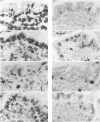
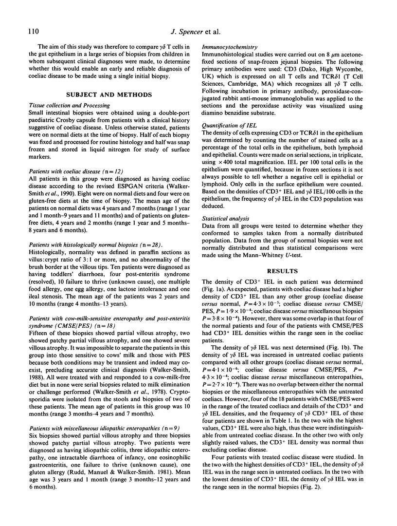
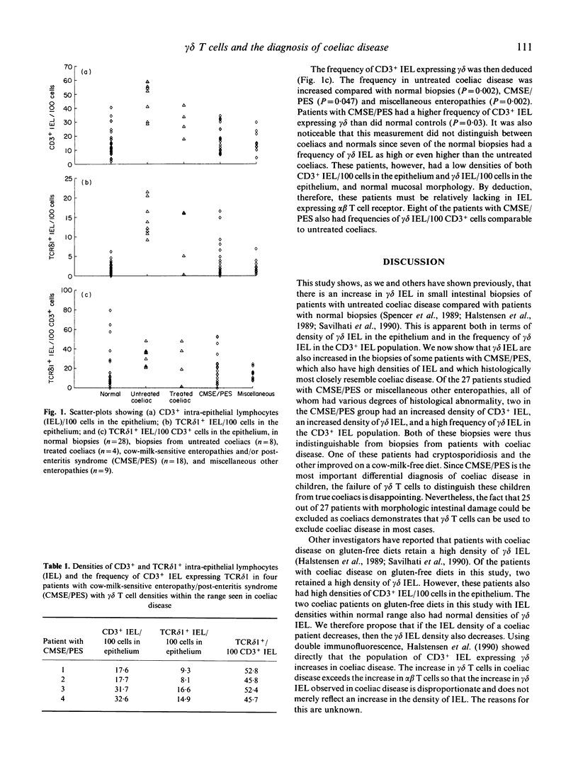
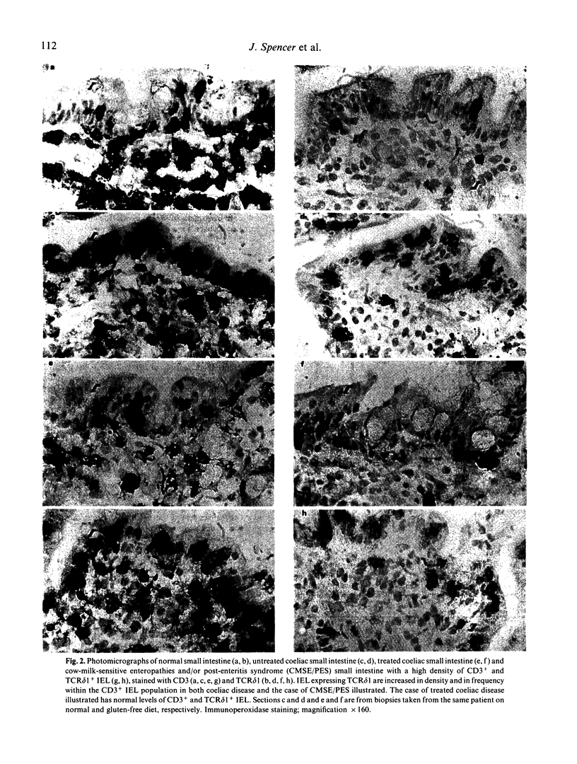
Gamma/delta T cells are increased in the gut epithelium of patients with coeliac disease compared with normal controls. The aim of this study was to determine whether the increase in gamma delta intraepithelial lymphocytes (IEL) is specific for coeliac disease, in which case it could be of diagnostic importance. Biopsies were obtained from children with no intestinal disease, coeliac disease, cow-milk-sensitive enteropathy/post-enteritis syndrome (CMSE PES) and miscellaneous other enteropathies (n = 67). Intraepithelial CD3+ and gamma delta T cells were identified in frozen sections using peroxidase immunohistochemistry. In normal biopsies there were 0-7 gamma delta IEL/100 cells in the epithelium. In untreated coeliac patients this increased to 9-22 gamma delta IEL/100 cells in the epithelium (P = 0.000004). Of 27 patients with morphologic intestinal damage which was not due to coeliac disease, four with CMSE/PES had gamma delta IEL/100 cells in the epithelium in the same range as the patients with coeliac disease. Of these, two had high densities of CD3+ IEL in the epithelium and were indistinguishable from patients with untreated coeliac disease. The other two could be excluded as possible coeliacs because their CD3+ IEL/100 epithelial cells were in the normal range. Thus an increase in gamma delta IEL is not specific for coeliac disease. However, enumeration of both of gamma delta IEL and CD3+ IEL densities will be useful in the exclusion of coeliac disease as a diagnosis in some children.
Full text
PDF
Images in this article
Selected References
These references are in PubMed. This may not be the complete list of references from this article.
- Chorzelski T. P., Beutner E. H., Sulej J., Tchorzewska H., Jablonska S., Kumar V., Kapuscinska A. IgA anti-endomysium antibody. A new immunological marker of dermatitis herpetiformis and coeliac disease. Br J Dermatol. 1984 Oct;111(4):395–402. doi: 10.1111/j.1365-2133.1984.tb06601.x. [DOI] [PubMed] [Google Scholar]
- Halstensen T. S., Scott H., Brandtzaeg P. Intraepithelial T cells of the TcR gamma/delta+ CD8- and V delta 1/J delta 1+ phenotypes are increased in coeliac disease. Scand J Immunol. 1989 Dec;30(6):665–672. doi: 10.1111/j.1365-3083.1989.tb02474.x. [DOI] [PubMed] [Google Scholar]
- Loft D. E., Marsh M. N., Crowe P. T. Rectal gluten challenge and diagnosis of coeliac disease. Lancet. 1990 Jun 2;335(8701):1293–1295. doi: 10.1016/0140-6736(90)91183-b. [DOI] [PubMed] [Google Scholar]
- Marsh M. N., Haeney M. R. Studies of intestinal lymphoid tissue. VI--Proliferative response of small intestinal epithelial lymphocytes distinguishes gluten- from non-gluten-induced enteropathy. J Clin Pathol. 1983 Feb;36(2):149–160. doi: 10.1136/jcp.36.2.149. [DOI] [PMC free article] [PubMed] [Google Scholar]
- Marsh M. N. Studies of intestinal lymphoid tissue. IV--The predictive value of raised mitotic indices among jejunal epithelial lymphocytes in the diagnosis of gluten-sensitive enteropathy. J Clin Pathol. 1982 May;35(5):517–525. doi: 10.1136/jcp.35.5.517. [DOI] [PMC free article] [PubMed] [Google Scholar]
- Revised criteria for diagnosis of coeliac disease. Report of Working Group of European Society of Paediatric Gastroenterology and Nutrition. Arch Dis Child. 1990 Aug;65(8):909–911. doi: 10.1136/adc.65.8.909. [DOI] [PMC free article] [PubMed] [Google Scholar]
- Rudd P., Manuel P., Walker-Smith J. Anaphylactic shock in an infant after feeding with a wheat rusk. A transient phenomenon. Postgrad Med J. 1981 Dec;57(674):794–795. doi: 10.1136/pgmj.57.674.794. [DOI] [PMC free article] [PubMed] [Google Scholar]
- Savilahti E., Viander M., Perkkiö M., Vainio E., Kalimo K., Reunala T. IgA antigliadin antibodies: a marker of mucosal damage in childhood coeliac disease. Lancet. 1983 Feb 12;1(8320):320–322. doi: 10.1016/s0140-6736(83)91627-6. [DOI] [PubMed] [Google Scholar]
- Spencer J., Isaacson P. G., Diss T. C., MacDonald T. T. Expression of disulfide-linked and non-disulfide-linked forms of the T cell receptor gamma/delta heterodimer in human intestinal intraepithelial lymphocytes. Eur J Immunol. 1989 Jul;19(7):1335–1338. doi: 10.1002/eji.1830190728. [DOI] [PubMed] [Google Scholar]
- Unsworth D. J., Walker-Smith J. A., Holborow E. J. Gliadin and reticulin antibodies in childhood coeliac disease. Lancet. 1983 Apr 16;1(8329):874–875. doi: 10.1016/s0140-6736(83)91411-3. [DOI] [PubMed] [Google Scholar]
- Walker-Smith J., Harrison M., Kilby A., Phillips A., France N. Cows' milk-sensitive enteropathy. Arch Dis Child. 1978 May;53(5):375–380. doi: 10.1136/adc.53.5.375. [DOI] [PMC free article] [PubMed] [Google Scholar]


