Abstract
Shigella flexneri possesses at least two putative high-affinity manganese acquisition systems, SitABCD and MntH. Mutations in the genes encoding the components of both of these systems were constructed in S. flexneri. The sitA mntH mutant showed reduced growth, relative to the wild type, in Luria broth (L broth) containing the divalent metal chelator ethylene diamino-o-dihydroxyphenyl acetic acid, and the addition of either iron or manganese restored growth to the level of the wild-type strain. Although the sitA mntH mutant was not defective in surviving exposure to superoxide generators, it was defective in surviving exposure to hydrogen peroxide. The sitA mntH mutant formed wild-type plaques on Henle cell monolayers but had a reduced ability to survive in activated macrophage lines. Expression of the S. flexneri sit and mntH promoters was higher when Shigella was in Henle cells than when it was in L broth. Expression of both the sit and mntH promoters was repressed by either iron or manganese, and this repression was partially dependent upon Fur and MntR, respectively. The mntH promoter, but not the sit promoter, exhibited OxyR-dependent induction in the presence of hydrogen peroxide.
Like most pathogens, the facultative intracellular bacterium that causes bacterial dysentery in humans, Shigella flexneri, requires iron for growth. Numerous studies have addressed the importance of high-affinity iron transport systems in the growth and pathogenesis of Shigella (reviewed in reference 34). However, far less is known about the importance of manganese acquisition in this and other pathogens, even though bacteria need to carefully control the transport of many metals across their membranes in order to maintain homeostasis. Additionally, the divalent metal iron and manganese cations may be interchangeable in some biological processes, as there are multiple similarities between the chelate structures of these ions. Finally, manganese, either as a cofactor in superoxide dismutases or alone as an nonenzymatic antioxidant, enhances oxidative stress survival in many bacteria. Thus, there has been a recent increased interest in understanding high-affinity manganese acquisition in bacteria (reviewed in reference 24).
Two types of high-affinity manganese acquisition systems have been reported for many bacterial species. MntH is a proton-dependent divalent cation transporter located in the cytoplasmic membrane (25, 29). Bacterial MntH proteins are homologous to the eukaryotic NRAMP (natural-resistance-associated macrophage protein) family of proteins which transport either Mn2+ or Fe2+ (7). Phagosome-associated NRAMP1 mediates resistance to infection by intracellular pathogens, presumably either by sequestering iron and manganese from the invading pathogen or by supplying metals for enzymes that mediate protection of the phagocyte against the reactive oxygen species generated to combat the infection (reviewed in references 3 and 9). NRAMP2 is widely expressed in eukaryotic cells and mediates divalent transition metal transport (12, 13).
High-affinity Mn2+ acquisition also can be mediated by ABC transport systems. These systems consist of a periplasmic ligand-binding protein (in gram-negative bacteria) or a lipoprotein (in gram-positive bacteria), two cytoplasmic membrane permeases, and two subunits of a peripheral cytoplasmic membrane protein with ATP binding motifs (8). The transported ligand binds to the periplasmic binding protein or lipoprotein and is transferred to cytoplasmic membrane permeases. Transport through the cytoplasmic membrane requires ATP hydrolysis by the associated ATPase. In S. flexneri, the ABC transport system Sit is predicted to transport manganese and iron (41). Similar manganese and manganese/iron ABC transport systems have been identified in numerous eubacteria and in a few members of the domain Archaea (8).
The role of the S. flexneri Sit system, which is found in all Shigella species, in high-affinity iron acquisition and virulence has been examined (41). The sit mutant showed reduced growth, relative to the wild-type strain, in Luria broth containing an iron chelator but formed wild-type plaques on Henle cell monolayers, indicating the bacterium was able to acquire iron and/or manganese in the host cell. This is similar to the phenotypes of S. flexneri strains carrying single mutations in genes encoding other high-affinity iron transport systems (41). However, double mutants with a defective Sit system and a second defective iron acquisition system (such as Iuc or Feo) formed slightly smaller plaques on Henle cell monolayers, and a mutant defective in all three systems (Sit, Feo, and Iuc) did not form plaques (41). All Shigella species also contain the predicted Mn2+ transporter MntH (47), but the previous study did not examine this transporter. The report presented here describes our initial investigation into the relative contributions of the Sit and MntH high-affinity metal acquisition systems to Shigella virulence and physiology and regulation of expression of these systems.
MATERIALS AND METHODS
Bacterial strains, plasmids, and growth conditions.
Bacterial strains and plasmids used in this work are listed in Table 1. Escherichia coli strains were routinely grown in Luria broth (L broth) or Luria agar (L agar). Shigella flexneri strains were grown in L broth or on tryptic soy broth agar plus 0.01% Congo red dye at 37°C. Several media were used to grow strains in reduced metal conditions: EZ-RDM medium (http://www.genome.wisc.edu/functional/protocols.htm) made without added iron or manganese or T medium (45) were used as base media and were supplemented with 0.2% to 0.4% glucose, 2 μg/ml nicotinic acid, and added metals as indicated. Alternatively, deferrated ethylene diamino-o-dihydroxyphenyl acetic acid (EDDA) was added to L broth (39). For oxidative stress regulation assays, bacteria were grown in M9 medium (42 mM Na2HPO4, 24 mM KH2PO4, 9 mM NaCl, 119 mM NH4Cl, 2 mM MgSO4, 0.1 mM CaCl2) containing 0.2% glucose and 2 μg/ml nicotinic acid under microaerobic conditions (full, capped tubes without shaking). Antibiotics were used at the following concentrations (per milliliter): 125 μg carbenicillin, 25 μg kanamycin, 15 μg chloramphenicol, 12.5 μg tetracycline, and 200 μg streptomycin.
TABLE 1.
Bacterial strains and plasmids used in this study
| Strain or plasmid | Characteristic(s) | Reference or source |
|---|---|---|
| E. coli strains | ||
| DH5α | endA1 hsdR17 supE44 thi-1 recA1 gyrA relA1 Δ(lacZYA-argF)U169 deoR [φ80dlacΔ(lacZ)M15] | 43 |
| SIP879 | MC4100 mntH::Mud1 aroB | 33 |
| SIP943 | MC4100 mntH::Mud1 aroB mntR | 33 |
| S. flexneri strains | ||
| SA100 | S. flexneri wild-type serotype 2a | 35 |
| SM100 | SA100 Strr | S. Seliger |
| SM166 | SM100 sitA::cam | 41 |
| UR002 | SM100 mntH::kan | This study |
| UR003 | SM100 sitA::cam mntH::kan | This study |
| UR009 | SM100 oxyR::kan | This study |
| UR010 | SM100 fur::Tn5 | This study |
| Plasmids | ||
| pLR29 | Promoterless GFP plasmid | 40 |
| pEG2 | sitA-gfp fusion on pLR29 | 40 |
| pRJ8 | mntH-gfp fusion on pLR29 | This study |
| pLAFR1 | Cosmid vector | 10 |
| pWKS30 | Low-copy-number cloning vector | 46 |
| pSIT | pLAFR1 carrying SA100 sit operon (pEG1) | 40 |
| pMNTH | pWKS30 carrying SA100 mntH | This study |
Recombinant DNA and PCR methods.
Plasmid DNA and chromosomal DNA were isolated using the QIAprep Spin Miniprep kit or the DNeasy tissue kit (QIAGEN, Santa Clarita, Calif.), respectively. Isolation of DNA fragments from agarose gels was performed using the QIAquick Gel Extraction kit (QIAGEN). All PCRs were carried out using either Taq (QIAGEN) or Pfu polymerase (Stratagene Cloning Systems, La Jolla, Calif.) according to the manufacturer's instructions. To clone the mntH gene, the gene was amplified from S. flexneri SM100 by PCR with primers mntHfor (5′GCGATAATCCCGATTGAAGA3′) and mntHrev (5′TTTCGCAAACCTTAAATGGC3′). The mntH fragment was ligated with pWKS30 (46) digested with SmaI and HincII to generate pMNTH.
Construction of Shigella mutants by P1 transduction.
The mntH and sitA mntH mutants were constructed by P1 transduction (30) of the mntH::kan mutation from E. coli MM2115 (25) into S. flexneri SM100 and SM166, respectively. The oxyR mutant and the fur mutant were constructed by P1 transduction of oxyR::kan from E. coli GS077 (48) and fur::Tn5 from S. flexneri SA211 (44), respectively, into SM100. Transductants were selected on Congo red agar containing the appropriate antibiotics and verified by PCR.
Oxidative stress assays.
Overnight cultures were diluted 1:50 in saline, and then 100 μl was spread on T agar and L agar plates. A Whatman no. 1 filter paper disk (8 mm in diameter) was added to the plates, and 10 μl of either hydrogen peroxide (1 M), paraquat (0.5 M), menadione (0.1 M), or phenazine methosulfate (0.1 M) was spotted onto the disk. The L agar and T medium plates were incubated for 24 h and 48 h, respectively, at 37°C, and zones of growth inhibition were measured.
Cell culture assays.
Monolayers of Henle cells (intestine 407 cells; American Type Culture Collection, Manassas, VA) were maintained in minimum essential medium (MEM) (Invitrogen, Carlsbad, CA) supplemented with 2 mM glutamine, 1× MEM nonessential amino acid solution (Invitrogen), and 10% fetal bovine serum (Invitrogen). Monolayers of the J774A.1 macrophage line (American Type Culture Collection) were routinely maintained in Dulbecco's modified Eagle's high-glucose medium (4.5 g glucose per liter, 4 mM l-glutamine, and 1 mM sodium pyruvate) (HyClone, Logan, UT) supplemented with 10% fetal bovine serum. The U937 human monoblastic macrophage-like cell line (American Type Culture Collection) was maintained in RPMI 1640 medium (with 25 mM HEPES and l-glutamine) (HyClone) supplemented with 1 mM sodium pyruvate, 1× MEM nonessential amino acids, and 10% fetal bovine serum. To induce macrophage differentiation, U937 cell cultures were supplemented with 200 ng phorbol myristate acetate per ml (Sigma Chemical, St. Louis, MO) for 7 days prior to Shigella infection and 200 ng lipopolysaccharide per ml (Sigma Chemicals) 1 day prior to Shigella infection (15, 28). All cell lines were grown in a 5% CO2 atmosphere at 37°C.
Plaque assays on Henle cells were done as described previously (31) using the modifications described by Hong et al. (16), and plaques were scored after 3 or 4 days. To assess the ability of Shigella to survive in macrophages, subconfluent monolayers of macrophage lines were infected with Shigella at a multiplicity of infection of 100 for 30 min and treated with gentamicin for 3 hours, after which time an intracellular multiplication assay was done as described previously (16).
For apoptosis assays, semiconfluent macrophage monolayers in 35-mm plates were infected with approximately 1 × 10⁸ bacteria as described for Henle cell invasions (16). After a 2-hour incubation period, apoptosis was assessed with the Vybrant apoptosis assay kit 2 (Invitrogen) according to the manufacturer's instructions.
Phenotypic microarray analysis.
Wild-type S. flexneri (SM100) and the sit mntH mutant (UR003) were sent to Biolog (Hayward, CA) for phenotypic microarray analysis (4). Two runs were performed for each strain.
Shigella sitA and mntH expression studies.
To construct the mntH-gfp reporter fusion, mntH primers mntHforP (5′GGCAACGATAATGGCAACTT3′) and mntHrevP (5′GCTCTAGACAGTAACCCGCCAATCAC3′) were used to amplify the Shigella mntH promoter from S. flexneri SM100. The PCR product was digested with XbaI and cloned into pLR29 (40) digested with XbaI and SmaI to generate pRJ8. sitA expression was measured using the plasmid-borne sitA-gfp fusion pEG2 (40). After growth under the appropriate conditions, fluorescence was quantitated by two methods: (i) using a Turner TD-700 fluorometer with a 486-nm excitation filter and a 510- to 700-nm emission filter and normalizing fluorescence to cell growth by dividing the fluorescence of the sample by the optical density at 650 nm or (ii) using a FACSCaliber (Becton Dickenson) fluorescence-activated cell sorter with an excitation at 488 nm to measure single-cell fluorescence. FACSCaliber settings were as follows: forward scatter of E01, side scatter of 505, and relative fluorescence between 515 and 545 nm of 798. Before fluorescence-activated cell sorting analysis, samples were fixed in 4% paraformaldehyde as described previously (40).
For quantitative reverse transcription-PCR (RT-PCR), total RNA was isolated from bacteria using the RNeasy Mini kit (QIAGEN), which included a DNase I treatment step to degrade DNA. Immediately, before RNA isolation, samples were stabilized by the addition of stabilizing buffer (95% ethanol, 5% phenol [pH 4.3]) for 5 min. Isolated RNA was treated again with DNase I (QIAGEN) to remove any residual contaminating DNA. cDNA was made from 200 ng total RNA using Superscript III (Invitrogen). Quantitative real-time PCR was performed on the cDNA samples using the Platinum SYBR green quantitative PCR kit (Invitrogen) and the Chromo4 continuous fluorescence detector with an Alpha unit DNA Engine thermocycler (Bio-Rad, Hercules, CA). Primers for the PCRs were as follows: for mntH, UR111 (5′TCAGCTACTGTG GGTTGTCG3′) and UR112 (5′GCCTGAACCCAATAGAACCA3′); for sitA, UR113 (5′TCTCAATGGGGTTCCAGAAG3′) and UR114 (5′AGCATTATCTGGCGACATCC3′); and for rrsA, UR117 (5′CACGATTACTAGCGATTCCGACTT3′) and UR118 (5′CGTCGTAGTCCGGATTGGA3′). Data analysis was done using the Opticon monitor software package (Bio-Rad). A standard curve was generated for each gene using 10-fold dilutions of S. flexneri SM100 chromosomal DNA, and the amount of cDNA for mntH or sit in each cDNA sample was extrapolated from the standard curve. Finally, the amount of mntH or sit gene expression was normalized to the housekeeping gene rrsA by dividing the relative amounts of mntH or sit cDNA by the relative amounts of rrsA cDNA in each sample.
RESULTS
Construction of strains UR002 and UR003.
Previous work in Shigella flexneri suggested that the Sit system, which was predicted to mediate high-affinity iron and/or manganese transport, contributed to iron acquisition in vitro and in cultured cells (41). However, the strains used in this study still contained the putative Mn2+ transporter MntH. To begin to elucidate the importance of manganese transport in Shigella, mutations were made in the S. flexneri mntH gene, singly and in combination with the sit mutation. These newly constructed mutants were designated UR002 (mntH::kan) and UR003 (sit::cam mntH::kan).
Contributions of the Sit and MntH systems to oxidative stress survival in Shigella.
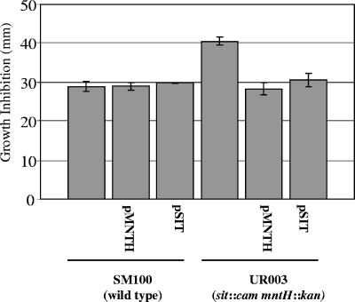
To examine the contributions of Sit and MntH to oxidative stress survival, we compared the zones of growth inhibition of S. flexneri sit, mntH, or sit mntH mutants in a disk diffusion assay with either hydrogen peroxide or various superoxide-generating compounds as oxidizing agents. Compared to the wild-type strain SM100, the sit and mntH single mutants showed similar sized zones of growth inhibition by all oxidizing agents (Table 2). Similarly, the sit mntH double mutant UR003 had zones of growth inhibition similar in size to those of the wild-type strain in the presence of the superoxide generators phenazine methosulfate and menadione (Table 2) and paraquat (data not shown). However, compared to the wild-type strain SM100, UR003 showed significantly larger zones of growth inhibition by hydrogen peroxide (Table 2). This increased sensitivity to hydrogen peroxide could be complemented by the addition of either the mntH gene on pMNTH or the sit gene on pSIT to the sit mntH mutant UR003 (Fig. 1). These results suggest that the S. flexneri MntH and Sit high-affinity metal transport systems have redundant function with respect to mediating bacterial survival in the presence of hydrogen peroxide.
TABLE 2.
Contributions of the Sit and MntH systems to oxidative stress survival in S. flexneri
| Strain | Zone of growth inhibition (mm)a on the following medium
|
|||||
|---|---|---|---|---|---|---|
| L agar with:
|
T agar with:
|
|||||
| H2O2b | PMSc | MDc | H2O2 | PMS | MD | |
| SM100 (wild type) | 22 ± 0 | 34 ± 2 | 22 ± 0 | 38 ± 3 | 47 ± 1 | 27 ± 4 |
| SM166 (sit::cam) | 20 ± 0 | 33 ± 3 | 20 ± 1 | 37 ± 1 | 46 ± 3 | 28 ± 1 |
| UR002 (mntH::kan) | 19 ± 1 | 33 ± 4 | 21 ± 2 | 34 ± 3 | 52 ± 4 | 29 ± 1 |
| UR003 (sit::cam mntH::kan) | 31 ± 4d | 35 ± 3 | 21 ± 1 | 54 ± 6d | 52 ± 3 | 27 ± 2 |
The data presented are the means ± standard errors of the means for three experiments.
10 μl of 1 M H2O2.
10 μl of 0.1 M phenazine methosulfate (PMS) or menadione (MD).
Significantly different (P < 0.05) compared to the value for the wild type.
FIG. 1.
Complementation of the hydrogen peroxide sensitivity of the Shigella flexneri sitA mntH mutant. S. flexneri strains (106 cells from overnight cultures) were spread on L agar, and an 8-mm-diameter disk containing 10 μl of 1 M H2O2 was placed in the middle of each plate. The zones of growth inhibition were measured after incubation at 37°C. The data presented are the means of three experiments, and the standard errors of the means (error bars) are indicated.
Contributions of the Sit and MntH systems to growth of S. flexneri in media containing EDDA.
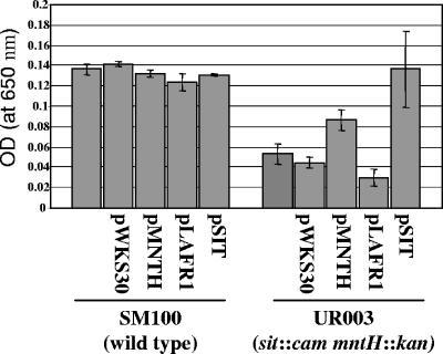
To examine the roles of Sit and MntH in the growth of Shigella in metal-limiting media, we compared the optical densities of cultures of wild-type S. flexneri to those of the sit mntH mutant after growth in EDDA-containing media. EDDA is a metal chelator with a much higher affinity for Fe3+ than for Mn2+, but when present at high levels, it chelates Mn2+ (26). After 12 h of growth in L broth containing 350 μM EDDA, growth of the sit mntH double mutant UR003 was significantly decreased relative to the wild-type strain SM100 (Fig. 2). Addition of the mntH gene on pMNTH or the sit gene on pSIT enhanced growth of UR003 in L broth containing EDDA (Fig. 2). There was no difference in the optical density between the strains after 12 h of growth in cultures grown without EDDA (data not shown).
FIG. 2.
The S. flexneri sitA mntH mutant has reduced growth in L broth containing EDDA. Overnight cultures of each strain were subcultured 1:1,000 into L broth containing 350 μM EDDA and grown at 37°C. The optical density (OD) of the cultures was measured after 12 h. The data presented are the means of three experiments, and the standard errors of the means (error bars) are indicated.
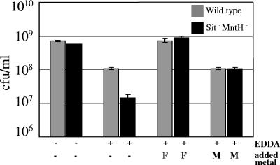
To determine whether the decreased growth of the S. flexneri sit mntH mutant in the presence of EDDA was due to depletion of iron or manganese in the medium, each of the metals was added to the EDDA-containing cultures (Fig. 3). Addition of FeCl3 to the cultures stimulated the growth of the sitA mntH mutant UR003 to wild-type level, which was comparable to that in cultures without EDDA (Fig. 3). This suggests that both wild-type Shigella and the sitA mntH mutant were iron starved in L broth containing EDDA. Addition of MnCl2 also abolished the difference in growth between the SM100 and UR003 strains; however, growth was not restored to the levels seen without EDDA (Fig. 3). This suggests that the sitA mntH mutant may also be slightly starved for manganese, since the addition of manganese restores the mutant's growth to the level of wild-type Shigella. Unlike MnCl2 and FeCl3, addition of CaCl2, MgCl2, or ZnCl2 did not enhance growth of the mutant (data not shown).
FIG. 3.
Addition of either iron or manganese to L broth containing EDDA restores growth of the S. flexneri sitA mntH mutant to wild-type levels. Overnight cultures of S. flexneri SM100 (wild type) and UR003 (sit::cam mntH::kan) were subcultured 1:1,000 into L broth with (+) or without (−) 350 μM EDDA, 350 μM ferric chloride (F), or 350 μM manganese chloride (M) as indicated. After 12 h of growth at 37°C, the number of bacteria per milliliter of culture was determined by viable plate counts. The data presented are the means of three experiments, and the standard errors of the means (error bars) are indicated.
Phenotypic microarray analysis of the S. flexneri sit mntH mutant.
In an attempt to identify other possible phenotypes associated with the absence of both the Sit and MntH high-affinity metal transport systems, the sit mntH mutant was analyzed using Phenotype MicroArrays (4). These arrays compared approximately 2,000 growth phenotypes in the wild-type S. flexneri and the sit mntH mutant. There were no differences in the metabolism of carbon (PM1 and PM2) or phosphate and sulfate (PM4). Likewise, there was no difference in the salt and osmotic tolerance (PM9) and no differences in pH and growth control (PM10). However, under certain growth conditions, there were several significant differences between the growth of the wild-type strain SM100 and sit mntH mutant UR003. Growth of UR003 was decreased, relative to that of SM100, in media containing gallic acid, 5,7-dichloro-8-hydroxyquinoline, or d-cycloserine (data not shown). In contrast, the sit mntH mutant UR003 grew better than the wild-type strain SM100 when xanthosine (PM3) or the dipeptide Val-Glu (PM8) was supplied as a nitrogen source (data not shown).
Contributions of the Sit and MntH systems to interactions with eukaryotic cells.
High-affinity iron acquisition is important for growth of Shigella in the eukaryotic intracellular environment (28, 41), but the importance of high-affinity manganese acquisition has not been examined. Thus, we tested the S. flexneri sit mntH mutant for growth in the Henle cell intracellular environment by examining its ability to form plaques on Henle cell monolayers. The sit mntH mutant UR003 formed plaques of the same number and size as the wild-type strain SM100 (data not shown). These data indicate that the Sit and MntH high-affinity transport systems are not required for growth in this eukaryotic cell type.
We also tested the S. flexneri sit mntH mutant for survival within macrophages by counting the number of viable intracellular bacteria after exposure to an activated macrophage line. The sit mntH mutant UR003 had a twofold decrease in survival in activated U937 macrophages relative to the wild-type strain SM100. Three hours postinfection, there were 30 ± 6 bacteria per infected macrophage in the UR003-infected macrophages, compared to 71 ± 10 bacteria in the SM100-infected macrophages. These data suggested that high-affinity manganese and/or iron acquisition may contribute to the ability of Shigella to survive in macrophages. Since induction of apoptosis is a mechanism by which Shigella survives uptake by macrophages (50), the decreased ability of the sit mntH mutant to survive in macrophages may be a result of the inability to activate apoptosis. We tested this hypothesis by comparing the ability of wild-type Shigella SM100 and the sit mntH mutant UR003 to induce apoptosis in macrophages. Both strains induced apoptosis in macrophages at the same levels (data not shown), suggesting that the decreased ability of the sit mntH mutant to survive in macrophages is not due to a defect in inducing apoptosis.
Regulation of Shigella sit and mntH expression.
Expression of the S. flexneri sit gene is induced in the eukaryotic intracellular environment (28, 40). Since the roles of MntH and Sit appear to be partially redundant, the regulation of expression of the genes that encode these high-affinity metal transporters in Shigella may also be similar. To test this hypothesis, we compared the regulation of the S. flexneri mntH and sit promoters using the mntH-gfp and sit-gfp transcriptional fusions, pRJ8 and pEG2, respectively. We infected Henle cells with S. flexneri strains carrying these fusions and measured the level of bacterial gene expression using flow cytometry. The relative amount of green fluorescent protein (GFP) per bacterial cell driven by the mntH and sit promoters increased from 76 ± 0 to 148 ± 6 (2-fold) and from 64 ± 1 to 778 ± 62 (12-fold), respectively, when Shigella was within the Henle cell.
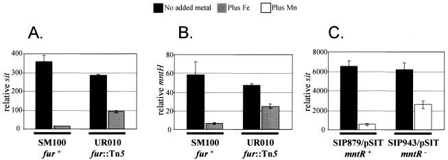
Previous work demonstrated that transcription from the Shigella sit promoter was repressed by either manganese or iron (41). We tested whether the mntH promoter was regulated by similar environmental stimuli by measuring expression of the mntH-gfp fusion in Shigella grown in minimal medium containing various metals. Addition of either FeSO4 or MnSO4 to the media repressed mntH expression fivefold and sixfold, respectively (Table 3). The Shigella mntH and sit promoters contain putative binding sites for the iron-responsive transcriptional repressor Fur and the manganese-responsive transcriptional repressor MntR. To examine the contributions of these proteins to Shigella mntH and sit gene regulation, we measured expression of these genes in strains containing mutations in the fur or mntR regulator genes using quantitative RT-PCR. In iron-replete media, expression of the sit and mntH genes was approximately sevenfold and fourfold greater, respectively, in the fur mutant UR010 than in the parent strain SM100 (Fig. 4A and B), suggesting that iron repression of mntH and sit expression is mediated by Fur. Since E. coli K-12 strains do not contain the sit genes, we transformed a previously isolated E. coli mntR mutant (33) with the Shigella sit genes on the low-copy-number cosmid pSIT to assess the role of MntR in manganese repression of sit gene expression. In manganese-replete media, expression of the sit gene was approximately fivefold greater in the mntR mutant SIP943 than in the parent strain SIP879 (Fig. 4C), suggesting that manganese repression of sit expression is mediated by MntR.
TABLE 3.
Repression of S. flexneri sit and mntH expression by manganese and iron
| Added metala | RFU/OD650b of SM100 carrying plasmid:
|
|
|---|---|---|
| pEG2 (sit-gfp) | pRJ8 (mntH-gfp) | |
| None | 2,614 | 731 |
| FeSO4 | 241 | 158 |
| MnSO4 | 132 | 124 |
| FeSO4 and MnSO4 | 82 | 89 |
Metal concentrations were 10 μM.
Fluorescence was quantitated by fluorometry (relative fluorescence units [RFU]) and was normalized to cell growth by dividing the fluorescence of the sample by the optical density at 650 nm (OD650). The data presented are the means of three experiments.
FIG. 4.
Contributions of Fur and MntR to repression of the S. flexneri sit and mntH promoters. (A and B) S. flexneri SM100 (wild type) and UR010 (fur::Tn5) were grown for 3 hours in EZ-RDM medium with or without 40 μM ferrous sulfate as indicated. (C) E. coli SIP879 (wild type) and SIP943 (mntR), both carrying pSIT, were grown for 5.5 h in EZ-RDM medium with or without 0.1 μM manganese sulfate as indicated. RNA was isolated from each sample and used to generate cDNAs, which were amplified using real-time PCR. The amount of sit and mntH gene expression was normalized to the housekeeping gene rrsA by dividing the relative amounts of sit or mntH cDNA by the relative amounts of rrsA cDNA in each sample. The data presented are the means of three experiments, and the standard errors of the means (error bars) are indicated.
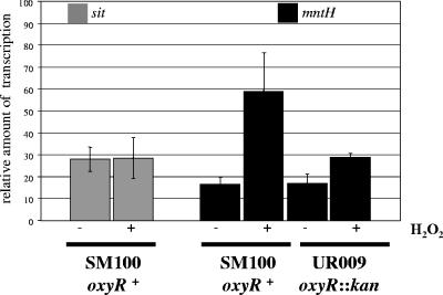
Since manganese enhances oxidative stress survival in many bacteria (24), the presence of oxidizing agents may induce the transcription of Shigella genes that encode high-affinity manganese transport systems. Thus, we measured expression of the mntH and sit genes in Shigella after exposure to hydrogen peroxide using quantitative RT-PCR. There was a threefold increase in the activity of the mntH promoter, but not the sit promoter, after exposure to 100 μM hydrogen peroxide (Fig. 5). The Shigella mntH promoter, but not the sit promoter, contains a putative binding site for the transcriptional activator OxyR, which activates gene expression in the presence of hydrogen peroxide. To examine the contribution of OxyR protein to regulation of Shigella mntH, we measured expression of mntH in S. flexneri UR009, which contains a mutation in the oxyR regulator gene. Induction of mntH expression by hydrogen peroxide was eliminated in strain UR009 (Fig. 5), suggesting that induction of mntH expression is mediated by OxyR.
FIG. 5.
OxyR-dependent hydrogen peroxide induction of the S. flexneri mntH promoter. S. flexneri SM100 (wild type) and UR009 (oxyR::kan) were grown microaerobically in M9 medium for 20 h. Hydrogen peroxide was added at a final concentration of 100 μM for 10 min. RNA was isolated from each sample and used to generate cDNAs which were amplified using real-time PCR. The amount of sit or mntH gene expression was normalized to the housekeeping gene rrsA by dividing the relative amounts of sit or mntH cDNA by the relative amounts of rrsA cDNA in each sample. The data presented are the means of three experiments, and the standard errors of the means (error bars) are indicated.
DISCUSSION
The roles of high-affinity iron transport systems in bacterial physiology and virulence have been studied in great detail; however, there has been less work focused on elucidating the contributions of high-affinity manganese transporters. Two Mn2+ transporter systems (the ABC transporter SitABCD and the NRAMP homologue MntH) have been identified and characterized in numerous physiologically diverse microorganisms (8, 24, 37). Although previous studies have examined the Shigella Sit system, a functional copy of MntH was still present in the strains used (27, 41). In the experiments reported here, we used S. flexneri mutants that lack both the Sit and MntH systems, which appear to have functional redundancy with respect to several phenotypes, to investigate the roles of these high-affinity metal transport systems in bacterial physiology and virulence.
Previous studies showed that the Shigella Sit system facilitated growth in medium that was limited for iron (27, 41). In our current experiments, addition of either FeCl3 or MnCl2 to L broth containing the metal chelator EDDA eliminated growth differences between the Shigella sit mntH mutant UR003 and the wild-type strain SM100. Furthermore, phenotypic microarray analysis showed that UR003 had reduced growth compared to wild-type SM100 in media containing 5,7-dichloro-8-hydroxyquinoline and gallic acid, which are metal chelators. Taken together, these data suggest that the Shigella MntH and Sit systems are transporters for both iron and manganese. The results of transport and/or growth assays with several bacterial species suggest that although many bacteria contain the Sit and/or MntH proteins, subtle differences in the amino acid sequences of the homologues in each species may result in altered affinities for metal cations so that the exact physiological roles of these systems may be distinct in different bacterial species (1, 2, 20, 23, 25, 29, 36, 42).
Animal studies with numerous intracellular and extracellular pathogens, including Salmonella enterica serovar Typhimurium, Yersinia pestis, Streptococcus mutans, Streptococcus pyogenes, and Staphylococcus aureus, have shown that high-affinity manganese transporters contribute to virulence (2, 5, 17, 20, 21, 25, 32). Although a role for the Shigella Sit system in intracellular growth has been demonstrated when other iron transporters are absent (41), in our current study elimination of both the Sit and MntH transporters did not affect plaque formation by Shigella on Henle cell monolayers. This suggests that either high-affinity manganese transport is not important for intracellular growth in epithelial cells or that there is another manganese transporter in Shigella that has not been identified. There is evidence for Mn2+ transport via other proteins distinct from MntH and Sit, such as the P-type ATPase in Lactobacillus plantarum (14) and the E. coli ZupT protein, which is related to the eukaryotic ZIP (ZRT, IRT-like protein) family of divalent metal ion transporters (11). Finally, in the absence of MntH and Sit, manganese transport in Shigella may proceed through high-affinity iron transport systems.
Although the S. flexneri sit mntH mutant was not defective in plaque formation on in vitro Henle cell monolayers, it is possible that Sit and/or MntH may be important for in vivo survival in human intestinal cells and/or at other stages of the infection process. Our studies showed that the S. flexneri sit mntH mutant did not survive as well as the wild-type strain in activated macrophage lines. This result coupled with the diminished ability of the Shigella sit mntH mutant to survive exposure to hydrogen peroxide suggests a role for high-affinity manganese transport systems in detoxification of reactive oxygen species produced by macrophages during the infection process. Similar results have been obtained in S. enterica serovar Typhimurium (5).
Several mechanisms could account for high-affinity manganese transporters facilitating oxidative stress survival. First, the transported Mn2+ could function as a nonenzymatic hydrogen peroxide detoxifier (reviewed in reference 18). Alternatively, Mn2+ may be a cofactor in an enzyme that detoxifies hydrogen peroxide. Direct detoxification of hydrogen peroxide is catalyzed by catalases. Although most of the identified catalases use iron as a redox cofactor, a manganese-dependent catalase (KatN) has been identified in S. enterica serovar Typhimurium (38). Shigella does not have a KatH homologue (47), but it is possible that there is another hydrogen peroxide-detoxifying enzyme in Shigella that does require manganese. Additionally, the superoxide dismutase SodA (MnSOD) requires Mn2+, and although the substrate for MnSOD is superoxide and not hydrogen peroxide, E. coli sodA mutants show a decreased ability to survive hydrogen peroxide (6).
The regulation of expression of the Shigella mntH gene and sit operon is consistent with the high-affinity metal acquisition phenotypes associated with the encoded gene products, namely, that gene expression is repressed by iron and manganese. Most of the iron and manganese repression is mediated through the transcriptional repressors Fur and MntR, respectively. In comparison to wild-type Shigella, there was significant derepression of gene expression in iron- or manganese-replete media in strains lacking functional Fur or MntR, respectively. This is similar to the regulation of these genes in other enteric bacteria including S. enterica serovar Typhimurium and E. coli (19, 20, 22, 33, 49).
Expression of the Shigella mntH gene increased in the presence of hydrogen peroxide; this induction was dependent on the transcriptional activator OxyR, which activates gene expression in the presence of hydrogen peroxide. Since manganese enhances bacterial survival after exposure to oxidizing agents, increased expression of high-affinity manganese transport genes in response to the presence of hydrogen peroxide would be beneficial. However, the sit operon was not induced by hydrogen peroxide in Shigella or in Salmonella (19). The sit operon in both species is located near the end of chromosomal genetic islands. Movement of these islands during evolution may have separated a putative OxyR binding site from the sit genes (41, 49). In avian E. coli, the sit operon is located on a large plasmid that contains the siderophore synthesis gene iucA. The 5′ end of the avian E. coli sit operon is located next to an insertion sequence; although Fur and MntH binding sites were identified, an OxyR binding site was not reported (42). Other unidentified regulatory elements in the promoters of the Shigella sit and mntH genes may also be different, since expression from the sit promoter increased more than expression from the mntH promoter did when Shigella was in Henle cells. Ikeda et al. have found that the sit genes are regulated by environmental signals other than metal levels and oxidative agents (19). Identifying additional signals and regulatory mechanisms that control expression of the Shigella sit and mntH genes and characterizing the rationale for the regulation may expose possible roles of these genes in the infection process.
Acknowledgments
We gratefully thank the following individuals for their generous help: Shelley Payne for critical reading of the manuscript, David Kehres for strain MG2114, Gisela Storz for strain GS077, and Silke Patzer for strains SIP879 and SIP943.
This work was supported by Public Health Service grant AI57511 awarded to L.R.-J. and by funding from the University of Richmond School of Arts and Sciences, the University of Richmond Quest Program, and the Virginia Federation of Independent Colleges.
Editor: D. L. Burns
REFERENCES
- 1.Agranoff, D. D., I. M. Monahan, J. A. Mangan, S. Krishna, and P. D. Butcher. 1999. Mycobacterium tuberculosis expresses a novel pH dependent divalent cation transporter belonging to the Nramp family. J. Exp. Med. 190:717-724. [DOI] [PMC free article] [PubMed] [Google Scholar]
- 2.Bearden, S. W., and R. D. Perry. 1999. The Yfe system of Yersinia pestis transports iron and manganese and is required for full virulence of plague. Mol. Microbiol. 32:403-414. [DOI] [PubMed] [Google Scholar]
- 3.Bellamy, R. 1999. The natural resistance-associated macrophage protein and susceptibility to intracellular pathogens. Microbes Infect. 1:23-27. [DOI] [PubMed] [Google Scholar]
- 4.Bouchner, B. R. 2003. New technologies to assess genotype-phenotype relationships. Nat. Rev. Genet. 4:309-314. [DOI] [PubMed] [Google Scholar]
- 5.Boyer, E., I. Bergevin, D. Malo, P. Gros, and M. F. Cellier. 2002. Acquisition of Mn(II) in addition to Fe(II) is required for full virulence of Salmonella enterica serovar Typhimurium. Infect. Immun. 70:6032-6042. [DOI] [PMC free article] [PubMed] [Google Scholar]
- 6.Carlioz, A., and D. Touati. 1986. Isolation of superoxide dismutase mutants in Escherichia coli: is superoxide dismutase necessary for aerobic life? EMBO J. 5:623-630. [DOI] [PMC free article] [PubMed] [Google Scholar]
- 7.Cellier, M., A. Belouchi, and P. Gros. 1996. Resistance to intracellular infections: comparative genomic analysis of Nramp. Trends Genet. 12:201-204. [DOI] [PubMed] [Google Scholar]
- 8.Claverys, J. P. 2001. A new family of high-affinity ABC manganese and zinc permeases. Res. Microbiol. 152:231-243. [DOI] [PubMed] [Google Scholar]
- 9.Forbes, J. R., and P. Gros. 2001. Divalent-metal transport by NRAMP proteins at the interface of host-pathogen interactions. Trends Microbiol. 9:397-403. [DOI] [PubMed] [Google Scholar]
- 10.Friedman, A. M., S. R. Long, S. E. Briwn, W. J. Buikema, and F. Ausubel. 1982. Construction of a broad host range cosmid cloning vector and its uses in the genetic analysis of Rhizobium meliloti. Gene 18:289-296. [DOI] [PubMed] [Google Scholar]
- 11.Grass, G., S. Franke, N. Taudte, D. H. Nies, L. M. Kucharski, M. E. Maguire, and C. Rensing. 2005. The metal permease ZupT from Escherichia coli is a transporter with a broad substrate spectrum. J. Bacteriol. 187:1604-1611. [DOI] [PMC free article] [PubMed] [Google Scholar]
- 12.Gruenheid, S., M. Cellier, S. Vidal, and P. Gros. 1995. Identification and characterization of a second mouse Nramp gene. Genomics 25:514-525. [DOI] [PubMed] [Google Scholar]
- 13.Gunshin, H., B. Mackenzie, U. V. Berger, Y. Gunshin, M. F. Romero, W. F. Boron, S. Nussberger, J. L. Gollan, and M. A. Hediger. 1997. Cloning and characterization of a mammalian proton-coupled metal-ion transporter. Nature 388:482-488. [DOI] [PubMed] [Google Scholar]
- 14.Hao, Z., S. Chen, and D. B. Wilson. 1999. Cloning, expression, and characterization of cadmium and manganese uptake genes from Lactobacillus plantarum. Appl. Environ. Microbiol. 65:4746-4752. [DOI] [PMC free article] [PubMed] [Google Scholar]
- 15.Harris, P. E. 1996. Human myeloid cell lines, p. 1-16. In L. A. Herzenberg, D. M. Wei, and C. Blackwell (ed.), Weir's handbook of experimental immunology. Blackwell Scientific Publications Ltd., Oxford, England.
- 16.Hong, M., Y. Gleason, E. E. Wyckoff, and S. M. Payne. 1998. Identification of two Shigella flexneri chromosomal loci involved in intercellular spreading. Infect. Immun. 66:4700-4710. [DOI] [PMC free article] [PubMed] [Google Scholar]
- 17.Horsburgh, M. J., S. J. Wharton, A. G. Cox, E. Ingham, S. Peacock, and S. J. Foster. 2002. MntR modulated expression of the Per regulon and superoxide resistance though control of manganese uptake. Mol. Microbiol. 44:1269-1286. [DOI] [PubMed] [Google Scholar]
- 18.Horsburgh, M. J., S. J. Wharton, M. Karavolos, and S. J. Foster. 2002. Manganese: elemental defence for a life with oxygen. Trends Microbiol. 10:496-501. [DOI] [PubMed] [Google Scholar]
- 19.Ikeda, J. S., A. Janakiraman, D. G. Kehres, M. E. Maguire, and J. M. Slauch. 2005. Transcriptional regulation of sitABCD of Salmonella enterica serovar Typhimurium by MntR and Fur. J. Bacteriol. 187:912-922. [DOI] [PMC free article] [PubMed] [Google Scholar]
- 20.Janakiraman, A., and J. M. Slauch. 2000. The putative iron transport system SitABCD encoded on SPI1 is required for full virulence of Salmonella typhimurium. Mol. Microbiol. 35:1146-1155. [DOI] [PubMed] [Google Scholar]
- 21.Janulczyk, R., S. Ricci, and L. Bjorck. 2003. MtsABC is important for manganese and iron transport, oxidative stress resistance, and virulence of Streptococcus pyogenes. Infect. Immun. 71:2656-2664. [DOI] [PMC free article] [PubMed] [Google Scholar]
- 22.Kehres, D. G., A. Janakiraman, J. M. Slauch, and M. E. Maguire. 2002. Regulation of Salmonella enterica serovar Typhimurium mntH transcription by H2O2, Fe2+, and Mn2+. J. Bacteriol. 184:3151-3158. [DOI] [PMC free article] [PubMed] [Google Scholar]
- 23.Kehres, D. G., A. Janakiraman, J. M. Slauch, and M. E. Maguire. 2002. SitABCD is the alkaline Mn2+ transporter of Salmonella enterica serovar Typhimurium. J. Bacteriol. 184:3159-3166. [DOI] [PMC free article] [PubMed] [Google Scholar]
- 24.Kehres, D. G., and M. E. Maguire. 2003. Emerging themes in manganese transport, biochemistry and pathogenesis in bacteria. FEMS Microbiol. Rev. 27:263-290. [DOI] [PubMed] [Google Scholar]
- 25.Kehres, D. G., M. L. Zaharik, B. B. Finlay, and M. E. Maguire. 2000. The NRAMP proteins of Salmonella typhimurium and Escherichia coli are selective manganese transporters involved in the response to reactive oxygen. Mol. Microbiol. 36:1085-1100. [DOI] [PubMed] [Google Scholar]
- 26.L'Eplattenier, F. L., I. Murase, and A. E. Martell. 1967. New multidentate ligands. VI. Chelating tendencies of N,N′-di(2-hydroxybenzyl) ethylene-N,N′-diacetic acid. J. Am. Chem. Soc. 89:837-843. [Google Scholar]
- 27.Liu, M., H. Liu, L. Sun, J. Dong, Y. Xue, S. Chen, and Q. Jin. 2005. Construction, detection and microarray analysis on the Shigella flexneri 2a sitC mutant. Sci. China Ser. C 48:228-240. [DOI] [PubMed] [Google Scholar]
- 28.Lucchini, S., H. Liu, Q. Jin, J. C. Hinton, and J. Yu. 2005. Transcriptional adaptation of Shigella flexneri during infection of macrophages and epithelial cells: insights into the strategies of a cytosolic bacterial pathogen. Infect. Immun. 73:88-102. [DOI] [PMC free article] [PubMed] [Google Scholar]
- 29.Makui, H., E. Roig, S. T. Cole, J. D. Helmann, P. Gros, and M. F. Cellier. 2000. Identification of the Escherichia coli K-12 Nramp orthologue (MntH) as a selective divalent metal ion transporter. Mol. Microbiol. 35:1065-1078. [DOI] [PubMed] [Google Scholar]
- 30.Miller, J. H. 1972. Experiments in molecular genetics. Cold Spring Harbor Laboratory, Cold Spring Harbor, N.Y.
- 31.Oaks, E. V., M. E. Wingfield, and S. B. Formal. 1985. Plaque formation by virulent Shigella flexneri. Infect. Immun. 48:124-129. [DOI] [PMC free article] [PubMed] [Google Scholar]
- 32.Paik, S., A. Brown, C. L. Munro, C. N. Cornelissen, and T. Kitten. 2003. The sloABCR operon of Streptococcus mutans encodes an Mn and Fe transport system required for endocarditis virulence and its Mn-dependent repressor. J. Bacteriol. 185:5967-5975. [DOI] [PMC free article] [PubMed] [Google Scholar]
- 33.Patzer, S. I., and K. Hantke. 2001. Dual repression by Fe2+-Fur and Mn2+-MntR of the mntH gene, encoding an NRAMP-like Mn2+ transporter in Escherichia coli. J. Bacteriol. 183:4806-4813. [DOI] [PMC free article] [PubMed] [Google Scholar]
- 34.Payne, S. M., and A. R. Mey. 2004. Pathogenic Escherichia coli, Shigella, and Salmonella, p. 199-218. In J. H. Crosa, A. R. Mey, and S. M. Payne (ed.), Iron transport in bacteria. American Society for Microbiology, Washington, D.C.
- 35.Payne, S. M., D. W. Niesel, S. S. Peixotto, and K. M. Lawlor. 1983. Expression of hydroxamate and phenolate siderophores by Shigella flexneri. J. Bacteriol. 155:949-955. [DOI] [PMC free article] [PubMed] [Google Scholar]
- 36.Platero, R. A., M. Jaureguy, F. J. Battistoni, and E. R. Fabiano. 2003. Mutations in sitB and sitD genes affect manganese-growth requirements in Sinorhizobium meliloti. FEMS Microbiol. Lett. 218:65-70. [DOI] [PubMed] [Google Scholar]
- 37.Richer, E., P. Courville, I. Bergevin, and M. F. Cellier. 2003. Horizontal gene transfer of “prototype” Nramp in bacteria. J. Mol. Evol. 57:363-376. [DOI] [PubMed] [Google Scholar]
- 38.Robbe-Saule, V., C. Coynault, M. Ibanez-Ruiz, D. Hermant, and F. Norel. 2001. Identification of a non-haem catalase in Salmonella and its regulation by RpoS (sigmaS). Mol. Microbiol. 39:1533-1545. [DOI] [PubMed] [Google Scholar]
- 39.Rogers, H. J. 1973. Iron-binding catechols and virulence in Escherichia coli. Infect. Immun. 7:438-444. [DOI] [PMC free article] [PubMed] [Google Scholar]
- 40.Runyen-Janecky, L. J., and S. M. Payne. 2002. Identification of chromosomal Shigella flexneri genes induced by the eukaryotic intracellular environment. Infect. Immun. 70:4379-4388. [DOI] [PMC free article] [PubMed] [Google Scholar]
- 41.Runyen-Janecky, L. J., S. A. Reeves, E. G. Gonzales, and S. M. Payne. 2003. Contribution of the Shigella flexneri Sit, Iuc, and Feo iron acquisition systems to iron acquisition in vitro and in cultured cells. Infect. Immun. 71:1919-1928. [DOI] [PMC free article] [PubMed] [Google Scholar]
- 42.Sabri, M., S. Leveille, and C. M. Dozois. 2006. A SitABCD homologue from an avian pathogenic Escherichia coli strain mediates transport of iron and manganese and resistance to hydrogen peroxide. Microbiology 152:745-758. [DOI] [PubMed] [Google Scholar]
- 43.Sambrook, J., E. F. Fritsch, and T. Maniatis. 1989. Molecular cloning: a laboratory manual, 2nd ed. Cold Spring Harbor Laboratory Press, Cold Spring Harbor, N.Y.
- 44.Schmitt, M. P., and S. M. Payne. 1988. Genetics and regulation of enterobactin genes in Shigella flexneri. J. Bacteriol. 170:5579-5587. [DOI] [PMC free article] [PubMed] [Google Scholar]
- 45.Simon, E. H., and I. Tessman. 1963. Thymidine requiring mutants of phage T4. Proc. Natl. Acad. Sci. USA 50:526-532. [DOI] [PMC free article] [PubMed] [Google Scholar]
- 46.Wang, R. F., and S. R. Kushner. 1991. Construction of versatile low-copy-number vectors for cloning, sequencing and gene expression in Escherichia coli. Gene 100:195-199. [PubMed] [Google Scholar]
- 47.Wei, J. M., B. Goldberg, V. Burland, M. M. Venkatesan, W. Deng, G. Fournier, G. F. Mayhew, G. Plunkett III, D. J. Rose, A. Darling, B. Mau, N. T. Perna, S. M. Payne, L. J. Runyen-Janecky, S. Zhou, D. C. Schwartz, and F. R. Blattner. 2003. Complete genome sequence and comparative genomics of Shigella flexneri serotype 2a strain 2457T. Infect. Immun. 71:2775-2786. [DOI] [PMC free article] [PubMed] [Google Scholar]
- 48.Zheng, M., X. Wang, L. J. Templeton, D. R. Smulski, R. A. LaRossa, and G. Storz. 2001. DNA microarray-mediated transcriptional profiling of the Escherichia coli response to hydrogen peroxide. J. Bacteriol. 183:4562-4570. [DOI] [PMC free article] [PubMed] [Google Scholar]
- 49.Zhou, D., W. D. Hardt, and J. E. Galan. 1999. Salmonella typhimurium encodes a putative iron transport system within the centisome 63 pathogenicity island. Infect. Immun. 67:1974-1981. [DOI] [PMC free article] [PubMed] [Google Scholar]
- 50.Zychlinsky, A., M. C. Prevost, and P. J. Sansonetti. 1992. Shigella flexneri induces apoptosis in infected macrophages. Nature 358:167-169. [DOI] [PubMed] [Google Scholar]


