Abstract
The practical application of gene therapy as a treatment for cystic fibrosis is limited by poor gene transfer efficiency with vectors applied to the apical surface of airway epithelia. Recently, folate receptor alpha (FRα), a glycosylphosphatidylinositol-linked surface protein, was reported to be a cellular receptor for the filoviruses. We found that polarized human airway epithelia expressed abundant FRα on their apical surface. In an attempt to target these apical receptors, we pseudotyped feline immunodeficiency virus (FIV)-based vectors by using envelope glycoproteins (GPs) from the filoviruses Marburg virus and Ebola virus. Importantly, primary cultures of well-differentiated human airway epithelia were transduced when filovirus GP-pseudotyped FIV was applied to the apical surface. Furthermore, by deleting a heavily O-glycosylated extracellular domain of the Ebola GP, we improved the titer of concentrated vector severalfold. To investigate the folate receptor dependence of gene transfer with the filovirus pseudotypes, we compared gene transfer efficiency in immortalized airway epithelium cell lines and primary cultures. By utilizing phosphatidylinositol-specific phospholipase C (PI-PLC) treatment and FRα-blocking antibodies, we demonstrated FRα-dependent and -independent entry by filovirus glycoprotein-pseudotyped FIV-based vectors in airway epithelia. Of particular interest, entry independent of FRα was observed in primary cultures of human airway epithelia. Understanding viral vector binding and entry pathways is fundamental for developing cystic fibrosis gene therapy applications.
Viral vector-mediated gene transfer to airway epithelial cells as therapy for diseases such as cystic fibrosis (CF) presents many challenges. The pulmonary epithelia and resident immune effector cells possess innate and adaptive defenses that evolved to prevent the invasion of microbes; these same defenses may act as barriers for gene transfer vectors (27). In addition, Moloney leukemia virus-based retroviral vectors are hampered by the low proliferation rate of adult airway epithelial cells (26). In an effort to overcome adverse immune responses to vector-encoded proteins and the transient nature of gene expression with nonintegrating vector systems, we utilize a vector system based on the nonprimate lentivirus, feline immunodeficiency virus (FIV) (28, 29).
The apical surface of airway epithelia is notably resistant to gene transfer with several vector systems and therefore presents additional challenges for CF gene therapy. This obstacle is generally attributed to the basolateral polarization of the receptors for several classes of viral vectors. For example, the receptors for serotype 2 and serotype 5 adenovirus (CAR) and AAV-2 (heparin sulfate proteoglycan) are predominantly expressed on the basolateral surface of airway epithelia (6, 25). In the case of enveloped viruses, the glycoproteins bind to specific receptors on the cell surface to initiate membrane fusion; these envelope-receptor interactions dictate cellular tropism. Furthermore, the receptors for many commonly used retroviral envelopes appear to be functionally expressed basolaterally in polarized epithelia (4). To overcome these barriers to gene transfer, an improved understanding of receptor biology and virus-cell interactions is essential. There have been significant advances in the understanding of encapsidated virus-receptor interactions; however, the cellular receptors for many of envelope glycoproteins available to pseudotype lentiviral vectors are unknown or poorly characterized.
Filoviral envelope glycoproteins have received attention as candidates for pseudotyping retrovirus to target a variety of cell types (31). Together Ebola virus (EBO) and Marburg virus (MRB) comprise the two members of the viral family Filoviridae. In contrast to other enveloped RNA viruses such as paramyxoviruses, both retroviruses and filoviruses have a single type 1 transmembrane structural protein that assembles into homotrimers and mediates both receptor binding and fusion (30). Sequence analysis suggests an evolutionary relationship between the envelope glycoproteins of filoviruses and retroviruses (19, 20, 30), with evidence that filoviruses can infect the host through an airborne mechanism (10). However, the origins of the viruses or how they are maintained in nature is presently unknown.
Interestingly, recent studies suggest that the folate receptor alpha (FRα) directs cellular entry of retroviruses pseudotyped with filoviral envelope glycoproteins (2). FRα, a glycosylphosphatidylinositol (GPI)-linked protein, was identified as a potential receptor for filoviral glycoproteins through utilization of an expression library in cells nonpermissive for viral entry. In Jurkat cells, FRα expression facilitated MRB- or EBO-pseudotyped Moloney leukemia virus entry. In addition, FR-blocking reagents inhibited transduction of HOS or HeLa cells (2). These data suggest that FRα provides one cellular entry pathway for wild-type filovirus as well as retroviruses pseudotyped with filoviral glycoproteins. Recently, the feasibility of pseudotyping human immunodeficiency virus-based lentiviral vectors with filovirus envelope glycoproteins to target airway epithelia has been demonstrated (14); however, FRα-dependent entry has not been investigated in this important model system.
Herein we report the expression of FRα in airway epithelia and the polarity of FRα expression in well-differentiated primary cultures of human airway epithelia. In addition, we investigate the role of FRα as a receptor for FIV-based lentivirus pseudotyped with filoviral envelope glycoproteins in airway epithelial cells.
MATERIALS AND METHODS
Culture of human airway epithelia.
Airway epithelia were isolated from trachea or bronchi and were grown at the air-liquid interface as described previously (13). All preparations used were well differentiated (>2 weeks old; resistance > 1,000 Ω-cm2). This study was approved by the Institutional Review Board at the University of Iowa. A549 and H441 cell lines are derived from human lung carcinomas, and IB3 and HBE cell lines are transformed human airway cells. The cell lines HT1080 (ATCC 12012), HOS (ATCC CRL-1543), IB3 (34), and KB (ATCC CCL-17) were maintained in Dulbecco's modified Eagle's medium (Gibco)-10% fetal bovine serum (FBS). A549 (ATCC CCL-185) cells were maintained in Dulbecco's modified Eagle's medium F12 (catalog no. 11320-033; Gibco)-10% FBS. H441 (ATCC HTB-174) cells were maintained in RPMI medium (Gibco)-10% FBS. HBE (5) cells were maintained in modified Eagle's medium (Gibco)-10% FBS. In addition, each medium was supplemented with penicillin (100 U/ml) and streptomycin (100 μg/ml). In the FRα-blocking studies, the cells were washed and maintained 72 h in RPMI medium lacking folic acid (Gibco; 27016-021) and 5% FBS prior to the addition of the blocking reagent.
Vector production.
The second-generation FIV vector system utilized in this study was reported previously (11, 29). The FIV vector construct expressed the β-galactosidase cDNA directed by the cytomegalovirus promoter. All of the envelope constructs reported in this study utilized the cytomegalovirus early gene promoter to direct transcription. Those envelopes include the vesicular stomatitis virus G protein (VSVG), the EBO (Zaire strain) envelope glycoprotein (pEZGP [9]), and the MRB (Musoke variant) envelope glycoprotein (pMBGP [30]). EBOΔO (pEZGP 309-489) has been previously described (9). All of the filoviral envelope constructs reported in this study were expressed from pcDNA3.1 (Invitrogen, Carlsbad, Calif.)-derived plasmids. Pseudotyped FIV vector particles were generated by transient transfection of plasmid DNA into 293T cells as described previously (11). FIV vector preparations were titered on HT1080 cells at limiting dilutions, and these titers were used to calculate the multiplicities of infection (MOIs). In addition, we found that the filoviral glycoprotein conferred enough stability to the lentiviral vector to withstand centrifuge concentration of greater than 1,000-fold (data not shown); however, we typically concentrated vector 250-fold by centrifugation for in vitro experiments.
RPA.
FRα mRNA levels were determined by RNase protection assay (RPA) as previously described (23). The FRα probe was a partial cDNA sequence cloned into pCR2.1-TOPO (Invitrogen). The human β-actin cDNA templates were obtained from Ambion (Austin, Tex.). The full-length probe for FRα and human actin were 541 and 315 bp, respectively. The expected protected fragment sizes were 413 and 245 bp, respectively. The RPA reaction was conducted by using an RPA III kit with the manufacturer's protocol (Ambion) and was quantified with a Molecular Dynamics Storm 620 PhosphorImager System and the ImageQuant software provided by the manufacturer.
FACS.
For fluorescence-activated cell sorter (FACS) analysis, approximately 106 cells were first incubated in suspension with FcγII αCD32-blocking antibody (14531; Stem Cell Technologies) on ice for 15 min. Then a monoclonal antibody against FRα (MOv18; a kind gift from Silvana Canevari [18]) or the appropriate immunoglobulin G1 (IgG1) isotype control (554121; Pharmingen) was added and incubated on ice for 30 min. Cells were washed three times with 3% FBS in 1× phosphate-buffered saline (PBS). A goat anti-mouse fluorescein isothiocyanate (FITC)-conjugated secondary antibody (31569; Pierce) was then added and again incubated on ice for 30 min. Finally, cells were washed as before and resuspended in 500 μl of 3% FBS in 1× PBS. Data were collected by using a FACScan flow cytometer (Becton Dickinson), and the data were analyzed using CellQuest software.
Western blot analysis.
Western blot analysis for verifying FRα protein expression was conducted by using standard techniques. Briefly, cell lysates were denatured for 5 min at 100°C in Laemmli sample buffer, electrophoresed on 10% polyacrylamide gels (161-1155; Bio-Rad) at 125 V, and transferred to pure nitrocellulose (162-0145; Bio-Rad) overnight at 200 mA. The membrane was probed with a monoclonal anti-human FRα primary antibody (MOv18; 3 mg/ml) at 1:1,000 and was detected by using goat anti-mouse IgG conjugated to alkaline phosphatase at a 1:1,000 dilution (A-1682; Sigma).
Immunohistochemistry and confocal microscopy.
Epithelial cells were rinsed with 1× PBS, fixed in 2% paraformaldehyde for 5 to 10 min, and rinsed with 1× PBS. The epithelial cells were then incubated for 30 min at 37°C with a monoclonal anti-human FRα antibody (MOv18) or the appropriate isotype control diluted 1:100 in Hank's buffer (Gibco). The cells were washed with 1× PBS and were incubated with an FITC-conjugated anti-mouse secondary antibody (F-4143; Sigma) diluted 1:100 in 1× PBS for 30 min at 37°C. The primary and secondary antibodies were always applied to both the apical and basolateral surfaces of nonpermeabilized cells. Images were captured with a Bio-Rad MRC-1024 Hercules laser scanning confocal microscope equipped with a Kr/Ar laser.
Viral vector administration.
Pseudotyped FIV vector was applied directly to immortalized cell lines for 4 h at 37°C. Following incubation with the vector, cells were rinsed in media and cultured for 4 days. Following the 4-day incubation, cells were harvested and β-galactosidase activity was quantified. Primary cultures of human airway epithelial cells were transduced with pseudotyped FIV vector by diluting vector preparations in media to achieve the desired MOI, and 100 μl of the solution was applied to the apical surface of airway epithelial cells. After incubation for 4 h at 37°C, the virus was removed and cells were further incubated at 37°C for 4 days. To infect airway epithelia with pseudotyped FIV vector from the basolateral side, the Millicell culture insert containing the airway epithelia was turned over and the virus was applied to the basolateral surface for 4 h in 100 μl of media. Following the 4-h infection, the virus was removed and the culture insert was turned upright and allowed to incubate at 37°C, 5% CO2, for 4 days.
β-Galactosidase quantification and AZT administration.
The Galacto-light chemiluminescent reporter assay (Tropix, Bedford, Mass.) was used to quantify β-galactosidase activity following the manufacturer's protocol. The relative light units were quantified with a luminometer (Monolight 3010; Pharmingen) and were standardized to total protein as determined by modified Lowry assay (23240; Pierce Biotechnology) by using the manufacturer's protocol. To verify that the β-galactosidase activity observed in the transduced cells was due to reverse transcription-dependent expression and not the result of pseudotransduction of β-galactosidase present in the vector preparations, cells were infected in the presence or absence of zidovudine (AZT). The cells were incubated with 50 μM AZT (GlaxoWellcome) for 24 h prior to infection and were maintained in the media following vector administration.
Administration of FRα blockers.
To cleave GPI-linked cell surface proteins, cells were pretreated with 2 U of phosphatidylinositol-specific phospholipase C (PI-PLC) (P-6466; Molecular Probes)/ml for 2 h at 37°C. Following enzyme treatment, viral vector challenge and β-galactosidase detection proceeded as described above. To specifically block FRα, cells were preincubated with a mouse monoclonal FRα-blocking antibody (IgG1) (HFBP 458; a generous gift of Wilbur Franklin [7]) or an isotype control antibody (554121; Pharmingen) for 15 min at room temperature. We diluted the purified blocking antibody or isotype control antibody 1:100 in Ultroser G (2-μg/ml final concentration). Following antibody treatment, viral vector challenge and β-galactosidase detection proceeded as described above.
Statistics.
Unless otherwise noted, all numerical data are presented as the mean plus or minus standard deviation. Statistical analysis was performed with a two-tailed, unpaired Student t test by using Microsoft Excel software.
RESULTS
Expression of FRα in primary cultures of human airway epithelial cells.
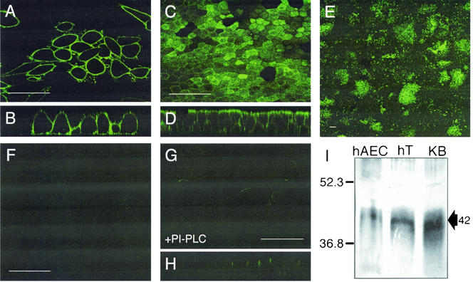
The identification of FRα as a mediator of filovirus cell entry offers the ability to investigate virus-host cell receptor interactions and pathways of infection. Chan and colleagues observed that PI-PLC and FRα antiserum inhibited entry of retrovirus pseudotyped with filoviral glycoproteins in a select group of cell types; however, the authors acknowledged that FRα may not facilitate virus entry into all cell types (2). We investigated FRα expression in primary cultures of well-differentiated human airway epithelia. To determine the polarity of expression, we immunostained the primary cultures with an FRα-specific monoclonal antibody under nonpermeabilizing conditions and imaged the cells with confocal microscopy. KB, a cell line known to express FRα at high levels, exhibited abundant cell surface levels of FRα (Fig. 1A) with no polarity of expression when viewed in vertical sections (Fig. 1B). Similarly, FRα protein expression was easily detected by immunostaining primary cultures of airway epithelia (Fig. 1C). When viewed in vertical sections, FRα was abundantly expressed at the apical surface (Fig. 1D). Interestingly, when viewed en face at a lower magnification, the distribution of FRα was heterogeneous (Fig. 1E). The reason for this expression pattern is not yet known; however, initial observations suggest that the pattern is not the result of cell-type-specific expression (e.g., ciliated versus nonciliated cells). Furthermore, the distribution was not affected by culturing cells under folate-free or excess-folate conditions (data not shown). No fluorescent signal was detected when an IgG1 isotype control primary antibody and the FITC-conjugated secondary antibody were used (Fig. 1F). As an added control to verify antibody specificity, the epithelia were pretreated with an enzyme that cleaves GPI linkages, i.e., PI-PLC. As shown, PI-PLC pretreatment removed detectable FRα expression as evaluated en face (Fig. 1G) or in vertical sections (Fig. 1H). Further confirmation of FRα expression was achieved by the detection of a 42-kDa band by Western blot analysis of primary airway cells, human trachea, and KB cell lysates (Fig. 1I). These data demonstrate that, in a polarized sheet of primary epithelia at a given time, not all cells express FRα but that within FRα-positive cells there is substantial expression at the apical surface.
FIG. 1.
FRα expression in primary cultures of human airway epithelia. Cells were fixed and incubated with an FRα-specific monoclonal antibody, followed by addition of an anti-mouse FITC-conjugated secondary antibody. KB cells were viewed by using confocal microscopy from an en face (A) or vertical (B) section. Primary cultures of airway epithelia were also viewed en face at high power (C) and from a vertical section (D), as well as en face at low power (E). To confirm antibody specificity, an isotype control primary antibody was used (F). Primary cultures of human airway cells were also imaged following PI-PLC treatment to confirm enzyme function (en face [G] and vertical [H] sections). (I) Western blot of indicated protein samples was conducted by using the same FRα-specific monoclonal antibody followed by an anti-mouse alkaline phosphatase-conjugated secondary antibody. The expected 42-kDa band is indicated with an arrow. hAEC, human airway epithelial cell; hT, human trachea; KB, KB cell line. Scale bars = 50 μm (A, C, F, and G) or 100 μm (E).
Pseudotyping FIV-based vectors with filoviral glycoproteins.
Abundant filovirus receptor is localized at the apical surface of airway epithelia; therefore, we hypothesized that pseudotyping FIV vector with filoviral glycoproteins would confer apical transduction properties. High viral titers facilitate in vitro experiments and are of prime concern when one is designing in vivo experiments. We routinely attain titers ranging from 108 to 109 transducing units (TU)/ml by pseudotyping FIV-based vectors with the MuLV amphotropic, VSV, or Ross River virus envelope glycoproteins following a 250-fold centrifuge concentration (12, 26, 28, 29). However, pseudotyping FIV-based vectors with filoviral envelope glycoproteins resulted in significantly lower viral titers. As shown in Table 1, when FIV was pseudotyped with the wild-type EBO and MRB glycoproteins, we achieved average titers of 5.5 × 10⁶ TU/ml and 2.5 × 10⁴ TU/ml, respectively, following a 250-fold centrifuge concentration. We engineered alterations in the envelope constructs designed to enhance filoviral glycoprotein incorporation into FIV virions and tested the effects on viral titer.
TABLE 1.
Modifications of the MRB and EBO envelope glycoproteins and the resultant titers of pseudotyped FIV vectorsa
| Construct name | Description of mutation | Increase (n-fold) | Mean ± SE | n |
|---|---|---|---|---|
| EBO WT | 5.5 × 10⁶ ± 3 × 10⁶ | 8 | ||
| EBOΔO | Deletion of EBO amino acids 309-489 inclusive | 73.98 | 4.1 × 10⁸ ± 1 × 10⁸ | 10 |
| MRB WT | 2.5 × 10⁴ ± 8 × 10³ | 6 | ||
| MRBΔO | Deletion of MRB amino acids 294-424 inclusive | 0.01 | 2.5 × 10² ± 2 × 10² | 4 |
| MRB C671A | Cysteine-to-alanine mutation at position 671 | 2.44 | 6.0 × 10⁴ ± 2 × 10⁴ | 7 |
| MRB C671S | Cysteine-to-serine mutation at position 671 | 0.58 | 1.4 × 10⁴ ± 9 × 10³ | 6 |
| MRB C673A | Cysteine-to-alanine mutation at position 673 | 1.18 | 2.9 × 10⁴ ± 1 × 10⁴ | 4 |
| MRB C673S | Cysteine-to-serine mutation at position 673 | 0.16 | 3.8 × 10³ ± 1 × 10³ | 4 |
| MRB F676stop | Phenylalanine to stop codon at position 676; isoleucine to lysine at position 675 | 2.43 | 6.0 × 10⁴ ± 5 × 10⁴ | 4 |
| MRB Y679stop | Tyrosine to stop codon at position 679 | 1.00 | 2.5 × 10⁴ ± 2 × 10⁴ | 6 |
Construct names and the descriptions of the mutations are indicated. WT, wild type. Titers are expressed as the mean (TU per milliliter) plus or minus standard error. The increases (n-fold) were calculated by normalizing the corresponding titer of FIV vector pseudotyped with the wild-type glycoprotein to 1. Significant increases of mutant glycoprotein-pseudotyped FIV vector titer over that of wild-type counterparts are indicated in boldface type.
Deletion of the O-glycosylated region from the extracellular domain of filoviral glycoproteins.
An initial strategy for enhancing filovirus glycoprotein-pseudotyped FIV vector titer was to delete an expansive region from the extracellular domain thought to be heavily O glycosylated. By deletion of this region, the efficiency of envelope protein synthesis and of transport to the cell surface is enhanced (9). This region may be functionally less important than the flanking regions of the protein simply because there is little sequence conservation in this region among all filoviral isolates. The deletion of amino acids 309 to 489 from the EBO glycoprotein (EBOΔO) resulted in a marked 74-fold increase in titer over the average titer obtained with the wild-type EBO glycoprotein (Table 1). Unfortunately, a comparable deletion in the extracellular domain of the MRB construct (MRBΔO) resulted in a dramatic loss of titer. Since potential differences between the EBO and MRB pseudotype transduction efficiencies were discovered to be of interest, multiple additional avenues were therefore pursued to increase MRB viral titer.
Mutating cytoplasmic tail acylation sites or generating cytoplasmic tail truncations of the MRB envelope glycoprotein.
Multiple studies have demonstrated that pseudotyping efficiency is influenced by the nature of the glycoprotein cytoplasmic domain (3, 16, 33). We designed alterations to the MRB envelope glycoprotein cytoplasmic domain intended to relieve steric interference or alter protein folding in such a way as to promote glycoprotein incorporation into the assembling virion. The MRB envelope glycoprotein contains two intracellular, potentially acylated cysteines that may interfere with efficient virion assembly. Each cysteine was mutated to either an alanine or a serine (Table 1). Encouragingly, the C671A mutation resulted in a greater-than-twofold increase in viral titer; however, the other point mutations resulted in no titer enhancement. The incorporation of a serine at either position significantly decreased viral titer (Table 1). In addition to these point mutations, we constructed two C-terminal deletions of the MRB envelope glycoprotein. Deleting the terminal 3 amino acids (Y679stop) had no effect on FIV vector titer compared to that of the wild-type glycoprotein; however, deleting the terminal 6 amino acids (F676stop) resulted in a greater-than-twofold increase in viral titer (Table 1). The latter construct introduces a lysine at position 675 for proper anchoring of the glycoprotein in the plasma membrane. Although the enhancements in titer were modest, these data demonstrate the potential of C-terminal mutagenesis of the MRB envelope glycoprotein to boost vector titer.
Replacing the cytoplasmic tail of the MRB envelope glycoprotein with the MuLV amphotropic or FIV envelope cytoplasmic tail.
Replacing the C terminus of the MRB envelope glycoprotein with that of another glycoprotein known to efficiently incorporate into budding FIV virions is an additional strategy that we pursued to enhance the viral titers with the MRB glycoprotein. The amphotropic (ampho) envelope glycoprotein from MuLV was a prime candidate for chimera construction for multiple reasons. Importantly, both the MRB and ampho glycoproteins are type 1 transmembrane proteins that form homotrimers when expressed on the cell surface (17, 30). In addition, similar strategies have proven effective for pseudotyping lentivirus vectors (21, 24). Using biochemical analyses and sequence homologies of the MRB and ampho glycoproteins (20), we chose to engineer the fusion site at MRB glycoprotein residue 670 and ampho 619 (termed MRB/ampho). In addition, we fused the MRB extracellular and transmembrane domains to an ampho intracellular domain with a mutation in the putative endocytosis signal (termed MRB/amphoY665A) (8) or a truncated ampho C terminus (termed MRB/amphoΔ650/675). Unfortunately, none of the MRB/ampho chimeric glycoproteins enhanced vector titers (data not shown).
In addition to MRB/ampho chimeric glycoproteins, we pursued a parallel approach by using the native FIV envelope glycoprotein sequence to generate MRB/FIVenv chimeric glycoproteins. We fused the MRB extracellular domain and transmembrane domain to the native FIV envelope intracellular domain. We hypothesized that the native C terminus of the envelope protein sequence would efficiently incorporate into the assembling vector. The chimera junction point is likely critical; therefore, we chose multiple fusion points ranging from amino acids 807 to 815 of the FIV envelope. However, none of the MRB/FIVenv chimeric constructs resulted in an increase of FIV vector titer (data not shown).
In summary, of the MRB glycoprotein mutations, only C671A and F676stop resulted in increased FIV vector titers (Table 1). However, these increases were modest and did not confer the titers conducive to multifaceted in vitro experiments with MOIs greater than 1. For this reason, the subsequent studies focused on vectors pseudotyped with the EBO or EBOΔO glycoprotein.
Apical transduction of human airway epithelia by filovirus glycoprotein-pseudotyped FIV.
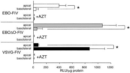
To test the polarity of vector transduction in primary cultures of human airway epithelia, FIV pseudotyped with wild-type EBO glycoprotein (EBO-FIV), EBO envelope glycoprotein with the deletion of amino acids 309 to 489 (EBOΔO-FIV), or VSV glycoprotein (VSVG-FIV) was applied to the apical or basolateral surface as indicated for 4 h at an MOI of ∼5 (Fig. 2). Following vector application, the cells were washed and incubated for 4 days before quantification of β-galactosidase expression as described in Materials and Methods. Both EBO-FIV and EBOΔO-FIV transduced airway epithelia from the apical surface at greater efficiency than from the basolateral surface. In contrast, VSVG-FIV transduced the basolateral surface more efficiently than the apical surface. In each case, pretreating the epithelia with AZT abolished β-galactosidase expression, indicating that the observed β-galactosidase activity is not the result of pseudotransduction. These data indicate that filovirus glycoprotein-pseudotyped FIV vectors preferentially transduce airway epithelia from the apical surface, thus providing indirect evidence in support of FRα as a receptor for vector entry.
FIG. 2.
Transduction levels of polarized airway cell lines with pseudotyped FIV vector. Primary cultures were transduced with pseudotyped FIV vector applied to the apical or basolateral surface. In addition, as a control for pseudotransduction, cells were pretreated with AZT for 24 h before vector application. Four days after initial vector incubation, cells were harvested and the β-galactosidase activity was quantified and normalized to total protein. n = 3 (samples from three independent human specimens). RLU, relative light units. ∗, P < 0.01.
Blocking transduction of filovirus glycoprotein-pseudotyped FIV with FRα inhibitors.
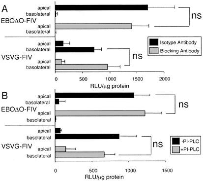
To examine the role of FRα as a receptor for filovirus glycoprotein-pseudotyped FIV, FRα-blocking or -cleaving experiments were conducted on primary cultures of well-differentiated human airway epithelia (Fig. 3). In each condition, FRα-specific blocking antibody or an isotype control antibody was applied to both the apical and basolateral surfaces of the airway epithelia (Fig. 3A). Following the incubation with the antibody, the vector was administered to the apical or basolateral surface as indicated (Fig. 3A). The ability of the viral vector to transduce cells was quantified by β-galactosidase activity normalized to total protein. The FRα-blocking antibody had no effect on the VSVG-FIV control vector. Contrary to expectation, application of the FRα-blocking antibody had no effect on transduction efficacy when EBOΔO-FIV was applied to either the apical or basolateral surface compared to the isotype antibody (Fig. 3A). Apical transduction of EBOΔO-FIV remained significantly higher than basolateral transduction in the presence or absence of the blocking antibody.
FIG. 3.
Transduction levels of polarized airway cell lines with pseudotyped FIV vector following FRα-blocking antibody or PI-PLC treatment. (A) Following pretreatment with an IgG1 isotype antibody (black bars) or an FRα-blocking antibody (gray bars), primary cultures were transduced with pseudotyped FIV vector applied to the apical or basolateral surface. (B) Following pretreatment with PI-PLC (gray bars) or without PI-PLC (black bars), primary cultures were transduced with pseudotyped FIV vector applied to the apical or basolateral surface at an MOI of 5. Four days after initial vector incubation, cells were harvested, analyzed by β-galactosidase assay, and normalized to total protein. n = 3 (samples from three independent human specimens). RLU, relative light units. ∗, P < 0.01.
To complement the FRα-blocking antibody studies, we pursued additional experiments utilizing the GPI-linkage cleaving enzyme, PI-PLC, to remove FRα from the cell surface. The ability of PI-PLC to cleave FRα from primary cells was evaluated by immunofluorescence (Fig. 1G and H). Cells were pretreated with PI-PLC, followed by incubation with EBOΔO-FIV or VSVG-FIV. Similar to the blocking antibody, PI-PLC did not reduce the transduction efficiency of EBOΔO-FIV or the VSVG-FIV control vector in primary cultures of airway epithelia (Fig. 3B). Again, apical transduction of EBOΔO-FIV remained significantly higher than basolateral transduction in the presence or absence of PI-PLC. These data indicate that FRα is not required as a receptor for EBOΔO-FIV in primary cultures of human airway epithelia.
Expression of FRα in immortalized airway epithelial cells.
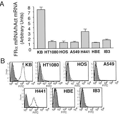
In light of this unexpected observation, we evaluated the potential role of FRα as a mediator of filoviral entry into multiple immortalized cell lines. We quantified the levels of FRα mRNA (Fig. 4A) and FRα protein (Fig. 4B) in control cell lines and airway epithelium-derived cell lines. KB is perhaps the most commonly utilized cell line for studying FRα in vitro and therefore, as expected, displayed abundant FRα mRNA and protein. Interestingly, our FIV vector-titering cell line, HT1080, exhibited minimal expression of FRα mRNA and protein (Fig. 4). Two cell lines utilized by Chan and colleagues (2), HeLa (not shown) and HOS, displayed relatively high and low FRα levels, respectively. Of the four airway-derived immortalized cell lines tested (H441, HBE, A549, and IB3) only H441 exhibited relatively high levels of FRα by either RPA or FACS analysis.
FIG. 4.
Relative FRα mRNA and protein levels from immortalized cell lines. (A) Total mRNA from the indicated cell lines was purified and analyzed by RPA by using FRα and human actin (hACT)-specific [α-32P]UTP-labeled antisense probes. Signal abundance was quantified with a PhosphorImager, and FRα expression was normalized to actin expression. n = 3. (B) To determine relative FRα protein abundance, protein lysates from the indicated cell lines were incubated with either an FRα-specific monoclonal antibody (open curve) or an isotype control (shaded curve). Samples were then incubated with an anti-mouse FITC-conjugated secondary antibody and were subjected to FACS analysis as described in Materials and Methods. n ≥ 2.
Transduction of immortalized airway epithelia by filovirus glycoprotein-pseudotyped FIV.
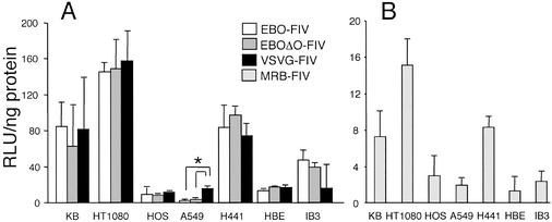
The indicated cell lines were transduced at an MOI of ∼5 with EBO-FIV, EBOΔO-FIV, or VSVG-FIV (Fig. 5A). Due to titer limitations, the cell lines were transduced with wild-type MRB envelope-pseudotyped FIV (MRB-FIV) at an MOI of ∼0.5 (Fig. 5B). As shown, HT1080 cells were consistently transduced with the greatest efficiency for all vectors (Fig. 5A and B) despite expressing only low levels of FRα (Fig. 4). KB cells and H441 cells expressed much higher levels of FRα than HT1080 cells but transduced at ∼50% the efficiency of HT1080 cells. IB3, HBE, HOS, and A549 were transduced at lower levels. EBO-FIV and EBOΔO-FIV transduced each cell line with similar efficiency, and the pattern of transduction between the cell lines was closely comparable with that of the MRB-FIV. Interestingly, the transduction efficiency of the VSVG-FIV was not significantly different from those of the EBO-FIV and EBOΔO-FIV except for the A549 cell line. VSVG-FIV transduced A549 cells at approximately fivefold-greater efficacy than EBO-FIV or EBOΔO-FIV.
FIG. 5.
Relative transduction levels of immortalized cell lines with pseudotyped FIV vector. The indicated cell lines were transduced with EBO-FIV, EBOΔO-FIV, or VSVG-FIV at an MOI of 5 (A) or MRB-FIV at an MOI of 0.5 (B). Four days after initial vector incubation, cells were harvested, analyzed by β-galactosidase assay, and normalized to total protein. MOIs were calculated by using vector titers determined on HT1080 cells. RLU, relative light units. n = 3. ∗, P < 0.01.
Blocking transduction of filovirus glycoprotein-pseudotyped FIV with FRα inhibitors.
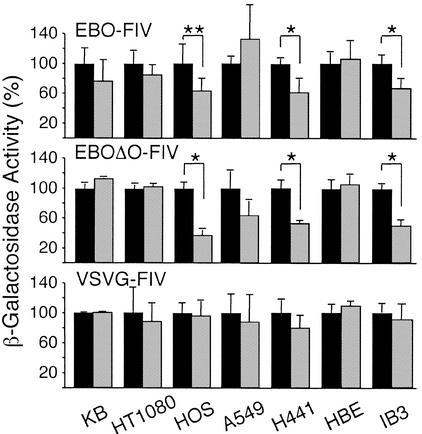
The contribution of FRα in facilitating filovirus glycoprotein-pseudotyped FIV binding and entry into airway-derived and control cell lines was further tested by pretreating cells with an FRα-specific blocking antibody or an isotype control antibody (Fig. 6). The ability of the viral vector to transduce cells was again quantified by β-galactosidase activity normalized to total protein. The transduction of KB and HT1080 cells was not significantly affected by pretreatment with the blocking antibody. These data suggest the existence of FRα-independent pathways for filoviral infection. Of the airway-derived cell lines, the blocking antibody successfully reduced transduction of the EBO-FIV and EBOΔO-FIV in H441 and IB3 cells but not in A549 or HBE cells. As previously observed by Chan et al., the FRα-blocking antibody successfully reduced the transduction efficiency in HOS cells (2). Importantly, EBO-FIV and EBOΔO-FIV were blocked to a similar extent in each cell line, suggesting that deleting the O-glycosylated region of the EBO glycoprotein does not alter its binding and fusion specificity or cell tropism.
FIG. 6.
Transduction levels of immortalized cell lines with pseudotyped FIV vector following FRα-blocking antibody treatment. Following pretreatment with an IgG1 isotype antibody (black bars) or an FRα-blocking antibody (gray bars), the indicated cell lines were transduced with EBO-FIV, EBOΔO-FIV, or VSVG-FIV at an MOI of 5. Four days after initial vector incubation, cells were harvested, analyzed by β-galactosidase assay, and normalized to total protein. The mean β-galactosidase activity following isotype antibody pretreatment for each cell line and vector administration is normalized to 100%. ∗, P < 0.01; ∗∗, P < 0.05.
In addition, each cell line was transduced with VSVG-FIV in the presence or absence of the FRα-blocking antibody (Fig. 6). No inhibitory effects on VSVG-FIV transduction were found for any cell line. In cells pretreated with AZT or lamivudine (not shown), β-galactosidase activity was dramatically reduced, indicating that the observed expression is not the result of pseudotransduction.
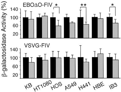
In a manner similar to that for the experiments in the primary cultures of airway epithelia, we utilized PI-PLC to remove FRα from the cell surface of the immortalized cell lines. The ability of PI-PLC to cleave FRα from immortalized cell lines was confirmed by FACS analysis (data not shown). In each case the enzyme treatment efficiently removed FRα. Cells were pretreated with PI-PLC, followed by incubation with EBOΔO-FIV or VSVG-FIV. PI-PLC treatment did not inhibit transduction of EBOΔO-FIV in KB, HT1080, A549, or HBE cells (Fig. 7). However, PI-PLC treatment did reduce the transduction efficiency of EBOΔO-FIV in HOS, H441, and IB3 cells (Fig. 7). In no cell line was the transduction efficiency of VSVG-FIV affected by pretreatment of PI-PLC. Together with the blocking antibody studies, these data further support the existence of FRα-independent entry pathways for filovirus pseudotypes in human airway epithelia (Fig. 3 and 6).
FIG. 7.
Transduction levels of airway- and non-airway-derived cell lines with pseudotyped FIV vector following PI-PLC treatment. Following pretreatment with PI-PLC (gray bars) or without PI-PLC (black bars), the indicated cell lines were transduced with EBOΔO-FIV or VSVG-FIV at an MOI of 5. Four days after initial vector incubation, cells were harvested, analyzed by β-galactosidase assay, and normalized to total protein. The mean β-galactosidase activity without PI-PLC pretreatment for each cell line and vector administration is normalized to 100%. ∗, P < 0.01; ∗∗, P < 0.05.
DISCUSSION
In this study we investigated the contribution of FRα to the transduction ability of a filovirus glycoprotein-pseudotyped FIV-based vector in airway epithelia and cell lines. Importantly, EBO-FIV and EBOΔO-FIV transduced well-differentiated polarized airway epithelia more efficiently when applied to the apical surface than when applied to the basolateral surface. This notable observation is unique among the many pseudotyped retroviral vectors reported to date and is consistent with previously published results (14). Kobinger et al. (14) demonstrated that EBO-pseudotyped human immunodeficiency virus transduced the apical surface of airway epithelia with greater efficacy than it did the basolateral surface. We demonstrated that the EBO envelope glycoprotein confers its apical transducing ability to a nonprimate lentiviral vector and tested its ability to utilize FRα as an avenue for cellular entry. Interestingly, we observed abundant expression of FRα at the apical surface by immunohistochemistry (Fig. 1). Indeed, GPI-linked proteins will typically sort to the apical surface of polarized epithelial cells (15). Based on this circumstantial evidence, one might expect FRα to contribute to binding and entry of filovirus glycoprotein-pseudotyped FIV vectors into primary cultures of human airway epithelia; however, we found that the folate-blocking antibody or PI-PLC treatments failed to inhibit the transduction of these cells.
As summarized in Table 2, we observed various levels of FRα expression in the cell lines tested. Furthermore, the relative levels of FRα protein or mRNA did not necessarily correlate with EBOΔO-FIV transduction efficiency and blocking FRα failed to inhibit transduction. Generally, we observed that PI-PLC or an FRα-blocking antibody was likeliest to perturb transduction in cell lines that already had low levels of transduction and low levels of FRα expression, such as with HOS or IB3 cells (Table 2). However, in no cell line did the blocking efforts reduce the transduction efficiencies by greater than twofold. Cell lines with ample levels of FRα or cells that were easily transduced with EBOΔO-FIV, such as KB and HT1080 cells, respectively, were typically not responsive to PI-PLC or blocking antibody treatments (Table 2).
TABLE 2.
Summary of FRα expression and EBOΔO transduction levels of airway- and non-airway-derived cell linesa
| Cell type | Tissue origin* | Detection assay result for:
|
Relative FRα level of:
|
Blockage by:
|
|||
|---|---|---|---|---|---|---|---|
| Galacto- lytes | Cell count | Protein | mRNA | PI- PLC | Blocking antibody | ||
| HT1080 | Fibroblast | +++ | +++ | Low | Low | No | No |
| HOS | Bone | + | NT | Low | Low | Yes | Yes |
| KB | Cervix | ++ | NT | High | High | No | No |
| H441 | Airway | ++ | + | High | High | Yes | Yes |
| HBE | Airway | + | ± | Low | Low | No | No |
| IB3 | Airway | + | ± | Low | Low | Yes | Yes |
| A549 | Airway | + | ± | Low | Low | No | No |
| HAE | Airway | + | ± | High** | Detected† | No | No |
Data from Fig. 1 through 8 are summarized for convenience. *, all cell lines are human derived. **, data acquired by immunofluorescence and Western blotting. †, data acquired by nonquantitative reverse transcriptase PCR. HAE, primary cultures of human airway epithelia. Plus signs represent qualitative comparisons as follows: +++, very abundant expression, ++, moderate expression; +, detectable expression; ±, expression detectable at limit of assay resolution; and NT, not tested.
One interesting outcome of our studies was the observation that the deletion of the O-glycosylation region from the EBO glycoprotein greatly increased the titers of FIV vector relative to results for the wild-type glycoprotein. In contrast, no titer benefit was observed in previous studies in which Simmons et al. (22) constructed EBO O-glycosylation deletion mutants. Therefore, the deletion site is likely critical. Biochemical analysis of the EBOΔO construct by Jeffers et al. (9) indicated that O-glycosylation deletion facilitates glycoprotein processing, incorporation into retrovirus particles, and viral transduction (9). Moreover, there may be an important therapeutic benefit of deleting the putative O-glycosylation domain; the serine-threonine-rich O-glycosylated region (or mucin-like domain) has been implicated as a pathogenic determinant of EBO (32). Yang and colleagues (32) observed that this region of the glycoprotein was required for vascular cytotoxicity and concluded that it may contribute to hemorrhage during an EBO infection. In addition, Simmons et al. (22) observed that the O-glycosylated region was necessary to induce loss of cell adherence. Therefore, by constructing the EBOΔO construct for pseudotyping, we may have added a safety benefit as well as achieving a dramatic boost in titer.
Interestingly, a similar deletion of the O-glycosylation region of the MRB glycoprotein did not yield a similar increase in titer. Clearly, the selection of which amino acids to delete in such an experiment is vital to producing a functional protein; therefore, more MRB deletion constructs will need to be tested. One potential untested possibility is that the mutations resulted in increased MRB envelope production but decreased the stability of the envelope complex, leading to decreased titer following concentration by centrifugation. As a whole, our efforts to increase the titers of MRB-FIV met with only limited success. Results similar to those for EBO-FIV and EBOΔO-FIV were evident; we observed that MRB-FIV transduces the apical surface of human airway epithelia with greater efficacy than it does the basolateral surface (data not shown); therefore, examining the differences in tropism and transduction efficiencies between EBO-FIV and MRB-FIV at equivalent MOIs is of interest.
We envision a model in which EBO-FIV has multiple avenues for binding and entry into different cell types. For example, C-type lectins have recently been demonstrated to confer enhanced cellular entry efficacy for EBO-pseudotyped retrovirus in hematopoietic cells (1). The contribution of DC-SIGN and L-SIGN in airway epithelial cells has not yet been investigated; however, the study conducted by Alvarez et al. (1) supports a model of cellular entry for EBO-FIV that is more complex than a single receptor. Cell lines such as HT1080 or KB may present a number of opportunities for EBO-FIV entry. In such cells, blocking FRα has little effect on transduction levels. Conversely, a cell line such as HOS or IB3 may offer fewer pathways for EBO-FIV entry; therefore, the individual contribution of FRα is much greater. Our data confirm the previous finding that the FRα acts as a cofactor contributing to filovirus entry into some but not all cell types (2).
In conclusion, our results do not eliminate the possibility that FRα may contribute to the binding and entry of filovirus glycoprotein-pseudotyped lentivirus into airway epithelia in vivo; however, its presence was not required to achieve transduction in primary cultures. These data also indicate that other unknown cellular factors are functioning as viral receptors for filovirus glycoprotein-pseudotyped lentivirus. These results confirm that filoviral glycoproteins are excellent candidates for pseudotyping lentiviral vectors to target the apical surface of airway epithelia. Although further challenges must be overcome for CF gene therapy, the availability of an integrating vector that transduces polarized airway epithelia cells from the apical surface will facilitate additional preclinical studies in vitro and in vivo.
Acknowledgments
We are grateful for the contributions of Phil Karp and to Pary Weber for culturing the human epithelial cells and to Sara Verbridge for her valued assistance. We thank Silvana Canevari and Wilbur Franklin for providing us with FRα antibodies. We are thankful for Curt Sigmund's provision of access to his PhosphorImager and the accompanying software and for Joe Zabner's critical review of the manuscript.
We acknowledge the support of the Gene Transfer Vector Core, Cell Culture Core, and Cell Morphology Core partially supported by the Cystic Fibrosis Foundation, NHLBI (PPG HL-51670), and the Center for Gene Therapy for Cystic Fibrosis (NIH P30 DK-54759). This work was supported by NIH grants RO1 HL-61460 (P.B.M.), PPG HL-51670 (P.B.M.), and NS-34568 (B.L.D.) and by the Cystic Fibrosis Foundation. P.L.S. was supported by an individual National Research Service Award (HL-67623).
REFERENCES
- 1.Alvarez, C. P., F. Lasala, J. Carrillo, O. Muñiz, A. L. Corbí, and R. Delgado. 2002. C-type lectins DC-SIGN and L-SIGN mediate cellular entry by Ebola virus in cis and in trans. J. Virol. 76:6841-6844. [DOI] [PMC free article] [PubMed] [Google Scholar]
- 2.Chan, S. Y., C. J. Empig, F. J. Welte, R. F. Speck, A. Schmaljohn, J. F. Kreisberg, and M. A. Goldsmith. 2001. Folate receptor-alpha is a cofactor for cellular entry by Marburg and Ebola viruses. Cell 106:117-126. [DOI] [PubMed] [Google Scholar]
- 3.Christodoulopoulos, I., and P. M. Cannon. 2001. Sequences in the cytoplasmic tail of the gibbon ape leukemia virus envelope protein that prevent its incorporation into lentivirus vectors. J. Virol. 75:4129-4138. [DOI] [PMC free article] [PubMed] [Google Scholar]
- 4.Compans, R. W. 1995. Virus entry and release in polarized epithelial cells. Curr. Top. Microbiol. Immunol. 202:209-219. [DOI] [PubMed] [Google Scholar]
- 5.Cozens, A. L., M. J. Yezzi, K. Kunzelmann, T. Ohrui, L. Chin, K. Eng, W. E. Finkbeiner, J. H. Widdicombe, and D. C. Gruenert. 1994. CFTR expression and chloride secretion in polarized immortal human bronchial epithelial cells. Am. J. Respir. Cell Mol. Biol. 10:38-47. [DOI] [PubMed] [Google Scholar]
- 6.Duan, D., Y. Yue, Z. Yan, P. B. McCray, Jr., and J. F. Engelhardt. 1998. Polarity influences the efficiency of recombinant adenoassociated virus infection in differentiated airway epithelia. Hum. Gene Ther. 9:2761-2776. [DOI] [PubMed] [Google Scholar]
- 7.Franklin, W. A., M. Waintrub, D. Edwards, K. Christensen, P. Prendegrast, J. Woods, P. A. Bunn, and J. F. Kolhouse. 1994. New anti-lung-cancer antibody cluster 12 reacts with human folate receptors present on adenocarcinoma. Int. J. Cancer Suppl. 8:89-95. [DOI] [PubMed] [Google Scholar]
- 8.Grange, M. P., V. Blot, L. Delamarre, I. Bouchaert, A. Rocca, A. Dautry-Varsat, and M. C. Dokhelar. 2000. Identification of two intracellular mechanisms leading to reduced expression of oncoretrovirus envelope glycoproteins at the cell surface. J. Virol. 74:11734-11743. [DOI] [PMC free article] [PubMed] [Google Scholar]
- 9.Jeffers, S. A., D. A. Sanders, and A. Sanchez. 2002. Covalent modifications of the Ebola virus glycoprotein. J. Virol. 76:12463-12472. [DOI] [PMC free article] [PubMed] [Google Scholar]
- 10.Johnson, E., N. Jaax, J. White, and P. Jahrling. 1995. Lethal experimental infections of rhesus monkeys by aerosolized Ebola virus. Int. J. Exp. Pathol. 76:227-236. [PMC free article] [PubMed] [Google Scholar]
- 11.Johnston, J. C., M. Gasmi, L. E. Lim, J. H. Elder, J.-K. Yee, D. J. Jolly, K. P. Campbell, B. L. Davidson, and S. L. Sauter. 1999. Minimum requirements for efficient transduction of dividing and nondividing cells by feline immunodeficiency virus vectors. J. Virol. 73:4991-5000. [DOI] [PMC free article] [PubMed] [Google Scholar]
- 12.Kang, Y., C. S. Stein, J. A. Heth, P. L. Sinn, A. K. Penisten, P. D. Staber, K. L. Ratliff, H. Shen, C. K. Barker, I. Martins, C. M. Sharkey, D. A. Sanders, P. B. McCray, Jr., and B. L. Davidson. 2002. In vivo gene transfer using a nonprimate lentiviral vector pseudotyped with Ross River virus glycoproteins. J. Virol. 76:9378-9388. [DOI] [PMC free article] [PubMed] [Google Scholar]
- 13.Karp, P. H., T. O. Moniger, S. P. Weber, T. S. Nesselhauf, J. L. Launspach, J. Zabner, and M. J. Welsh. 2002. An in vitro model of differentiated human airway epithelia. Methods Mol. Biol. 188:115-137. [DOI] [PubMed] [Google Scholar]
- 14.Kobinger, G. P., D. J. Weiner, Q. C. Yu, and J. M. Wilson. 2001. Filovirus-pseudotyped lentiviral vector can efficiently and stably transduce airway epithelia in vivo. Nat. Biotechnol. 19:225-230. [DOI] [PubMed] [Google Scholar]
- 15.Lisanti, M. P., I. W. Caras, M. A. Davitz, and E. Rodriguez-Boulan. 1989. A glycophospholipid membrane anchor acts as an apical targeting signal in polarized epithelial cells. J. Cell Biol. 109:2145-2156. [DOI] [PMC free article] [PubMed] [Google Scholar]
- 16.Mammano, F., F. Salvatori, S. Indraccolo, A. De Rossi, L. Chieco-Bianchi, and H. G. Göttlinger. 1997. Truncation of the human immunodeficiency virus type 1 envelope glycoprotein allows efficient pseudotyping of Moloney murine leukemia virus particles and gene transfer into CD4+ cells. J. Virol. 71:3341-3345. [DOI] [PMC free article] [PubMed] [Google Scholar]
- 17.Miller, A. D. 1996. Cell-surface receptors for retroviruses and implications for gene transfer. Proc. Natl. Acad. Sci. USA 93:11407-11413. [DOI] [PMC free article] [PubMed] [Google Scholar]
- 18.Miotti, S., S. Canevari, S. Menard, D. Mezzanzanica, G. Porro, S. M. Pupa, M. Regazzoni, E. Tagliabue, and M. I. Colnaghi. 1987. Characterization of human ovarian carcinoma-associated antigens defined by novel monoclonal antibodies with tumor-restricted specificity. Int. J. Cancer 39:297-303. [DOI] [PubMed] [Google Scholar]
- 19.Poumbourios, P., R. J. Center, K. A. Wilson, B. E. Kemp, and B. Kobe. 1999. Evolutionary conservation of the membrane fusion machine. IUBMB Life 48:151-156. [DOI] [PubMed] [Google Scholar]
- 20.Sanchez, A., M. P. Kiley, B. P. Holloway, and D. D. Auperin. 1993. Sequence analysis of the Ebola virus genome: organization, genetic elements, and comparison with the genome of Marburg virus. Virus Res. 29:215-240. [DOI] [PubMed] [Google Scholar]
- 21.Sandrin, V., B. Boson, P. Salmon, W. Gay, D. Negre, R. Le Grand, D. Trono, and F. L. Cosset. 2002. Lentiviral vectors pseudotyped with a modified RD114 envelope glycoprotein show increased stability in sera and augmented transduction of primary lymphocytes and CD34+ cells derived from human and nonhuman primates. Blood 100:823-832. [DOI] [PubMed] [Google Scholar]
- 22.Simmons, G., R. J. Wool-Lewis, F. Baribaud, R. C. Netter, and P. Bates. 2002. Ebola virus glycoproteins induce global surface protein down-modulation and loss of cell adherence. J. Virol. 76:2518-2528. [DOI] [PMC free article] [PubMed] [Google Scholar]
- 23.Sinn, P. L., X. Zhang, and C. D. Sigmund. 1999. JG cell expression and partial regulation of a human renin genomic transgene driven by a minimal renin promoter. Am. J. Physiol. 277:F634-F642. [DOI] [PubMed] [Google Scholar]
- 24.Stitz, J., C. J. Buchholz, M. Engelstadter, W. Uckert, U. Bloemer, I. Schmitt, and K. Cichutek. 2000. Lentiviral vectors pseudotyped with envelope glycoproteins derived from gibbon ape leukemia virus and murine leukemia virus 10A1. Virology 273:16-20. [DOI] [PubMed] [Google Scholar]
- 25.Walters, R. W., T. Grunst, J. M. Bergelson, R. W. Finberg, M. J. Welsh, and J. Zabner. 1999. Basolateral localization of fiber receptors limits adenovirus infection from the apical surface of airway epithelia. J. Biol. Chem. 274:10219-10226. [DOI] [PubMed] [Google Scholar]
- 26.Wang, G., B. L. Davidson, P. Melchert, V. A. Slepushkin, H. H. G. van Es, M. Bodner, D. J. Jolly, and P. B. McCray, Jr. 1998. Influence of cell polarity on retrovirus-mediated gene transfer to differentiated human airway epithelia. J. Virol. 72:9818-9826. [DOI] [PMC free article] [PubMed] [Google Scholar]
- 27.Wang, G., P. L. Sinn, and P. B. McCray, Jr. 2000. Development of retroviral vectors for gene transfer to airway epithelia. Curr. Opin. Mol. Ther. 2:497-506. [PubMed] [Google Scholar]
- 28.Wang, G., P. L. Sinn, J. Zabner, and P. B. McCray, Jr. 2002. Gene transfer to airway epithelia using feline immunodeficiency virus-based lentivirus vectors. Methods Enzymol. 346:500-514. [DOI] [PubMed] [Google Scholar]
- 29.Wang, G., V. Slepushkin, J. Zabner, S. Keshavjee, J. C. Johnston, S. L. Sauter, D. J. Jolly, T. W. Dubensky, Jr., B. L. Davidson, and P. B. McCray, Jr. 1999. Feline immunodeficiency virus vectors persistently transduce nondividing airway epithelia and correct the cystic fibrosis defect. J. Clin. Investig. 104:R55-R62. [DOI] [PMC free article] [PubMed] [Google Scholar]
- 30.Will, C., E. Mühlberger, D. Linder, W. Slenczka, H.-D. Klenk, and H. Feldmann. 1993. Marburg virus gene 4 encodes the virion membrane protein, a type I transmembrane glycoprotein. J. Virol. 67:1203-1210. [DOI] [PMC free article] [PubMed] [Google Scholar]
- 31.Wool-Lewis, R. J., and P. Bates. 1998. Characterization of Ebola virus entry by using pseudotyped viruses: identification of receptor-deficient cell lines. J. Virol. 72:3155-3160. [DOI] [PMC free article] [PubMed] [Google Scholar]
- 32.Yang, Z. Y., H. J. Duckers, N. J. Sullivan, A. Sanchez, E. G. Nabel, and G. J. Nabel. 2000. Identification of the Ebola virus glycoprotein as the main viral determinant of vascular cell cytotoxicity and injury. Nat. Med. 6:886-889. [DOI] [PubMed] [Google Scholar]
- 33.Zeilfelder, U., and V. Bosch. 2001. Properties of wild-type, C-terminally truncated, and chimeric maedi-visna virus glycoprotein and putative pseudotyping of retroviral vector particles. J. Virol. 75:548-555. [DOI] [PMC free article] [PubMed] [Google Scholar]
- 34.Zeitlin, P. L., L. Lu, J. Rhim, G. Cutting, G. Stetten, K. A. Kieffer, R. Craig, and W. B. Guggino. 1991. A cystic fibrosis bronchial epithelial cell line: immortalization by adeno-12-SV40 infection. Am. J. Respir. Cell Mol. Biol. 4:313-319. [DOI] [PubMed] [Google Scholar]


