Abstract
In Escherichia coli, a relatively low frequency of recombination exchanges (FRE) is predetermined by the activity of RecA protein, as modulated by a complex regulatory program involving both autoregulation and other factors. The RecA protein of Pseudomonas aeruginosa (RecAPa) exhibits a more robust recombinase activity than its E. coli counterpart (RecAEc). Low-level expression of RecAPa in E. coli cells results in hyperrecombination (an increase of FRE) even in the presence of RecAEc. This genetic effect is supported by the biochemical finding that the RecAPa protein is more efficient in filament formation than RecA K72R, a mutant protein with RecAEc-like DNA-binding ability. Expression of RecAPa also partially suppresses the effects of recF, recO, and recR mutations. In concordance with the latter, RecAPa filaments initiate recombination equally from both the 5′ and 3′ ends. Besides, these filaments exhibit more resistance to disassembly from the 5′ ends that makes the ends potentially appropriate for initiation of strand exchange. These comparative genetic and biochemical characteristics reveal that multiple levels are used by bacteria for a programmed regulation of their recombination activities.
RecA protein, a central enzyme of homologous recombination and recombinational DNA repair in bacteria, plays a pivotal role in genome reproduction and the maintenance of genome integrity (13, 20-22, 29, 44). The RecA protein of Escherichia coli (RecAEc) was the first member found of a larger family that includes the nearly ubiquitous RecA proteins in bacterial species, the Rad51 and Dmc1 proteins of eukaryotes, and the archaeal RadA proteins (8, 44).
The in vivo activity of bacterial RecA recombinase is most readily monitored during conjugation. When a fragment of donor DNA is injected into a recipient cell during bacterial conjugation, it is integrated either in its entirety (that is without internal recombination exchanges) or in parts (with additional exchanges inside the fragment). The former integration is stimulated by the two outer ends of the donor fragment (“ends-out” events). The integration of donor fragments accompanied by additional exchanges is stimulated by inner single-stranded (ss) or double-stranded (ds) ends (“ends-in” events) and is classically represented as that proceeding through single-strand gap repair (SSGR) and double-strand break repair (DSBR) mechanisms, respectively (for a review, see reference 14). The ends-out events are quantitatively characterized by genetic parameters, such as the yield of recombinants, which is usually normalized per number of donors or transconjugants. The ends-in events can be quantitatively described by the linkage frequency between multiple donor markers. This can be expressed as the frequency of recombination exchanges (FRE) per DNA unit length (for reviews, see references 24 and 25).
E. coli possesses two major recombination systems called the RecBC (later called the RecBCD) and RecF pathways (11), which appear to act on different types of lesions (2, 14). The RecBCD pathway predominates in the DSBR mechanism, while SSG appear to be repaired by the RecF pathway. During conjugation, the RecF pathway is readily observed only in a recBC sbcB sbcC background (11, 12). This involves inactivation, respectively, of the RecBCD helicase-ss endonuclease (3), the (3′→5′)-directed ss exonuclease I (39, 40), and the ATP-dependent dsDNA exonuclease SbcCD (12).
As previously suggested (2, 14, 24), ends-in exchanges are stimulated by daughter strand gaps. These can occur during replication and range from 240 to 730 bases per replication fork in vivo (17). The gaps are constantly presented during conjugation, both during replication of the donor DNA, when the single-stranded DNA (ssDNA) of the incoming donor fragment is converted to a duplex form via the synthesis of Okazaki fragments (28), and in recipient DNA during normal chromosome duplication. In vivo, the ssDNA is presumably complexed with ssDNA-binding protein (SSB). Consequently, to use ssDNA gaps as substrates for recombination, RecA protein has to displace SSB and polymerize on ssDNA. Under normal conditions, such events are very infrequent and recombination events are initiated mainly by proximal and distal ends of the donor DNA fragment (54). In fact, during conjugational recombination, the FRE value is only one exchange per 20 min of the E. coli chromosome (9, 25, 60). However, FRE can be elevated as much as 26-fold under special conditions of hyperrecombination that include SOS derepression, MutS inactivation, and RecA mutational activation (25). Note that the hyperrecombination does not increase, at least noticeably, the yield of conjugal recombinants, because all events with low or increased FRE proceed on the same set of chromosomes inside the same number of recipient cells (5, 25).
The RecF, RecO, and RecR proteins assist RecA in competition with SSB for binding to ssDNA and in some specific recombination functions as well. First, RecO physically interacts with both RecR and SSB (49, 56). The RecOR complex binds to SSB-covered ssDNA and facilitates formation of a RecA filament proficient in strand exchange (57). Second, the RecO protein promotes the annealing of complementary ssDNAs complexed with SSB. The RecR protein inactivates the annealing function of RecO but stimulates its mediator function in RecA-promoted strand exchange (18). Third, in the presence of ATP, RecF and RecR proteins physically interact and bind to double-stranded DNA (dsDNA). With enough protein, RecFR can coat duplex DNA uniformly (59). Fourth, RecA filament assembles on linear ssDNA by extension in the 5′-to-3′ direction (41, 49). The filament also disassembles with the same polarity (6), but the RecOR proteins stabilize RecA filaments even at the 5′ ends of linear ssDNA (49) and thus facilitate RecA-mediated D-loop formation at these ends (7). Fifth, recombination-directed replication (a break copy mechanism) is stimulated in vitro by the RecO and RecR proteins and inhibited by the presence of RecF (7, 61). Sixth, the RecF protein physically interacts with the RecX protein to protect RecA from the inhibitory activity of RecX during RecA filament extension (30).
The yield of conjugational recombinants can be greatly affected in cells lacking the recF, recO, and recR genes (16, 19, 32). Interestingly, each of the two parameters characterizing the ends-out and ends-in events, the yield of recombinants and FRE, has been found to be similar for both the RecBCD and RecF pathways (9, 16).
Pseudomonas aeruginosa is an aerobic gram-negative bacterium commonly found free living in moist environments. It is also an opportunistic pathogen of plants, animals, and humans. The RecA protein of P. aeruginosa (RecAPa) is similar to RecAEc, with amino acid changes at 101 positions (overall 71% identity and 86% similarity). A distinguishing characteristic of the RecAPa protein is its constitutive hyperrecombinogenic (hyper-rec) activity during conjugation when introduced into E. coli cells. This enhancement of recombination is SOS independent (5). In E. coli, expression of RecAPa increases FRE by six- to eightfold (37). RecAPa displaces both the E. coli SSB and P. aeruginosa SSB from ssDNA more efficiently than RecAEc does (4). RecAPa forms more salt-stable (4, 37) and temperature-stable (10) presynaptic structures. It possesses a greater affinity for ssDNA than RecAEc does (4). It promotes faster joint molecule formation in DNA strand exchange reactions but is less effective in generating the final products of DNA strand exchange (37). The propensity of RecAPa to initiate but not complete strand exchange is a characteristic of recombinase nucleoprotein filaments with enhanced DNA pairing and/or duplex DNA binding activities (21, 27). Once a strand exchange reaction is initiated, additional interactions between the recombinase filament and the same or different duplex DNAs can impede the DNA rotation needed to extend the heteroduplex DNA in the nascent joint molecule (43, 52, 53).
Here, we continue the analysis of RecAPa function by focusing on two additional characteristics of RecAPa that are thought to be important for the molecular mechanism of recombination in general and hyperrecombination in particular. These include the RecAPa ability (i) to form hyperreactive presynaptic filament in E. coli in a high background of native RecAEc protein and (ii) to suppress, at least partially, defects of RecF, RecO, and RecR proteins during presynaptic complex formation.
MATERIALS AND METHODS
E. coli strains and plasmids.
Donor strain KL227 (HfrP4x metB) and recipient strains AB1157 (thr-1 leuB6 proA2 hisG4 argE3 thi-1 supE44 rpsL31) of the RecBCD recombination pathway, JC7623 (as AB1157 but recBC sbcCD) of the RecF pathway, and recombination-deficient JC10289 (as AB1157 but Δ[recA-srlR306]::Tn10 = ΔrecA306) were from A. J. Clark's collection. Recombination-deficient strains JC7623-F, JC7623-O, and JC7623-R were constructed by P1 transduction to transfer recO1504::Tn5 (36), recR252::Tn10-9 (33), and recF349(del) (47) mutant genes, respectively, into strain JC7623. Plasmids pRecAEc (pUC19-recA1.1) and pRecAPa (pUC19-PA189.1) contain recAEc and recAPa genes, respectively, together with their native operator-promoter regions as described previously (37). Plasmid P200 F′-lac was used to standardize conjugation abilities of recipient strains.
DNA.
Circular M13mp8 ssDNA was from New England Biolabs. Its nucleotide molar concentration was determined by absorbance at 260 nm using an extinction coefficient of 6.5 × 10³ M−1 cm−1. The etheno-modified calf thymus ssDNA was as described previously (1). Its nucleotide molar concentration was determined by absorbance at 260 nm using an extinction coefficient of 8.325 × 10³ M−1 cm−1. M13mp8.1037(+) circular ssDNA and supercoiled M13mp8 dsDNA were from M. Cox's lab.
Other reagents.
ATP was from Sigma. All other reagents used in the study were research grade and were commercially available.
Proteins.
The RecAEc and RecAPa proteins were purified as described previously (37). RecA K72R protein was from M. Cox's lab. Concentrations of RecAEc and RecA K72R were determined by absorbance at 280 nm, using an extinction coefficient of 2.23 × 10⁴ M−1 cm−1. The RecAPa protein concentration was determined with the help of a protein Coomassie Plus protein assay reagent kit (Pierce) using the RecAEc protein as a standard. E. coli SSB was from Sigma, USB. The concentration was determined by absorbance at 280 nm, using an extinction coefficient of 2.83 × 10⁴ M−1 cm−1. Lactate dehydrogenase, phosphoenolpyruvate, and pyruvate kinase were from Sigma.
Conjugation.
Conjugation was carried out essentially as described previously (25). Both Hfr and F− strains were grown, crossed, and selected for recombinants at 37°C in mineral salts 56/2 medium supplied with all necessary growth factors at pH 7.5. The ratio between donors and recipients in mating mixtures was 1:10, 2 × 10⁷ to 4 × 10⁷ donors and 2 × 10⁸ to 4 × 10⁸ recipients per 1 ml. The yield of Thr+ Strr recombinants in all independent crosses (5 to 7% relative to donors) was normalized by the mating ability of each recipient used. The latter was determined by the yield of transconjugants F′-lac+ in crosses between the recipients and donor P200 F′-lac.
FRE value calculations.
FRE value calculations were carried out as described previously (5, 25). Quantitative estimations of FRE alterations (ΔFRE) promoted by the P. aeruginosa recA (recAPa) gene relative to the FRE value promoted by the E. coli recA (recAEc) gene were done by use of the following formula: ΔFRE = ln(2μ1 − 1)/(2μ2 − 1), where μ2 is the linkage of selected thr+ and unselected leu+ markers in a cross with strain AB1157 and μ1 is similar linkage in the cross analyzed. Donor KL227 transfers leu+ and thr+ as a proximal and distal marker, respectively. Calculations of uncertainty in determinations of relative values of FRE were done as deviations from the average values by making use of the Excel 97 program with formula (= 2 × standard deviation) and by inputting the values from independent repeats of three experiments.
Determination of intracellular RecAEc and RecAPa amounts.
The intracellular amounts of RecAEc and RecAPa were determined in all recipients used for FRE analyses and presented relative to the amount of RecAEc in strain AB1157. E. coli cells were grown up to mid-log phase in LB medium at 37°C.
A cell pellet containing 5 × 10⁷ cells was lysed by boiling with sodium dodecyl sulfate, electrophoresed through sodium dodecyl sulfate-10% polyacrylamide gels. The RecAEc and RecAPa amounts were detected by immunoblotting using polyclonal chicken antibodies to these proteins (Genetel Lab) in a standard procedure (46). Primary antibody binding was visualized with secondary antibodies coupled to horseradish peroxidase (Genetel Lab). The blots were then stained with diaminobenzidine (Sigma) and scanned, and the amounts of proteins were documented by the use of the TotalLab program. The data of two independent experiments were averaged. Polyclonal antibodies raised against RecAEc and RecAPa showed a strong cross-reactivity though, in principle, their specificities were observed with the amount of RecA proteins lower than 0.03 μg (data not shown). Although the molecular weights of RecAEc (37,842) and RecAPa (36,877) are close enough, they form well-separated bands in the 10% polyacrylamide gels used in the analysis. This fact allowed us to use an equimolar mixture of antibodies against RecAEc and RecAPa to reveal the intracellular amounts of RecAEc and RecAPa in recipients analyzed relative to the amount of RecAEc in strain AB1157. Figure 1 shows that this equimolar mixture of antibodies visualizes in equal manner both RecAEc and RecAPa proteins presented in equal amount (0.0068 μg). This control means that this mixture can be successfully used for quantitative determination of the intracellular amounts of RecAEc and RecAPa in different strains.
FIG. 1.
Visualization and quantitation of the RecAEc and RecAPa proteins by immunoblotting with a mixture of polyclonal antibodies raised against RecAEc and RecAPa. For controls, lanes RecAPa and RecAEc contain equal amounts of pure RecA proteins. Other lanes show the data from a typical experiment comparing the RecAPa and RecAEc amounts in different strains when protein was extracted from the same number of cells (for details, see Materials and Methods).
ssDNA-dependent ATP hydrolysis.
ssDNA-dependent ATP hydrolysis reactions were carried out at 37°C in TMD buffer (25 mM Tris-HCl [pH 7.5], 10 mM MgCl2 and 1 mM dithiothreitol) containing 2 μM RecA, 2 mM ATP with its regenerating system (5 mM phosphoenolpyruvate and 30 units ml−1 pyruvate kinase), coupling system (3 mM NADH and 30 units ml−1 lactate dehydrogenase), and circular M13mp8 ssDNA and SSB as indicated.
RecA-mediated D-loop formation.
RecA-mediated D-loop formation was carried out between circular supercoiled dsDNA and linear ssDNA with homologous regions at the 3′ or 5′ end. The agarose gel assay was used to visualize joint molecule formation. The reactions were carried out in TMD buffer containing 3 μM RecA, 6 μM linear ssDNA [either M13mp8.1037(+)PstI or M13mp8.1037(+)EcoRI], 6 μM supercoiled M13mp8 dsDNA, 2 mM ATP with its regenerating system, and 1.4 μM SSB. The reaction mixture was prepared at 0°C, and the reaction was initiated by the temperature shift to 37°C for the time indicated.
Fluorescence assay of RecA/ATP/ɛDNA complex formation under conditions of poly(dT) challenge.
Experiments were performed at 37°C in TMD buffer containing 2.5 μM RecA, 2 mM ATP with its regenerating system, and 3 μM ɛDNA. After RecAPa and RecAEc complexes were preformed and the increase of fluorescence was established, 50 μM poly(dT) was added to initiate the quenching of fluorescent signal (time zero) as a result of poly(dT) challenge.
RESULTS
Low levels of RecAPa significantly increase the frequency of recombinational exchanges even in the presence of wild-type RecAEc.
As mentioned above, FRE is a genetic parameter that calculates ends-in events and thus characterizes the aggressiveness of RecA protein in the initiation of recombination. As expected (25), in the conjugational cross KL227 × AB1157, the absolute value of FRE was about one exchange per each 20-min region of the E. coli chromosome, as measured by thr-leu linkage (Table 1).
TABLE 1.
Intracellular amounts of RecAEc or RecAPa protein in different recipients and relative change in FRE values measured in crosses between donor KL227 (HfrR4) and recipients
| Recipient | Relevant genotype
|
Relative intracellular amt (mean ± SD) ofb:
|
Recombination parameter
|
|||
|---|---|---|---|---|---|---|
| recA | Other rec genea | RecAEc | RecAPa | Linkage (thr+-leu+) (mean ± SD) | ΔFRE | |
| AB1157 | recAEc+ | wt | 1.0 | 0.0 | 0.924 ± 0.018 (800)c | 1 |
| JC10289 | ΔrecAEc | wt | 0.0 | 0.0 | NDd | ND |
| JC10289/pRecAPa | ΔrecAEc + recAPa+ | wt | 0.0 | 0.27 ± 0.05 | 0.636 ± 0.028 (1,000) | 7.9 |
| AB1157/pRecAPa | recAEc + recAPa+ | wt | 0.67 ± 0.09 | 0.20 ± 0.02 | 0.718 ± 0.015 (1,200) | 5.0 |
| JC7623 | recAEc+ | recBC sbcBC | 0.90 ± 0.20 | 0.0 | 0.919 ± 0.014 (1,200) | 1.1 |
| JC7623/pRecAPa | recAEc + recAPa+ | recBC sbcBC | 0.84 ± 0.05 | 0.32 ± 0.05 | 0.715 ± 0.018 (600) | 5.1 |
wt means that other rec genes are of wild type or that the recipient has the RecBCD pathway of recombination; recBC sbcBC recipients have the RecF pathway.
Intracellular RecAEc and RecAPa amounts were determined relative to the amount of RecAEc in strain AB1157. The data of two independent experiments were averaged. The data are given after subtraction of the background (0.003 ± 0.002).
Numbers in parentheses are the numbers of clones analyzed (for details, see Materials and Methods).
ND, not determined.
Plasmid pRecAPa confers ampicillin resistance on its host and contains the recAPa gene under the control of its own promoter. This gene is weakly expressed in E. coli. In fact, immunoblotting using an equimolar mixture of polyclonal antibodies to the RecAEc and RecAPa proteins (see Materials and Methods) showed that the level of RecAPa in JC10289/pRecAPa cells is only 30% of the RecAEc concentration found in strain AB1157 (compare lanes AB1157, JC10289, and JC10289/pRecAPa in Fig. 1 and their quantitation presented in Table 1). However, this relatively small amount of RecAPa provided a 7.9-fold increase in recombination frequency as measured by ΔFRE (Table 1). Moreover, the same low-level expression of RecAPa in a background including wild-type RecAEc resulted in a fivefold enhancement of recombination when measured separately for both the RecBCD and RecF pathways (see data for AB1157/pRecAPa and JC7623/pRecAPa, respectively, in Fig. 1 and Table 1).
In principle, recombination may have occurred between the recAEc and recAPa genes (located in the chromosome and plasmid, respectively) in these strains, resulting in exchange of location of the genes or production of chimeric recAEc/recAPa genes with novel properties. In order to be sure that we used conjugation recipients with the reported configuration of the recAEc and recAPa genes, the following control experiments were carried out. Two recipient strains used in mating, AB1157/pRecAPa and JC7623/pRecAPa, were diluted 1 to 10 in 56/2 growth medium without ampicillin (which served as a plasmid selection factor) in order to allow multiplication of cells lacking the pRecAPa plasmid. The recombination parameters of these novel populations of AB1157 and JC7623 recipients were determined in crosses with donor KL227 and compared with those presented in Table 1. After 6 to 8 generations in the medium without ampicillin, the recombination frequency (FRE) was restored to near wild-type levels in both AB1157 and JC7623. The ΔFRE declined from 5.0 to 3.4 after 4 generations for strain AB1157 and from 5.1 to 2.2 after 3 generations for strain JC7623. The ΔFRE changed to 1.1 after an additional 3 or 4 generations for both AB1157 and JC7623 when 45 to 47 clones from 50 analyzed for each culture had lost the Ampr phenotype because of the loss of pRecAPa plasmids. These nearly complete restorations of FRE values after the loss of pRecAPa plasmids by recipients show that both AB1157/pRecAPa and JC7623/pRecAPa cultures used in the experiments were not contaminated to any significant degree by recombinant variants present in the E. coli chromosome.
The data presented in this section indicate the following. (i) The RecAPa protein exhibits a more robust recombinase activity, as measured by decreased genetic linkage in vivo, than the RecAEc protein does. (ii) This property does not depend on the recombination pathway used. (iii) The RecAPa protein is much more efficient in initiation of additional recombination exchanges even in the presence of RecAEc. The latter statement is supported by the following biochemical observation.
Unlike RecAEc protein, RecAPa effectively competes with RecA K72R in presynaptic complex formation.
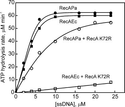
Figure 2 shows the ssDNA concentration dependence of the ATPase activity produced by different presynaptic complexes of the type RecA/ATP/circular M13 ssDNA, all formed in the presence of sufficient SSB to remove the ssDNA secondary structures. Two control and two test complexes were compared. The controls included presynaptic complexes with pure RecAPa and RecAEc; the test complexes contained protein mixtures of either RecAEc and RecA K72R or RecAPa and RecA K72R taken in equal proportion. The Lys-to-Arg substitution in the RecA K72R protein occurs in a well-conserved nucleotide binding region. The protein binds but does not hydrolyze ATP, though it is still able to promote the fundamental DNA pairing reaction (42). Moreover, this mutant protein forms mixed presynaptic filaments with RecAEc, competing on an equal basis with wild-type RecAEc for binding sites in ssDNA (50, 51).
FIG. 2.
ssDNA-dependent ATP hydrolysis of four presynaptic complexes formed by RecAPa, RecAEc, RecAPa plus RecA K72R, and RecAEc plus RecA K72R at increasing concentrations of circular ssDNA. SSB was used at a concentration providing one SSB monomer per 10 nucleotides of ssDNA. Each point on the curves represents an individual sample and shows a steady-state rate measured over a period of about 15 min.
Each point of the curves presented in Fig. 2 is the rate of ATP hydrolysis obtained 15 min after initiation of the reaction by mixing RecA(s), ATP with its regenerating system, and closed circular ssDNA of phage M13mp8 in buffer (pH 7.5) at 37°C. For pure RecAEc and RecAPa, the ATP hydrolysis rate increased with ssDNA concentration to a maximum of 60 to 63 μM min−1 with an apparent stoichiometry of about three bases per RecA subunit. As expected (50), RecA K72R did not hydrolyze ATP, and the hydrolysis rate observed for an equal mixture of RecAEc and RecA K72R proteins increased only slightly with increasing ssDNA concentration and had an approximately 10-fold-lower acceleration. This resulted from formation of mixed RecA filaments and inhibition of ATP hydrolysis because of the dimension of cooperative interactions between RecAEc protomers in filaments when RecA K72R monomers are included (50). Quite another situation was found for the mixture of RecAPa plus RecA K72R (equal amounts of proteins). The ATP hydrolysis rate increased with ssDNA concentration, reaching 87% of the maximal value described for pure RecAPa. Thus, the excess ssDNA in the reaction mixture resulted in formation of presynaptic complexes with ATPase activity that was closer to that of pure RecAPa filaments.
The data indicate that RecAPa protein is much more active in competition with RecA K72R for presynaptic filament formation. Because the capacity of the mutant RecA K72R protein to bind ssDNA is not changed significantly relative to that of RecAEc (42), the data reveal the RecAPa advantages in recombination initiation in comparison with RecAEc.
RecAPa protein partially suppresses the genetic consequences of recF, recO, and recR mutations.
Inactivation of the recF, recO, or recR gene results in a substantial decrease in the yield of recombinants during conjugation in E. coli cells relying on the RecF pathway. On the other hand, as shown in Table 1, relatively low levels of RecAPa protein promote hyperrecombination in E. coli cells utilizing the RecF pathway. We decided to determine whether the presence of RecAPa could compensate for the loss of recF, recO, or recR function.
Table 2 presents the relative yield of Thr+ Strr recombinants (RYR) obtained in three sets of crosses (each repeated twice) between donor strain KL227 or KL227 recF349, as indicated, and four different recipient strains with different RecF pathway genotypes including (i) classical strain JC7623 recBC sbcCD (a basal cross); (ii) JC7623/pRecAPa, in which hyperrecombination by RecAPa protein is promoted in the RecAEc background (the first testing cross); (iii) JC7623-0, JC7623-R, or JC7623-F recipients deficient in general recombination because of defects in the recO, recR, or recF gene, respectively (the second test); and (iv) JC7623-0/pRecAPa, JC7623-R/pRecAPa, or JC7623-F/pRecAPa recipients deficient in RecAEc-dependent recombination while potentially proficient in recombination promoted by RecAPa (the third test). The yield of recombinants in all three testing crosses was normalized to that in the basal cross. The result of the first test showed that addition of a small portion of RecAPa protein (30%) to the RecAEc protein in the cells, which rendered them hyper-rec as measured by FRE (Table 1), did not change RYR significantly, if any (Table 2). The second test showed that both insertion mutations, recO1504 and recR252, and the deletion mutation recF349 lowered the yield of recombinants by 40 to 50, 140 to 180, and 200 to 300 times, respectively, a result that is in satisfactory agreement with the data in the literature (33, 36, 47). The third test cross revealed average increases of 15-, 12.5-, and 7.5-fold in recombination frequency when RecAPa was provided in backgrounds deficient in the recO, recR, and recF genes, respectively.
TABLE 2.
Partial suppression of recombination deficiency that resulted from recO, recR, or recF mutations in the RecF pathway of recombination by RecAPa protein as observed via the relative yield of Thr+ Strr recombinants in crosses between donor KL227 (HfrR4-type) and recipients originating from JC7623 recBC sbcCD
| Recipient or recombination | Relevant genotype
|
RYRa
|
||
|---|---|---|---|---|
| recA+ | Other rec geneb | Expt 1 | Expt 2 | |
| JC7623 | recAEc+ | 1.0 (7.1)c | 1.0 (3.4) | |
| JC7623/pRecAPa | recAEc+ + recAPa+ | 0.96 | 1.0 | |
| JC7623-O | recAEc+ | recO1504 | 0.024 | 0.020 |
| JC7623-O/pRecAPa | recAEc+ + recAPa+ | recO1504 | 0.30 | 0.35 |
| Deficiencyd (recO+/recO1504) | 41.6 | 50.0 | ||
| Suppressione (recAEc+ recAPa+ recO1504/recAEc+ recO1504) | 12.5 | 17.5 | ||
| JC7623 | recAEc+ | 1.0 (5.2) | 1.0 (5.0) | |
| JC7623/pRecAPa | recAEc+ + recAPa+ | 0.85 | 1.0 | |
| JC7623-R | recAEc+ | recR252 | 0.0067 | 0.0056 |
| JC7623-R/pRecAPa | recAEc+ + recAPa+ | recR252 | 0.108 | 0.05 |
| Deficiency (recR+/recR252) | 145 | 178 | ||
| Suppression (recAEc+ recAPa+ recR252/recAEc+ recR252) | 16.1 | 8.9 | ||
| JC7623 | recAEc+ | 1.0 (4.3) | 1.0 (4.4) | |
| JC7623/pRecAPa | recAEc+ + recAPa+ | 0.95 | 1.05 | |
| JC7623-Ff | recAEc+ | recF349 | 0.0033 | 0.0049 |
| JC7623-F/pRecAPaf | recAEc+ + recAPa+ | recF349 | 0.029 | 0.03 |
| Deficiency (recF+/recF349) | 300 | 204 | ||
| Suppression (recAEc+ recAPa+ recF349/recAEc+ recF349) | 8.8 | 6.1 | ||
RYR means the yield of Thr+ Strr recombinants relative to that found in crosses with JC7623, which is identical to the yield of Thr+ Strr recombinants for strain AB1157 (cells with the RecBCD pathway). The yield of recombinants in all independent crosses was normalized by the mating ability of each recipient used.
The full names of mutations used were as follows: recO1504::Tn5, recR252::Tn10-9, and recF349(del).
Numbers in parentheses show the yield of Thr+ Strr recombinants as a percentage of the donors in the conjugation mixture.
Deficiency of recombination is a decrease in the RYR that resulted from the recO, recR, or recF insertion or deletion mutations used in the study.
Suppression of the deficiency mentioned above resulted from introduction into recipients the pRecAPa plasmid, which expressed the RecAPa protein.
In crosses with JC7623 recF349, Hfr KL227 recF349 was used. It was necessary to exclude the possibility of transferring the recF+ allele by the donor during a 60-min mating used in the study.
These in vivo data indicate that relative to RecAEc protein, the hyper-rec RecAPa protein is less dependent on the E. coli RecO, RecR, and RecF proteins in initiation of recombination events in the RecF pathway.
As known (7), the RecOR protein complex facilitates RecA-mediated D-loop formation at the 5′ ends of linear ssDNA. If RecAPa were able to compensate at least partially for the inactivity of the RecOR complex, one could expect the RecAPa function to be more active at 5′ ends of ssDNA. This was the case.
RecAPa protein promotes D-loop formation with equal efficiency at both the 3′ and 5′ ends of linear ssDNA, even in the presence of SSB.
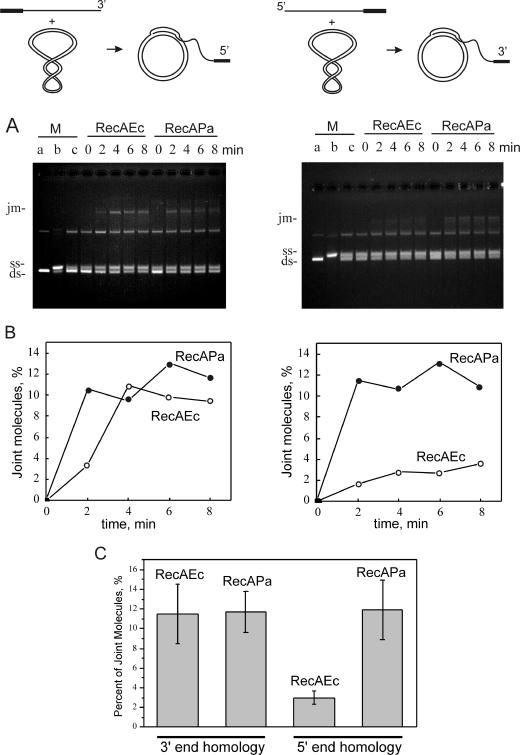
To determine the extent of any end bias in the D-loop formation promoted by the RecAPa and RecAEc presynaptic complexes, an in vitro joint molecule assay was used. The assay monitors homologous pairing between linear M13mp8 ssDNA and supercoiled dsDNA of the same phage (7). A 7-kb region of homology between these substrates was restricted to either the 3′ or 5′ end of the linear ssDNA by insertion at its 5′ or 3′ end, respectively, of a 1,037-nucleotide segment heterologous to the M13mp8 DNA (see the scheme at the top of Fig. 3).
FIG. 3.
Time course of initiation of D-loop formation between circular supercoiled dsDNA and linear ssDNA with a region of homology at either the 3′ or 5′ end in a comparison of the characteristics of RecAPa and RecAEc in promotion of the reaction. The DNA substrates and the reactions are illustrated by schemes presented at the top of the figure. M13mp8.1037(+) circular ssDNA was linearized with either EcoRI or PstI to place the 1,037-nucleotide insert (filled rectangle) at the 5′ or 3′ end, respectively. The supercoiled M13mp8 dsDNA has 7,229 bp of homology to the 3′ end of EcoRI-digested M13mp8.1037(+) linear ssDNA or to the 5′ end of PstI-digested M13mp8.1037(+) linear ssDNA. The agarose gel assay was used to visualize joint molecule formation as described in Materials and Methods. (A) Agarose gel presentation of joint molecule formation. Lanes a, b, and c contain markers (M) of dsDNA, ssDNA, and dsDNA plus ssDNA, respectively. The positions of joint molecules (jm), linear ssDNA (ss), and closed circular dsDNA (ds) are shown to the left of the gels. (B) Quantitation of the data presented in panel A by use of the Kodak DS1D image analysis software. (C) Values calculated from the data presented in panel B and two other independent experiments. The bands obtained in the D-loop reactions proceeding 4, 6, and 8 min after initiation were summarized because they looked similar.
D-loop formation between the linear ssDNA and supercoiled dsDNA was carried out in the presence of SSB at 37°C. The time course of appearance of the 3′- or 5′-end-directed joint molecules promoted by RecAEc or RecAPa proteins is presented in Fig. 3A. Quantitation of these events is presented in Fig. 3B. Consistent with previous observations (37), joint molecule formation proceeded faster for RecAPa than for RecAEc, though both reached a maximum within 4 min and sustained that level for some time thereafter.
The values presented in Fig. 3C are the quantitative estimations of maximum joint molecule formation observed in Fig. 3A and analyzed in Fig. 3B (reactions after 4, 6, and 8 min), averaged from three independent experiments as the percentage of the total DNA (ssDNA plus dsDNA plus joint molecules) participating in the reaction. When the linear ssDNA was homologous at its 3′ end with its supercoiled partner, joint molecules were formed with equal efficiency in the reactions promoted by the RecAEc and RecAPa proteins. However, when the homology was limited to the 5′ ends, the efficiency of reactions promoted by RecAEc was reduced about fourfold, in good agreement with observations described earlier (7, 15). In contrast, the RecAPa protein promoted the formation of joint molecules with equal proficiency at both the 5′ and 3′ ends of the linear ssDNA.
This finding is in good accordance with the capacity of RecAPa protein to form relatively stable presynaptic filaments on linear ssDNA which might persist at the 5′ ends of the ssDNA (6, 7, 40).
RecAPa protein filaments disassemble at only half the rate of RecAEc protein filaments.
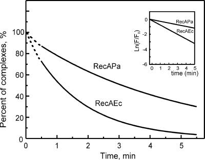
As documented previously (6), the RecAEc presynaptic complex disassembles on linear ssDNA in the 5′-to-3′ direction. To compare the rates of disassembly of the RecAPa and RecAEc filaments physically and measure the rate quantitatively, the change of fluorescence of RecA protein complexed with etheno-DNA (ɛDNA) was used. RecA binding to ɛDNA results in a proportional increase of fluorescence upon complex formation (34) and vice versa in a fluorescence decrease when the complex disassembles. To form the presynaptic RecA/ATP/ɛDNA complex so that no more than one ssDNA was bound per RecA filament, we limited the amount of ɛDNA in the reaction mixture. The binding of calf thymus ɛDNA used in the study to RecAEc or RecAPa proteins resulted in an increase of the specific fluorescence by a factor of 3.25 (data not shown). These complexes were formed and then challenged with excess poly(dT) to bind free RecA and any dissociated RecA molecules, and the resulting decline in fluorescence was monitored.
As shown in Fig. 4, the time-dependent drop in fluorescence proceeds exponentially for both the RecAEc and RecAPa presynaptic complexes, as expected for a first-order disassembly process. When plotted as ln (F/F0) versus time (see inset to Fig. 4), where F is the fluorescence at a given time and F0 is the original fluorescence (at time zero), both the RecAPa and RecAEc are converted into linear functions. The ratio of the half-time period of the drop in fluorescence, RecAEc/ RecAPa, is 1:2.7. This means that the RecAPa filaments are more than twice as resistant to poly(dT) challenge as the RecAEc filaments are.
FIG. 4.
Time-course decrease in fluorescence after the addition of excess poly(dT) to the RecA/ATP/ɛDNA complex in a comparison of disassembly of RecAPa and RecAEc complexes. The presynaptic complex RecA/ATP/ɛDNA was formed through the primary DNA binding site of RecA (excess RecA compared to ɛDNA). Experiments were performed as described in Materials and Methods. After RecAPa and RecAEc complexes were preformed, the increase of fluorescence was established. Excess poly(dT) was then added to initiate the quenching of fluorescent signal (time zero) as result of the poly(dT) challenge. The measurements were started 0.5 min later. The broken lines are the extrapolation of data to the moment of poly(dT) addition to the reaction mixture. (Inset) The same data but presented in semilogarithmic scale. F/F0 is a portion of fluorescence F of the RecAEc or RecAPa presynaptic complexes still existing to the time indicated; F0 is the fluorescence at time zero.
The data indicate that the rate of RecAPa disassembly from the 5′ end of ssDNA proceeds much slower than that of RecAEc.
DISCUSSION
Genetic analysis of the dependence of FRE values on RecA protein (either RecAEc or RecAPa or both proteins) mediating homologous recombination in E. coli supported a previous observation (37) that the RecAPa protein promotes the ends-in type of recombination events more frequently (eight times) during conjugation. Note that ends-in but not ends-out events appear to be involved in the FRE increase because otherwise a significant increase of recombinant yield could also be observed, but this was not the case. Also, the analysis showed that even a small portion of intracellular RecA presented in E. coli by RecAPa, which forms only one third of normal RecAEc concentration (Fig. 1) (that is usually about 10,000 molecules per cell [35, 48]) gave a fivefold increase of FRE, which was reproduced in both RecBCD and RecF genetic pathways of recombination (Table 1). It means that RecAPa, being in E. coli, successfully predominates over RecAEc in formation of such presynaptic complexes which are able to initiate additional ends-in recombination events that result in hyperrecombination. Two questions arise. (i) What is the RecA composition of such a presynaptic complex? (ii) What events of DNA metabolism stimulate the ends-in events?
In an attempt to answer the first question, we compared the ATPase activities of presynaptic complexes formed by two protein mixtures: RecAPa plus RecA K72R and RecAEc plus RecA K72R, where all three constituents were presented in equimolar concentrations. Because the RecA K72R protein can only bind but not hydrolyze ATP (42, 52), its participation in presynaptic complex formation might result in a decrease of the ATPase activity of the latter. This expectation was found for RecAEc/RecA K72R complexes, while the decrease was much lower for RecAPa/RecA K72R complexes (Fig. 2). In these experiments, RecA K72R mimics the behavior of RecAEc; thus, this in vitro test allows the conclusion that in full concordance with in vivo analysis, RecAPa also predominates over RecAEc in presynaptic complex formation.
In principle, RecAPa can form mixed filaments with RecAEc though the interfaces of their protomers differ in nine positions (5, 55). This statement follows from a high recombination activity of the RecAX45 chimera containing one interface from RecAEc and the other complementary interface from RecAPa (5, 38). Since RecAPa has more affinity for ssDNA (4, 37) and forms more stable filaments (Fig. 4) than RecAEc does, the mixed RecAPa/RecAEc presynaptic complexes appeared to be strong enough to realize hyperrecombination with ΔFRE of 5 (Table 1) though less efficiently than pure RecAPa filaments (ΔFRE = 7.9 [Table 1]).
To answer the second question, we should consider two types of ends-in events: ss- or dsDNA breaks and take into account that (i) the former can be converted into the latter in a special enzymatic reaction (23) and (ii) the latter form the basis of recombination-directed replication (13, 20). However, for hyperrecombination when the number of ends-in events should increase 5, 8, or 26 times (5, 25), it seems reasonable to suggest SSGR (14) as the main mechanism in realization of hyper-rec ends-in events. In fact, RecAPa effectively displaces SSB from ssDNA (4, 37) and thus from ssDNA gaps in donor and recipient DNA during conjugation, which results in the formation of presynaptic complexes initiating the SSGR process.
According to a common view, the SSGR mechanism is directed by the RecF pathway (2, 14). However, in the case of RecAPa, the full activity of this pathway appeared to be unnecessary. Genetic analysis of the RecF pathway activity, measured through the yield of conjugal recombinants, on RecAPa- or RecAEc-mediated recombination showed that even deficiency of this pathway can be compensated, at least partially, by recombinogenic activity of the RecAPa protein. This in vivo finding is in good agreement with the role of the RecOR protein complex in stabilization of RecAEc filament recombination activity at its 5′ end (7) and with the abilities of the RecAPa filament (i) to initiate recombination from the 5′ and 3′ ends in an equal manner (Fig. 3) and (ii) to be more stable at the 5′ end (Fig. 4). These observations are in concordance with the SSGR model predicted by Cromie and Leach (14) and the in vivo evidence for the abilities of both 3′ and 5′ ssDNA ends to stimulate recombination (40).
Recombinational characteristics of RecA803 (31) and RecA730 (26) (= RecA1211 [58]) proteins bearing amino acid substitutions V37M and E38K, respectively, were described earlier. Like RecAPa, these point mutant RecAEc proteins can partially complement the deficiency of RecF protein. Moreover, genetic analysis has revealed twofold and eightfold increases of FRE values for recA803 and recA730 (= recA1211) mutants, respectively (25; V. A. Lanzov and A. J. Clark, unpublished results). Interestingly, RecAPa protein also has amino acid residue substitutions in the same positions (V37I and E38P). In fact, all residues in positions from 31 to 39 in RecAPa are changed relative to those in RecAEc. On the whole, this observation provides indirect evidence that loops 29 and 44 of the RecA protein structure elucidated by Story et al. (55) might be responsible for regulation of RecA interactions with the RecFOR proteins.
Qualitatively, some biochemical characteristics of RecA803 and especially RecA730, which are thought to be responsible for recombination, look like those of RecAPa that are improved or enhanced relative to those of RecAEc. The important difference between RecAPa and RecA730 proteins involves their relation to SOS induction. RecA730 promotes hyperrecombination mainly (but not fully) in an SOS-dependent manner (25), while RecAPa generally does this independently of the SOS response. Nevertheless, the existence of these point mutant RecAEc proteins help us to understand that the weak interconnections between RecAPa and RecFOR do not reflect only the different origins of the proteins (the former is from P. aeruginosa, while the latter is from E. coli) but rather are based in the stronger recombinogenic activity of the RecAPa protein. In fact, the RecFOR system of proteins exists in P. aeruginosa and in many other bacteria (45). However, to date, little is known about the interchangeability of RecFOR systems from different sources. It seems reasonable to suggest that the inherent recombinase function of RecAPa has been more completely unmasked by evolution to provide efficient DNA repair as appropriate for a respiratory pathogen subjected to high levels of oxidative damage.
Given its close structural relationship to RecAEc, RecAPa provides an excellent system with which to investigate the structural basis of recombinase function. We anticipate that continued efforts to compare the two proteins, focusing on key amino acid residues already defined as important to the enhanced function of RecAPa, will yield new insights.
Acknowledgments
This work was supported by a Fogarty International Research Collaboration Award (2 R03 TW001318-04; to M.M.C.) and by grants from the Russian Foundation for Basic Research (05-04-48266; to V.A.L.), the Russian Ministry of Education and Science (RNP 2.2.1.1.4663), and the National Institutes of Health (GM32335; to M.M.C.).
We thank Leonid M. Firsov (PNPI) for fruitful discussions of some experiments. D.M.B., I.V.B., and Y.V.K. acknowledge with gratitude M. Cox's research group for providing assistance with the portion of the work carried out in the United States.
REFERENCES
- 1.Alexseyev, A. A., D. M. Baitin, S. Kuramitsu, T. Ogawa, H. Ogawa, and V. A. Lanzov. 1997. A recombinational defect in the C-terminal domain of Escherichia coli RecA2278-5 protein is compensated by protein binding to ATP. Mol. Microbiol. 23:255-265. [DOI] [PubMed] [Google Scholar]
- 2.Amundsen, S. K., and G. R. Smith. 2003. Interchangeable parts of the Escherichia coli recombination machinery. Cell 112:741-744. [DOI] [PubMed] [Google Scholar]
- 3.Arnold, D. A., and S. C. Kowalczykowski. 1999. RecBCD helicase/nuclease. Encyclopedia of life sciences. Nature Publishing Group, London, United Kingdom. [Online.] http://www.els.net.
- 4.Baitin, D. M., E. N. Zaitsev, and V. A. Lanzov. 2003. Hyper-recombinogenic RecA protein from Pseudomonas aeruginosa with enhanced activity of its primary DNA binding site. J. Mol. Biol. 328:1-7. [DOI] [PubMed] [Google Scholar]
- 5.Bakhlanova, I. V., T. Ogawa, and V. A. Lanzov. 2001. Recombinogenic activity of chimeric recA genes (Pseudomonas aeruginosa/Escherichia coli): a search for RecA protein regions responsible for this activity. Genetics 159:7-15. [DOI] [PMC free article] [PubMed] [Google Scholar]
- 6.Bork, J. M., M. M. Cox, and R. B. Inman. 2001. RecA protein filaments disassemble in the 5′ to 3′ direction on single-stranded DNA. J. Biol. Chem. 276:45740-45743. [DOI] [PubMed] [Google Scholar]
- 7.Bork, J. M., M. M. Cox, and R. B. Inman. 2001. The RecOR proteins modulate RecA protein function at 5′ ends of single-stranded DNA. EMBO J. 20:7313-7322. [DOI] [PMC free article] [PubMed] [Google Scholar]
- 8.Brendel, V., L. Brocchieri, S. J. Sandler, A. J. Clark, and S. Karlin. 1997. Evolutionary comparisons of RecA-like proteins across all major kingdoms of living organisms. J. Mol. Evol. 44:528-541. [DOI] [PubMed] [Google Scholar]
- 9.Bresler, S. E., S. V. Krivonogov, and V. A. Lanzov. 1978. Scale of the genetic map and genetic control of recombination after conjugation in Escherichia coli K-12. Mol. Gen. Genet. 166:337-346. [DOI] [PubMed] [Google Scholar]
- 10.Chervyakova, D., A. Kagansky, M. Petukhov, and V. Lanzov. 2001. [L29M] substitution in the interface of subunit-subunit interactions enhances Escherichia coli RecA protein properties important for its recombinogenic activity. J. Mol. Biol. 314:923-935. [DOI] [PubMed] [Google Scholar]
- 11.Clark, A. J. 1971. Toward a metabolic interpretation of genetic recombination of E. coli and its phages. Annu. Rev. Microbiol. 25:437-464. [DOI] [PubMed] [Google Scholar]
- 12.Connelly, J. C., E. S. Deleau, E. A. Okely, and D. R. F. Leach. 1997. Overexpression, purification, and characterization of the SbcCD protein from Escherichia coli. J. Biol. Chem. 272:19819-19826. [DOI] [PubMed] [Google Scholar]
- 13.Cox, M. M. 2002. The nonmutagenic repair of broken replication forks via recombination. Mutat. Res. 510:107-120. [DOI] [PubMed] [Google Scholar]
- 14.Cromie, G. A., and D. R. Leach. 2000. Control of crossing over. Mol. Cell 6:815-826. [DOI] [PubMed] [Google Scholar]
- 15.Dutreix, M., B. J. Rao, and C. M. Radding. 1991. The effects on strand exchange of 5′ versus 3′ ends of single-stranded DNA in RecA nucleoprotein filaments. J. Mol. Biol. 219:645-654. [DOI] [PubMed] [Google Scholar]
- 16.Horii, Z., and A. J. Clark. 1973. Genetic analysis of the RecF pathway to genetic recombination in Escherichia coli K12: isolation and characterization of mutants. J. Mol. Biol. 80:327-344. [DOI] [PubMed] [Google Scholar]
- 17.Inman, R. B., and M. Schnos. 1971. Structure of branch points in replicating DNA: presence of single-stranded connections in lambda DNA branch points. J. Mol. Biol. 56:319-325. [DOI] [PubMed] [Google Scholar]
- 18.Kantake, N., M. V. Madiraju, T. Sugiyama, and S. C. Kowalczykowski. 2002. Escherichia coli RecO protein anneals ssDNA complexed with its cognate ssDNA-binding protein: a common step in genetic recombination. Proc. Natl. Acad. Sci. USA 99:15327-15332. [DOI] [PMC free article] [PubMed] [Google Scholar]
- 19.Kolodner, R., R. A. Fishel, and M. Howard. 1985. Genetic recombination of bacterial plasmid DNA: effect of RecF pathway mutations on plasmid recombination in Escherichia coli. J. Bacteriol. 163:1060-1066. [DOI] [PMC free article] [PubMed] [Google Scholar]
- 20.Kowalczykowski, S. C. 2000. Initiation of genetic recombination and recombination-dependent replication. Trends Biochem. Sci. 25:156-165. [DOI] [PubMed] [Google Scholar]
- 21.Kowalczykowski, S. C., D. A. Dixon, A. K. Eggleston, S. D. Lauder, and W. M. Rehrauer. 1994. Biochemistry of homologous recombination in Escherichia coli. Microbiol. Rev. 58:401-465. [DOI] [PMC free article] [PubMed] [Google Scholar]
- 22.Kuzminov, A. 1999. Recombinational repair of DNA damage in Escherichia coli and bacteriophage lambda. Microbiol. Mol. Biol. Rev. 63:751-813. [DOI] [PMC free article] [PubMed] [Google Scholar]
- 23.Kuzminov, A. 2001. Single-strand interruptions in replicating chromosomes cause double-strand breaks. Proc. Natl. Acad. Sci. USA 98:8241-8246. [DOI] [PMC free article] [PubMed] [Google Scholar]
- 24.Lanzov, V. A. 2002. Hyper-recombination in Escherichia coli with and without SOS response, p. 21-38. In M. Ruiz-Rubio, E. Alejandre-Duran, and T. Roldan-Arjona (ed.), Recent research development in DNA repair and mutagenesis. Research Signpost, Kerala, India.
- 25.Lanzov, V. A., I. V. Bakhlanova, and A. J. Clark. 2003. Conjugational hyperrecombination achieved by derepressing the LexA regulon, altering the properties of RecA protein and inactivating mismatch repair in Escherichia coli K-12. Genetics 163:1243-1254. [DOI] [PMC free article] [PubMed] [Google Scholar]
- 26.Lavery, P. E., and S. C. Kowalczykowski. 1992. Biochemical basis of the constitutive repressor cleavage activity of recA730 protein. A comparison to recA441 and recA803 proteins. J. Biol. Chem. 267:20648-20658. [PubMed] [Google Scholar]
- 27.Lavery, P. E., and S. C. Kowalczykowski. 1990. Properties of recA441 protein-catalyzed DNA strand exchange can be attributed to an enhanced ability to compete with SSB protein. J. Biol. Chem. 265:4004-4010. [PubMed] [Google Scholar]
- 28.Lawley, T. D., G. S. Gordon, A. Wright, and D. E. Taylor. 2002. Bacterial conjugative transfer: visualization of successful mating pairs and plasmid establishment in live Escherichia coli. Mol. Microbiol. 44:947-956. [DOI] [PubMed] [Google Scholar]
- 29.Lusetti, S. L., and M. M. Cox. 2002. The bacterial RecA protein and the recombinational DNA repair of stalled replication forks. Annu. Rev. Biochem. 71:71-100. [DOI] [PubMed] [Google Scholar]
- 30.Lusetti, S. L., M. D. Hobbs, E. A. Stohl, S. Chitteni-Pattu, R. B. Inman, H. S. Seifert, and M. M. Cox. 2006. The RecF protein antagonizes RecX function via direct interaction. Mol. Cell 21:41-50. [DOI] [PMC free article] [PubMed] [Google Scholar]
- 31.Madiraju, M. V., P. E. Lavery, S. C. Kowalczykowski, and A. J. Clark. 1992. Enzymatic properties of the RecA803 protein, a partial suppressor of recF mutations. Biochemistry 31:10529-10535. [DOI] [PubMed] [Google Scholar]
- 32.Mahdi, A. A., and R. G. Lloyd. 1989. Identification of the recR locus of Escherichia coli K-12 and analysis of its role in recombination and DNA repair. Mol. Gen. Genet. 216:503-510. [DOI] [PubMed] [Google Scholar]
- 33.Mahdi, A. A., and R. G. Lloyd. 1989. The recR locus of Escherichia coli K-12: molecular cloning, DNA sequencing and identification of the gene product. Nucleic Acids Res. 17:6781-6794. [DOI] [PMC free article] [PubMed] [Google Scholar]
- 34.Menetski, J. P., and S. C. Kowalczykowski. 1985. Interaction of recA protein with single-stranded DNA. Quantitative aspects of binding affinity modulation by nucleotide cofactors. J. Mol. Biol. 181:281-295. [DOI] [PubMed] [Google Scholar]
- 35.Moreau, P. L. 1987. Effects of overproduction of single-stranded DNA-binding protein on RecA protein-dependent processes in Escherichia coli. J. Mol. Biol. 194:621-634. [DOI] [PubMed] [Google Scholar]
- 36.Morrison, P. T., S. T. Lovett, L. E. Gilson, and R. Kolodner. 1989. Molecular analysis of the Escherichia coli recO gene. J. Bacteriol. 171:3641-3649. [DOI] [PMC free article] [PubMed] [Google Scholar]
- 37.Namsaraev, E. A., D. Baitin, I. V. Bakhlanova, A. A. Alexseyev, H. Ogawa, and V. A. Lanzov. 1998. Biochemical basis of hyper-recombinogenic activity of Pseudomonas aeruginosa RecA protein in Escherichia coli cells. Mol. Microbiol. 27:727-738. [DOI] [PubMed] [Google Scholar]
- 38.Ogawa, T., A. Shinohara, H. Ogawa, and J. Tomizawa. 1992. Functional structures of the recA protein found by chimera analysis. J. Mol. Biol. 226:651-660. [DOI] [PubMed] [Google Scholar]
- 39.Prasher, D. C., L. Conarro, and S. R. Kushner. 1983. Amplification and purification of exonuclease I from Escherichia coli K12. J. Biol. Chem. 258:6340-6343. [PubMed] [Google Scholar]
- 40.Razavy, H., S. K. Szigety, and S. M. Rosenberg. 1996. Evidence for both 3′ and 5′ single-strand DNA ends in intermediates in chi-stimulated recombination in vivo. Genetics 142:333-339. [DOI] [PMC free article] [PubMed] [Google Scholar]
- 41.Register, J. C., III, and J. Griffith. 1985. The direction of RecA protein assembly onto single strand DNA is the same as the direction of strand assimilation during strand exchange. J. Biol. Chem. 260:12308-12312. [PubMed] [Google Scholar]
- 42.Rehrauer, W. M., and S. C. Kowalczykowski. 1993. Alteration of the nucleoside triphosphate (NTP) catalytic domain within Escherichia coli recA protein attenuates NTP hydrolysis but not joint molecule formation. J. Biol. Chem. 268:1292-1297. [PubMed] [Google Scholar]
- 43.Rice, K. P., A. L. Eggler, P. Sung, and M. M. Cox. 2001. DNA pairing and strand exchange by the Escherichia coli RecA and yeast Rad51 proteins without ATP hydrolysis: on the importance of not getting stuck. J. Biol. Chem. 276:38570-38581. [DOI] [PubMed] [Google Scholar]
- 44.Roca, A. I., and M. M. Cox. 1997. RecA protein: structure, function, and role in recombinational DNA repair. Prog. Nucleic Acid Res. Mol. Biol. 56:129-223. [DOI] [PubMed] [Google Scholar]
- 45.Rocha, E. P., E. Cornet, and B. Michel. 2005. Comparative and evolutionary analysis of the bacterial homologous recombination system. PloS Genet. 1:e15. [DOI] [PMC free article] [PubMed] [Google Scholar]
- 46.Sambrook, J., E. F. Fritsch, and T. Maniatis. 1989. Molecular cloning: a laboratory manual, 2nd ed. Cold Spring Harbor Laboratory, Cold Spring Harbor, N.Y.
- 47.Sandler, S. J., and A. J. Clark. 1993. Use of high and low level overexpression plasmids to test mutant alleles of the recF gene of Escherichia coli K-12 for partial activity. Genetics 135:643-654. [DOI] [PMC free article] [PubMed] [Google Scholar]
- 48.Sassanfar, M., and J. W. Roberts. 1990. Nature of the SOS-inducing signal in Escherichia coli. The involvement of DNA replication. J. Mol. Biol. 212:79-96. [DOI] [PubMed] [Google Scholar]
- 49.Shan, Q., J. M. Bork, B. L. Webb, R. B. Inman, and M. M. Cox. 1997. RecA protein filaments: end-dependent dissociation from ssDNA and stabilization by RecO and RecR proteins. J. Mol. Biol. 265:519-540. [DOI] [PubMed] [Google Scholar]
- 50.Shan, Q., and M. M. Cox. 1997. RecA filament dynamics during DNA strand exchange reactions. J. Biol. Chem. 272:11063-11073. [DOI] [PubMed] [Google Scholar]
- 51.Shan, Q., and M. M. Cox. 1996. RecA protein dynamics in the interior of RecA nucleoprotein filaments. J. Mol. Biol. 257:756-774. [DOI] [PubMed] [Google Scholar]
- 52.Shan, Q., M. M. Cox, and R. B. Inman. 1996. DNA strand exchange promoted by RecA K72R. Two reaction phases with different Mg2+ requirements. J. Biol. Chem. 271:5712-5724. [DOI] [PubMed] [Google Scholar]
- 53.Sigurdsson, S., K. Trujillo, B. Song, S. Stratton, and P. Sung. 2001. Basis for avid homologous DNA strand exchange by human Rad51 and RPA. J. Biol. Chem. 276:8798-8806. [DOI] [PubMed] [Google Scholar]
- 54.Smith, G. R. 1991. Conjugational recombination in E. coli: myths and mechanisms. Cell 64:19-27. [DOI] [PubMed] [Google Scholar]
- 55.Story, R. M., I. T. Weber, and T. A. Steitz. 1992. The structure of the E. coli recA protein monomer and polymer. Nature 355:318-325. [DOI] [PubMed] [Google Scholar]
- 56.Umezu, K., N. W. Chi, and R. D. Kolodner. 1993. Biochemical interaction of the Escherichia coli RecF, RecO, and RecR proteins with RecA protein and single-stranded DNA binding protein. Proc. Natl. Acad. Sci. USA 90:3875-3879. [DOI] [PMC free article] [PubMed] [Google Scholar]
- 57.Umezu, K., and R. D. Kolodner. 1994. Protein interactions in genetic recombination in Escherichia coli. Interactions involving RecO and RecR overcome the inhibition of RecA by single-stranded DNA-binding protein. J. Biol. Chem. 269:30005-30013. [PubMed] [Google Scholar]
- 58.Wang, W. B., M. Sassanfar, I. Tessman, J. W. Roberts, and E. S. Tessman. 1988. Activation of protease-constitutive RecA proteins of Escherichia coli by all of the common nucleoside triphosphates. J. Bacteriol. 170:4816-4822. [DOI] [PMC free article] [PubMed] [Google Scholar]
- 59.Webb, B. L., M. M. Cox, and R. B. Inman. 1995. An interaction between the Escherichia coli RecF and RecR proteins dependent on ATP and double-stranded DNA. J. Biol. Chem. 270:31397-31404. [DOI] [PubMed] [Google Scholar]
- 60.Wood, T. H., and R. H. Walmsley. 1969. Conjugation in Escherichia coli K-12 and its modification by irradiation. Biophys. J. 9:391-420. [DOI] [PMC free article] [PubMed] [Google Scholar]
- 61.Xu, L., and K. J. Marians. 2003. PriA mediates DNA replication pathway choice at recombination intermediates. Mol. Cell 11:817-826. [DOI] [PubMed] [Google Scholar]


