Abstract
Core members of the MBD protein family (MeCP2, MBD1, MBD2 and MBD4) share a methyl-CpG-binding domain that has a specific affinity for methylated CpG sites in double-stranded DNA. By multimerizing the MDB domain of Mbd1, we engineered a poly-MBD protein that displays methyl-CpG-specific binding in vitro with a dissociation constant that is >50-fold higher than that of a monomeric MBD. Poly-MBD proteins also localize to methylated foci in cells and can deliver a functional domain to reporter constructs in vivo. We propose that poly-MBD proteins are sensitive reagents for the detection of DNA methylation levels in isolated native DNA and for cytological detection of chromosomal CpG methylation.
INTRODUCTION
Proteins that have been demonstrated to bind specifically to methylated DNA belong to two characterized families. The methyl-CpG-binding domain (MBD) family comprises five polypeptides in mammals, four of which MBD1, MBD2, MBD4 and MeCP2 have been shown to preferentially bind a symmetrically methylated CpG motif (1–3). These proteins share the MBD which confers specific DNA binding (4). The unrelated Kaiso protein family binds to DNA sequences containing methyl-CpGs via a distinctive zinc finger motif (5,6). In keeping with evidence that DNA methylation is a signal for transcriptional repression, MeCP2, MBD1, MBD2 and Kaiso all repress transcription in vitro and associate with co-repressors (1,5,7–10).
The aim of the present study was to harness the affinity of the MBD to create a superior reagent for the detection of methyl-CpG in vitro and in vivo. Assessment of global levels of DNA methylation currently relies on mass spectrometry (11), ‘nearest neighbour’ methods (12), enzymatic methyl-group acceptance assays (13) or HPLC analysis (14). These quantitative measures are complemented by the use of antibodies against 5-methylcytosine (m5C), which in addition to quantification permit visualization of m5C distribution within DNA or chromosomes (15,16). A disadvantage of anti-m5C antibodies is that the epitope is best exposed when DNA is single-stranded and DNA must therefore normally be denatured before detection. The MBD, on the other hand, is specific for native double-stranded DNA. Another potential benefit of the MBD reagent would be its specificity for m5C in the context of a symmetrically methylated CpG sequence (4).
To test the MBD reagent, we designed a series of artificial methyl-CpG-binding proteins based on multimerization of the MBD of Mbd1. The resulting polypeptides bound strongly to methylated DNA with high specificity and could compete with other methyl-CpG-binding proteins for DNA binding in vitro. When expressed in vivo, the poly-MBD protein localized to the methyl-CpG-rich heterochromatic foci of mouse cells and could recruit a functional domain specifically to methylated DNA sequences. Poly-MBD proteins proved to be sensitive and specific reagents for the detection of methyl-CpGs in immobilized DNA and cytological preparations.
MATERIALS AND METHODS
Plasmids
To generate constructs encoding the poly-MBD with varying numbers of MBDs, two PCR fragments (PCR-A and PCR-B) were amplified from each of the templates pFlag-mMBD1 or pFlag-mMBD1-R22A (17) to produce the wild-type and R22A control constructs, respectively. PCR-A was amplified using primers 5′-GCGAATTCGATGCCAAAAAAGAAGAGAAAGGTAGATATCATGGCTGAGTCCTGGCAGGACTGCC-3′ and 5′-GCTCTAGACACGTGTTAAGCGTAGTCTGGGACGTCGTATGGGTACTTGGGAATGGGATGGCATAAGG-3′. It encodes a N-terminal nuclear localization signal (NLS), cDNA from mouse Mbd1 corresponding to amino acids 1–75 and a C-terminal HA-tag. In addition, it has a 5′ EcoRI site, a EcoRV site just downstream of the NLS, a PmlI site just downstream of the HA-tag and a 3′ XbaI site. PCR-B was amplified using primers 5′-AAGGTAGATATCATGGCTGAGTCCTGGCAGGACTGCC-3′ and 5′-CTAGACACGTGAGAACCACCACCACCAGAACCACCACCACCCTTGGGAATGGGATGGCATAAGG-3′. It encodes a mouse Mbd1 fragment corresponding to amino acids 1–75 followed by a flexible linker [(Gly4-Ser)2] with a 5′ EcoRV and a 3′ PmlI site. The pTRE-1xMBD construct was made by inserting EcoRI/XbaI digested PCR-A into EcoRI/XbaI opened pTRE (Clontech). Poly-MBD constructs were made by inserting PmlI/EcoRV digested PCR-B into an EcoRV cut MBD construct. A PCR fragment encoding the activation domain of VP16 was inserted between the NLS and the first MBD using the EcoRV site, creating pTRE-NxMBD-VP16. For expression, the inserts were excised using EcoRI/XbaI and transferred into pET30b+ (pET-NxMBD, bacterial expression), pcDNA3 (p-NxMBD, mammalian expression) or pLZRS-MS (pLZRS-NxMBD, retroviral delivery). The retroviral shuttle vector has been described previously (18). Reporter plasmids (pRL-TK and pGL2-control) are from Promega.
Cell lines and retroviral infection
DNA methylation deficient Dnmt1 n/n (19), p53−/− mouse embryonic fibroblasts [described in (17)] were maintained in DMEM (Gibco) supplemented with 15% bovine calf serum, non-essential amino acids, sodium pyruvate and antibiotics (Gibco). The presence of a homozygous p53 mutation allows survival of Dnmt1-deficient cells (20). Phoenix packaging cells were grown in DMEM and wild-type mouse fibroblasts in MEM alpha, all supplemented with 10% bovine calf serum and antibiotics (Gibco). Production of virus particles and infection of adherent cells was performed as described (http://www.stanford.edu/group/nolan/index.html). Cell lines expressing N-MBD proteins were made by retroviral infection.
Transfection and reporter gene activity
Reporter constructs were either M.SssI or mock methylated and co-transfected with p-NxMBD expression constructs plus a non-modified internal transfection control. The amount of expression construct was kept constant by adding empty vector (pcDNA3). Cells were transfected using Lipofectamine (Invitrogen) or JetPEI (QBiogene) according to the manufacturer's instructions. Reporter gene activity and activity of the internal transfection control was measured 40–48 h after transfection using the Dual Luciferase system (Promega) according to the manufacturer's instructions. Each transfection was performed in triplicate and repeated at least twice. Reporter activity is expressed relative to the internal control activity to correct for differences in transfection efficiency.
Recombinant proteins
Recombinant His6-tagged NxMBD proteins were purified from 750 ml induced BL21(DE3) cultures on Ni-NTA agarose (Qiagen) using denaturation and on column renaturation cycles in accordance with the manufacturer's instructions. Recombinant GST-MeCP2 was made as described previously (4). Nuclear extracts were made from mouse fibroblasts as described previously (21).
Bandshifts
Binding reactions including purified His-tagged NxMBD protein, recombinant MeCP2 or nuclear extract in 20 mM HEPES, pH 7.9, 3 mM MgCl2, 10% glycerol, 1 mM dithiothreitol, 100 mM KCl and 0.05 µg/µl sonicated Escherichia coli DNA (Sigma) were pre-incubated 10 min at room temperature before the addition of 25 fmol end-labelled double-stranded probe. For supershift reactions anti-His6 (Santa Cruz, G-18) or anti-MBD3 (Santa Cruz, C-18) was added. After a further 25 min incubation at room temperature, the reactions were loaded on to either 6% polyacrylamide/0.5× TBE gels and run for 2 h at 240 V (4°C) or 1.3% agarose/0.5× TBE gels and run at 6 V/cm (4°C). Polyacrylamide gels were dried on to 3 mm Whatman paper and agarose gels on to DE81 (Whatman). Radioactivity was detected using a phosphor-screen and a Storm 840. All oligonucleotide probes (3ME, 2ME, 1ME, 0ME) used for bandshift are based on the sequence 5′-ATCAGACGTTCGCCGGCGGATTGGCTTGGCTGCGAAGAAGATA-3′ and the complementary strand. In 3ME all the underlined CpGs are symmetrically methylated, in 2ME C17 and C33 are methylated and in 1ME C17 is methylated whereas all CpG sites in 0ME are left unmethylated. The oligonucleotides were annealed in 10 mM Tris–HCl, pH 8, 1 mM EDTA and 50 mM NaCl. The CG11 probe is described previously (22), the methylated version is methylated at all CpG dinucleotides by M.Sss I.
Immunostaining
Cells were fixed in 4% paraformaldehyde in PBS for 20 min at room temperature followed by permeabilization in 0.2% Triton X-100 in PBS. Slides were blocked in 3% BSA/PBS before incubation with primary antibody (anti-HA, Santa Cruz F-7, 1:1000) for 60 min. After washing and incubation with Alexa-594 coupled anti-mouse antibody (1:1000, Molecular Probes), slides were washed and mounted in Vectashield with DAPI (Vector). Images were obtained using a Zeiss microscope fitted with a CCD camera and processed using Adobe PhotoShop or a Delta Vision deconvolution microscope and SoftWorx software.
Staining purified DNA using NxMBD proteins
Native double-stranded mock- or M.SssI-methylated lambda phage DNA was slot-blotted on to Nytran Supercharge membrane (Schleicher & Schuell) and immobilized by UV crosslinking. Alternatively, DNA was denatured by incubation at 95°C for 10 min, transferred and immobilized on Optitran membrane (Schleicher & Schuell). The filters were blocked 2 h at room temperature or ON at 4°C and incubated with purified recombinant NxMBD proteins (10 µg/ml) for 2 h at room temperature. After washing, the bound protein was detected by incubation with first anti-HA antibody (1:500, F-7, Santa Cruz), then horseradish peroxidase coupled anti-mouse antibody (1: 5000, Amersham). For detection using anti-m5C antibody, the NxMBD and anti-HA incubations were substituted by a 1 h incubation with anti-m5C antibody (1:500, Eurogentec). All incubations were in TBS-T supplemented with 5% skimmed milk powder. The blots were developed by enhanced chemiluminescence and exposed to film.
Staining cells using NxMBD proteins
Attached mouse fibroblasts were permeabilized for 2 min on ice in 0.2% Triton X-100 in PBS and fixed 15 min at room temperature in 4% paraformaldehyde in PBS. The cells were incubated with purified recombinant NxMBD protein diluted in PBS/10% goat serum for 2 h, washed and fixed again for 15 min with 4% paraformaldehyde in PBS. The NxMBD protein was detected by incubation with first anti-HA antibody (1:500, F-7, Santa Cruz), then Alexa-594 coupled anti-mouse antibody (1:1000, Molecular probes). The cells were counterstained with DAPI and mounted in Vectashield (Vector). The cells were examined using a DeltaVision deconvolution microscope with SoftWorx software. Metaphase spreads were obtained from mouse fibroblasts arrested 2 h with 0.1 µg/ml colcimid. Trypsinized cells were swollen 10 min in 75 mM KCl, and adjusted to 0.1% Tween-20 before spinning 20 000 cells on to slides [cytospin 3 (Shandon), 800 r.p.m., 4 min]. After air drying, the slides were equilibrated with KCM buffer (10 mM Tris–HCl, pH 8, 120 mM KCl, 20 mM NaCl, 0.5 mM EDTA and 0.1% Triton X-100) before incubating with purified recombinant NxMBD protein diluted in KCM plus 10% goat serum for 1–2 h. After washing, the cells were fixed in 3% formaldehyde in KCM. The NxMBD protein was detected by incubation with first anti-HA antibody (1:500, F-7, Santa Cruz), then Alexa-594 coupled anti-mouse antibody (1:1000, Molecular probes). The slides were fixed in 3% formaldehyde in KCM followed by counterstaining and mounting in Vectashield with DAPI (Vector). The chromosomes were examined as above.
RESULTS AND DISCUSSION
Multimerization of MBDs increases their affinity for methylated DNA
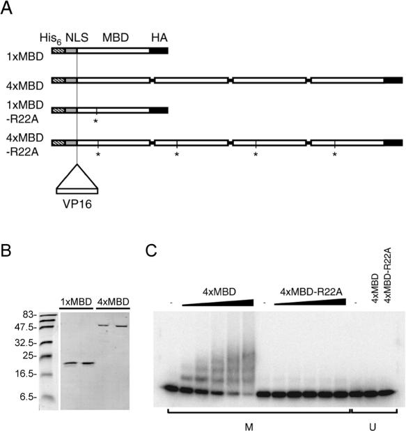
To create a high-affinity methyl-CpG-binding protein, we chose the MBD of Mbd1 as it is well characterized biochemically and structurally and is known to recognize a single methylated CpG (1,23). Moreover, the transcriptional repression domain and the recently identified DNA-binding CXXC domain of Mbd1 could be easily excluded from the construct as both map to regions distant from the MBD (17,24). To increase the binding affinity, multiple copies of the MBD were linked together, connected by flexible linker peptides, to generate ‘NxMBD’ polypeptides, where N is the number of MBDs. Increased binding of a poly-MBD protein to a methylated DNA molecule is expected for two reasons. (i) Each protein molecule has a higher concentration of DNA-binding sites than does a single MBD and, therefore, the probability of a stable interaction resulting from a DNA–protein collision is increased. Also, when a complex dissociates, the local concentration of MBDs is higher than for one MBD, favouring rebinding. (ii) The poly-MBD can interact with several sites on a DNA molecule with multiple mCpGs, thereby increasing the stability of the complex. Multimerization of a DNA-binding domain has previously been shown to create a high-affinity multi-AT-hook protein (25). In addition, the poly-MBD protein included an N-terminal NLS and was equipped with a C-terminal HA-tag for detection purposes (Figure 1A). Negative control proteins in which each MBD carried the R22A point mutation that disrupts DNA binding (23) were created in parallel (NxMBD–R22A).
Figure 1.
A multimerized-MBD protein binds to methylated DNA. (A) Diagram of engineered wild-type (NxMBD) and mutant (NxMBD–R22A) proteins with 1 or 4 MBDs. Asterisks mark the position of the inactivating point mutation (R22A). The NLS is in grey, the MBD in white and the HA-tag in black. An N-terminal His6-tag, which is present in bacterially expressed proteins, is hatched. The insertion point of the VP16-activation domain (VP16) is indicated. (B) A coomassie-stained polyacrylamide gel showing purified 4xMBD (4×) and 1xMBD (1×). (C) Increasing amounts (2.5, 5, 10, 25, 50 ng) of wild-type (4xMBD) or negative control (4xMBD–R22A) tetrameric MBD proteins were incubated with M.SssI-methylated (M) or non-methylated (U) CG11 probe that contains 27 CpG sites (22). The complexes were resolved on 1.3% agarose gels. The ladder of bands is due to complexes that contain multiple protein molecules on a single DNA molecule.
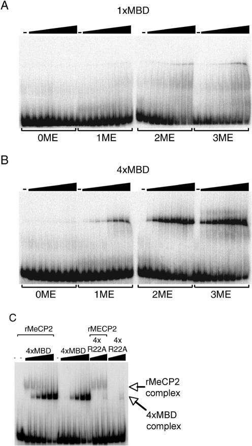
Recombinant His6-tagged 1xMBD and 4xMBD proteins were expressed in bacteria, purified to homogeneity (Figure 1B) and tested for in vitro DNA-binding. Bandshift analysis demonstrated that the wild-type proteins specifically form complexes with a methylated probe containing 27 methylated CpGs, but no binding of the mutant control proteins was observed (Figure 1C). The ladder of complexes reflects the varying number of proteins associated with each DNA probe molecule (4). The hypothesis that the multimerization of this domain would enhance its binding affinity was tested by titrating identical weights of monomeric (1xMBD) or tetrameric (4xMBD) proteins (Figure 2A and B) into binding reactions that contain oligonucleotide probes with 0–3 methylated CpGs. Based on the amounts of each protein required to complex ∼50% of the probe, we calculated the dissociation constants for 1xMBD and 4xMBD proteins (Table 1). The results showed that 4xMBD has a 50–80-fold higher affinity for substrates containing 1, 2 or 3 methylated CpGs than does 1xMBD. The affinity of 4xMBD for the probe with 3 methyl-CpG moieties was ∼20 nM. To test whether the multimeric protein competes with a wild-type methyl-CpG-binding protein for binding to methylated DNA, increasing amounts of 4xMBD were added to bandshift reactions that contained purified recombinant MeCP2. As shown in Figure 2C, DNA–MeCP2 complexes were competed away by the purified wild-type 4xMBD protein, whereas the mutant R22A protein did not compete. A similar result was obtained using nuclear extracts as a source of MBPs (data not shown).
Figure 2.
Multimerization of the MBD increases the affinity for methylated DNA. Increasing amounts (2.5, 5, 10, 15, 25, 50, 75, 100 ng) of wild-type 1xMBD (A) or 4xMBD protein (B) were incubated with duplex oligonucleotide probes containing either 0, 1, 2 or 3 methyl-CpGs (0ME, 1ME, 2ME and 3ME, respectively). The complexes were resolved on 6% polyacrylamide gels. (C) Multimeric MBD protein can displace the methyl-CpG-binding protein MeCP2 from methylated DNA in vitro. Increasing amounts of wild-type 4xMBD (2.5, 10, 25, 100, 250 ng) or control 4xMBD–R22A (2.5, 10, 25, 100, 250 ng) tetrameric protein were added to reactions containing 150 ng recombinant GST–MeCP2 fusion protein (rMeCP2) and incubated with the 2ME oligonucleotide probe (see above). Both MBD proteins and MeCP2 were present in the reactions before the addition of the labelled probe. The absence of multiple bands as seen with the CG11 probe (Figure 1) may be explained by the small number of CpGs in 2ME and 3ME probes (2 or 3 compared to 27 in CG11) and the use of polyacrylamide rather than agarose gel for fractionation of complexes.
Table 1.
Binding constants (μM) for 1xMBD and 4xMBD with probes that have 1, 2 or 3 methyl-CpG moieties (1ME, 2ME and 3ME)
| Protein | 1ME | 2ME | 3ME |
|---|---|---|---|
| 4xMBD | 0.5 µMa | 0.05 µMa | 0.02 µMa |
| 1xMBD | 30 µMa | 3 µMa | 2 µMa |
| 4x/1x ratio | 55 | 59 | 81 |
aBinding constants were deduced from bandshift data shown in Figure 2 in which probe (25 fmol) was incubated in 100 mM salt with 3–300 nmol 1xMBD or 1–100 nmol 3xMBD (see Materials and Methods).
Detection of methylated DNA by NxMBD proteins in vitro
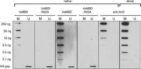
We examined the sensitivity of methyl-CpG-binding in vitro by incubating recombinant 1xMBD and 4xMBD plus their mutant counterparts with nylon membranes carrying either M.SssI-methylated or non-methylated bacteriophage lambda DNA. The results showed that 4xMBD was able to detect 0.1 ng of immobilized double-stranded DNA, whereas 1xMBD was more than 20-fold less sensitive in this assay (Figure 3). Binding to methylated DNA was lost in the R22A mutant proteins. No background staining of non-methylated DNA (up to 250 ng) was detected with the poly-MBD protein probes. For comparison, we probed equivalent slot-blots with a commercially available anti-m5C antibody. The antibody was ∼100-fold less sensitive than 4xMBD when probed against native double-stranded methylated DNA, but gave a comparable signal level when the immobilized DNA was denatured. Denaturation abolished binding of the MBD proteins to methylated DNA (data not shown).
Figure 3.
Recombinant 4xMBD proteins is a sensitive detection reagent for methylated CpG in immobilized DNA. CpG methylated (M) or unmethylated (U) lambda phage DNA was immobilized on membranes and detected using the indicated NxMBD proteins (10 µg/ml) followed by anti-HA and HRP-anti-mouse antibody, or with anti-m5C antibody (1:500) followed by HRP-anti-mouse antibody. The amount of DNA in each slot is indicated at the left. Native DNA (native) was blotted on to nitrocellulose membrane and denatured (denat) DNA was blotted on to nylon membranes. The bottom right slot on each filter contained 50 ng of HA-tagged 1xMBD as a positive control.
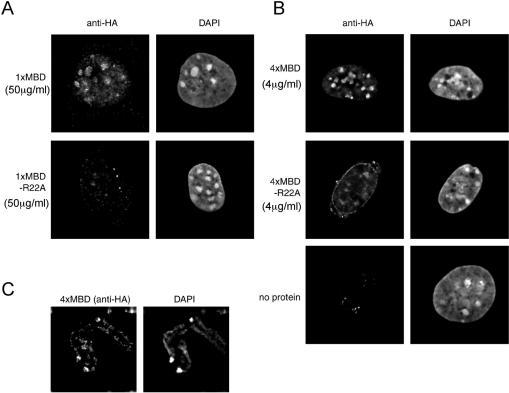
We then asked whether the artificial MBD proteins were able to access densely methylated heterochromatic foci in fixed mouse fibroblasts. After incubation with NxMBD proteins, bound proteins were immobilized by fixation to prevent washout during the subsequent procedures. Incubation with the wild-type protein resulted in characteristic staining of DAPI-positive heterochromatin (Figure 4A and B). The increased affinity resulting from multimerization was apparent, as 4 µg/ml of the tetrameric 4xMBD protein stained more strongly than 50 µg/ml of monomeric 1xMBD (Figure 4A and B). The 4xMBD protein also stained metaphase chromosomes at centromeric regions and along the chromosome arms (Figure 4C). No signal on metaphase chromosomes was detected using 1xMBD or the 1x or 4xMBD–R22A negative control proteins. Taken together with the slot-blot data (Figure 3), these results indicate that 4xMBD is a sensitive and specific reagent for the detection of CpG methylation of double-stranded genomic DNA in the genome. Multimerization of the MBD does not compromise the specificity of the MBD, as non-methylated DNA is not detectably recognized.
Figure 4.
The 4xMBD protein is a sensitive stain for methyl-CpG-rich regions in fixed cells. Permeabilized and fixed mouse fibroblasts were incubated with 50 µg/ml 1xMBD (A) or 4 µg/ml 4xMBD (B), or their R22A mutant forms, and visualized with anti-HA plus Alexa-594-conjugated anti-mouse antibodies. Specific staining is seen at heterochromatic DAPI bright spots with the wild-type probe but not with their mutant counterparts. Weak staining by the 4xMBD–R22A mutant protein is sometimes detected, but does not co-localize with DAPI bright spots. Staining shows improved resolution with 4xMBD compared to 1xMBD and requires >10-fold less protein. (C) Metaphase spreads from mouse fibroblasts were incubated with 4 µg/ml 4xMBD and visualized with anti-HA plus Alexa-594-conjugated anti-mouse antibodies. DNA is counterstained with DAPI. Intense staining of pericentromeric satellite DNA is apparent plus weaker staining of the chromosome arms.
In vivo binding of multimeric DNA to methyl-CpG sites
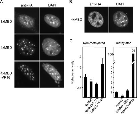
Following the in vitro studies described above, we analysed the in vivo binding characteristics of NxMBD proteins. The proteins were expressed in mouse fibroblasts and detected using an anti-HA antibody. As expected, the wild-type proteins co-localized with methyl-CpG-dense heterochromatic foci (Figure 5A). 4xMBD showed significantly more intense staining than 1xMBD, in agreement with the in vitro analysis. The negative control proteins (R22A) did not co-localize with the heterochomatic foci (data not shown). As a further control for the specificity of in vivo binding, we found that heterochromatic localization of wild-type 4xMBD proteins was lost in cells that lack Dnmt1 (17) and, hence, have greatly reduced levels of CpG methylation (Figure 5B).
Figure 5.
Wild-type NxMBD proteins localize to CpG-rich heterochromatin in a DNA methylation dependent manner in vivo and can deliver a functional domain to a methylated reporter gene. The indicated NxMBD proteins were expressed in wild-type (A) or DNA methylation deficient Dnmt1-null (B) mouse fibroblasts that were fixed and stained using an anti-HA antibody. Left panels show anti-HA signals and right panels show DNA counterstained with DAPI. (A) Co-localization of wild-type 1xMBD, 4xMBD and the 4xMBD–VP16 fusion proteins with heterochromatic foci in interphase cells. (B) Dispersed nuclear localization of 1xMBD and 4xMBD in Dnmt1-null cells that lack DNA methylation. (C) The 4xMBD–VP16 fusion protein specifically activates a methylated reporter construct in vivo. Non-methylated or methylated reporter constructs were co-transfected alone, or with 4xMBD (500 ng), 4xMBD–R22A (500 ng) or a 4xMBD–VP16 fusion construct (250 ng). The average relative reporter activity for a triplicate experiment is shown. The error bars represent ±SD.
To determine whether 4xMBD could recruit a functional domain to non-heterochromatic methylated sites in vivo, the 4xMBD protein was fused to the VP16-activation domain and expressed in cells that also received reporter constructs. As shown in Figure 5C, the 4xMBD–VP16 fusion caused a ∼100-fold increase in expression from a methylated reporter, whereas unfused 4xMBD protein had no effect. A slight induction (∼2-fold) of the non-methylated construct by 4xMBD–VP16 was observed, which may be due to non-specific effects of its strong activation domain. Staining of cells transfected with the 4xMBD–VP16 construct demonstrated that the overall localization of the fusion protein is indistinguishable from that of non-fusion 4xMBD (Figure 5A). We conclude that MBD is available to interact with euchromatic methylated sites and can recruit a functional domain to these sites in vivo.
CONCLUSIONS
We demonstrate here that the 4xMBD polypeptide has a high affinity for methyl-CpG sites in vitro and in vivo. Multimerization significantly amplifies the affinity without loss of specificity as measured by interaction with (i) methylated DNA in solution, (ii) DNA immobilized on membranes and (iii) chromosomal DNA in cytological preparations. These methods consistently suggest a ∼100-fold enhancement of binding due to tetramerization compared with the monomeric MBD. In spite of this improved binding, we saw little evidence for loss of specificity, as non-methylated DNA was negative for interaction with the 4xMBD polypeptide by all assays. Available antibodies raised against the m5C base require DNA to be denatured by heat or alkali in order to fully expose the epitope. An advantage of the poly-MBD proteins is that they recognize methyl-CpG in duplex DNA and therefore do not require cytological preparations to be subjected to harsh denaturing conditions that may compromise specimen integrity. Also, 4xMBD can be produced in the laboratory simply and relatively cheaply by expression in bacteria. Poly-MBD proteins can therefore be considered as an alternative to anti-m5C antibodies as sensitive and specific reagents for the detection of methyl-CpG sites in genomic DNA.
Acknowledgments
We thank Ton van Amstel for help using the Delta Vision microscope. This work was funded by the Wellcome Trust, UK. Funding to pay the Open Access publication charges for this article was provided by the Wellcome Trust.
Conflict of interest statement. None declared.
REFERENCES
- 1.Hendrich B., Bird A. Identification and characterization of a family of mammalian methyl-CpG binding proteins. Mol. Cell. Biol. 1998;18:6538–6547. doi: 10.1128/mcb.18.11.6538. [DOI] [PMC free article] [PubMed] [Google Scholar]
- 2.Cross S.H., Meehan R.R., Nan X., Bird A. A component of the transcriptional repressor MeCP1 is related to mammalian DNA methyltransferase and trithorax-like protein. Nature Genet. 1997;16:256–259. doi: 10.1038/ng0797-256. [DOI] [PubMed] [Google Scholar]
- 3.Lewis J.D., Meehan R.R., Henzel W.J., Maurer-Fogy I., Jeppesen P., Klein F., Bird A. Purification, sequence and cellular localisation of a novel chromosomal protein that binds to methylated DNA. Cell. 1992;69:905–914. doi: 10.1016/0092-8674(92)90610-o. [DOI] [PubMed] [Google Scholar]
- 4.Nan X., Meehan R.R., Bird A. Dissection of the methyl-CpG binding domain from the chromosomal protein MeCP2. Nucleic Acids Res. 1993;21:4886–4892. doi: 10.1093/nar/21.21.4886. [DOI] [PMC free article] [PubMed] [Google Scholar]
- 5.Prokhortchouk A., Hendrich B., Jorgensen H., Ruzov A., Wilm M., Georgiev G., Bird A., Prokhortchouk E. The p120 catenin partner Kaiso is a DNA methylation-dependent transcriptional repressor. Genes Dev. 2001;15:1613–1618. doi: 10.1101/gad.198501. [DOI] [PMC free article] [PubMed] [Google Scholar]
- 6.Filion G.J., Zhenilo S., Salozhin S., Yamada D., Prokhortchouk E., Defossez P.A. A family of human zinc finger proteins that bind methylated DNA and repress transcription. Mol. Cell. Biol. 2006;26:169–181. doi: 10.1128/MCB.26.1.169-181.2006. [DOI] [PMC free article] [PubMed] [Google Scholar]
- 7.Wade P.A., Jones P.L., Vermaak D., Wolffe A.P. A multiple subunit Mi-2 histone deacetylase from Xenopus laevis cofractionates with an associated Snf2 superfamily ATPase. Curr. Biol. 1998;8:843–846. doi: 10.1016/s0960-9822(98)70328-8. [DOI] [PubMed] [Google Scholar]
- 8.Nan X., Ng H.-H., Johnson C.A., Laherty C.D., Turner B.M., Eisenman R.N., Bird A. Transcriptional repression by the methyl-CpG-binding protein MeCP2 involves a histone deacetylase complex. Nature. 1998;393:386–389. doi: 10.1038/30764. [DOI] [PubMed] [Google Scholar]
- 9.Yoon H.G., Chan D.W., Reynolds A.B., Qin J., Wong J. N-CoR mediates DNA methylation-dependent repression through a methyl CpG binding protein Kaiso. Mol. Cell. 2003;12:723–734. doi: 10.1016/j.molcel.2003.08.008. [DOI] [PubMed] [Google Scholar]
- 10.Fujita N., Watanabe S., Ichimura T., Ohkuma Y., Chiba T., Saya H., Nakao M. MCAF mediates MBD1-dependent transcriptional repression. Mol. Cell. Biol. 2003;23:2834–2843. doi: 10.1128/MCB.23.8.2834-2843.2003. [DOI] [PMC free article] [PubMed] [Google Scholar]
- 11.Singer J., Schnute W.C.J., Schively J.E., Todd C.W., Riggs A.D. Sensitive detection of 5-methylcytosine and quantitation of the 5-methylcytosine/cytosine ratio in DNA by gas chromatography-mass spectrometry using multiple specific ion monitoring. Anal. Biochem. 1979;94:297–301. doi: 10.1016/0003-2697(79)90363-4. [DOI] [PubMed] [Google Scholar]
- 12.Ramsahoye B.H. Nearest-neighbor analysis. Methods Mol. Biol. 2002;200:9–15. doi: 10.1385/1-59259-182-5:009. [DOI] [PubMed] [Google Scholar]
- 13.Balaghi M., Wagner C. DNA methylation in folate deficiency: use of CpG methylase. Biochem. Biophys. Res. Commun. 1993;193:1184–1190. doi: 10.1006/bbrc.1993.1750. [DOI] [PubMed] [Google Scholar]
- 14.Ramsahoye B.H. Measurement of genome-wide DNA cytosine-5 methylation by reversed-phase high-pressure liquid chromatography. Methods Mol. Biol. 2002;200:17–27. doi: 10.1385/1-59259-182-5:017. [DOI] [PubMed] [Google Scholar]
- 15.Miller O.L., Schnoedl W., Allen J., Erlanger B.F. Immunofluorescent localization of 5MC. Nature. 1974;251:636–637. doi: 10.1038/251636a0. [DOI] [PubMed] [Google Scholar]
- 16.Barbin A., Montpellier C., Kokalj-Vokac N., Gibaud A., Niveleau A., Malfoy B., Dutrillaux B., Bourgeois C.A. New sites of methylcytosine-rich DNA detected on metaphase chromosomes. Hum. Genet. 1994;94:684–692. doi: 10.1007/BF00206964. [DOI] [PubMed] [Google Scholar]
- 17.Jorgensen H.F., Ben-Porath I., Bird A.P. Mbd1 is recruited to both methylated and nonmethylated CpGs via distinct DNA binding domains. Mol. Cell. Biol. 2004;24:3387–3395. doi: 10.1128/MCB.24.8.3387-3395.2004. [DOI] [PMC free article] [PubMed] [Google Scholar]
- 18.Kinsella T.M., Nolan G.P. Episomal vectors rapidly and stably produce high-titer recombinant retrovirus. Hum. Gene. Ther. 1996;7:1405–1413. doi: 10.1089/hum.1996.7.12-1405. [DOI] [PubMed] [Google Scholar]
- 19.Li E., Bestor T.H., Jaenisch R. Targeted mutation of the DNA methyltransferase gene results in embryonic lethality. Cell. 1992;69:915–926. doi: 10.1016/0092-8674(92)90611-f. [DOI] [PubMed] [Google Scholar]
- 20.Jackson-Grusby L., Beard C., Possemat R., Tudor M., Fambrough D., Csankovszki G., Dausman J., Lee P., Wilson C., Lander E., et al. Loss of genomic methylation causes p53-dependent apoptosis and epigenetic deregulation. Nature Genet. 2001;27:31–39. doi: 10.1038/83730. [DOI] [PubMed] [Google Scholar]
- 21.Nielsen S.J., Praestegaard M., Jorgensen H.F., Clark B.F. Different Sp1 family members differentially affect transcription from the human elongation factor 1 A-1 gene promoter. Biochem. J. 1998;333:511–517. doi: 10.1042/bj3330511. [DOI] [PMC free article] [PubMed] [Google Scholar]
- 22.Meehan R.R., Lewis J.D., McKay S., Kleiner E.L., Bird A.P. Identification of a mammalian protein that binds specifically to DNA containing methylated CpGs. Cell. 1989;58:499–507. doi: 10.1016/0092-8674(89)90430-3. [DOI] [PubMed] [Google Scholar]
- 23.Ohki I., Shimotake N., Fujita N., Jee J., Ikegami T., Nakao M., Shirakawa M. Solution structure of the methyl-CpG binding domain of human MBD1 in complex with methylated DNA. Cell. 2001;105:487–497. doi: 10.1016/s0092-8674(01)00324-5. [DOI] [PubMed] [Google Scholar]
- 24.Ng H.-H., Jeppesen P., Bird A. Active repression of methylated genes by the chromosomal protein MBD1. Mol. Cell. Biol. 2000;20:1394–1406. doi: 10.1128/mcb.20.4.1394-1406.2000. [DOI] [PMC free article] [PubMed] [Google Scholar]
- 25.Strick R., Laemmli U.K. SARs are cis DNA elements of chromosome dynamics: synthesis of a SAR repressor protein. Cell. 1995;83:1137–1148. doi: 10.1016/0092-8674(95)90140-x. [DOI] [PubMed] [Google Scholar]


