Abstract
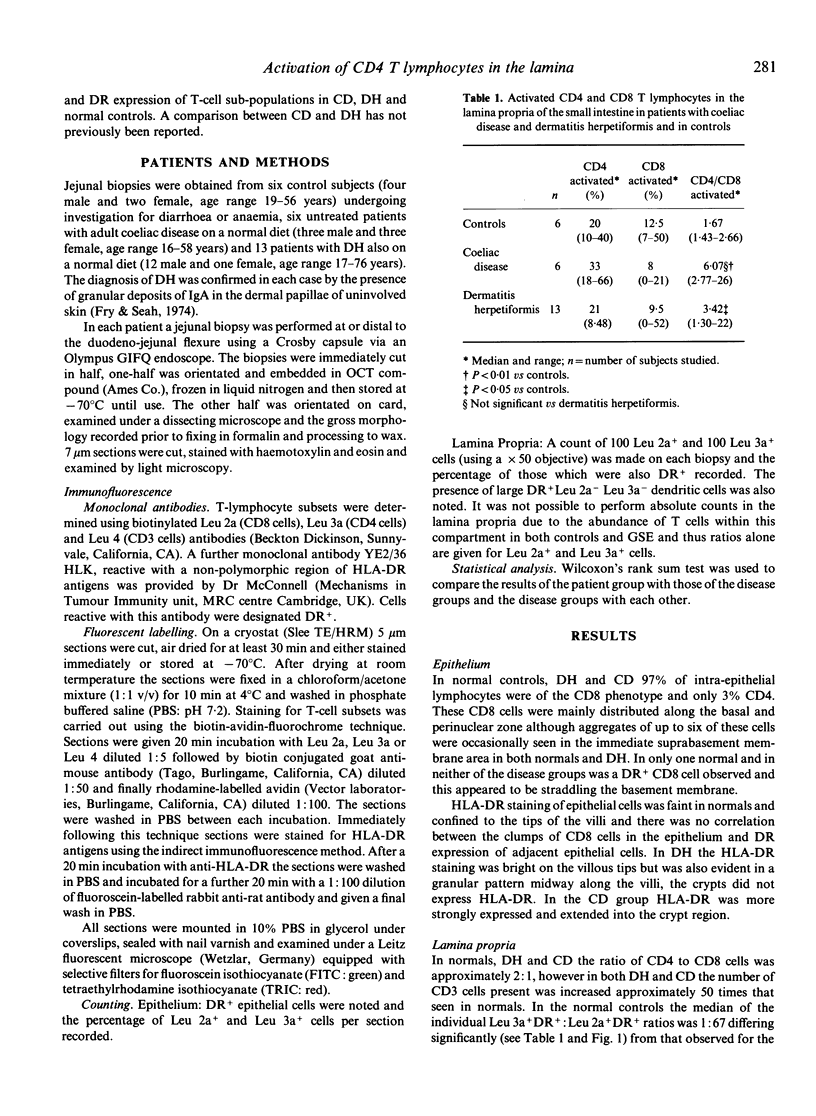
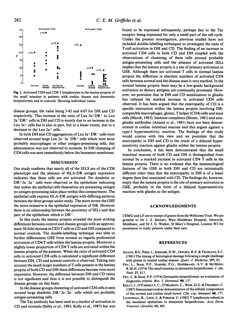
The distribution and activation of T-lymphocyte subsets in the small intestinal mucosa of coeliac disease and dermatitis herpetiformis subjects on a normal diet has been studied and compared to normal controls. Double-labelling immunofluorescence techniques with monoclonal antibodies were used on cryostat tissue sections. Intestinal epithelial cells demonstrated staining for HLA-DR, the intensity being proportional to the degree of enteropathy. In both patients and controls nearly all (97%) intra-epithelial lymphocytes were of the CD8 subset and not activated as judged by HLA-DR expression. In the lamina propria there was an approximate 50-fold increase in T cells in the patients as compared with the controls. Whilst the ratio of total CD4 to total CD8 cells was unchanged, the CD4 subset was preferentially activated in the patients. Thus in the normal controls the median ratio of activated CD4 cells to activated CD8 cells was 1.67 whilst for dermatitis herpetiformis and coeliac disease it was 3.42 and 6.07 respectively. These findings suggest that the lamina propria is a site of vigorous T-cell activity in gluten-sensitive individuals and is consistent with the view that the enteropathy of dermatitis herpetiformis and coeliac disease is the result of a delayed-type hypersensitivity against gliadin.
Full text
PDF


Selected References
These references are in PubMed. This may not be the complete list of references from this article.
- Anand B. S., Piris J., Jerrome D. W., Offord R. E., Truelove S. C. The timing of histological damage following a single challenge with gluten in treated coeliac disease. Q J Med. 1981;50(197):83–94. [PubMed] [Google Scholar]
- Fry L., Seah P. P. Dermatitis herpetiformis: an evaluation of diagnostic criteria. Br J Dermatol. 1974 Feb;90(2):137–146. doi: 10.1111/j.1365-2133.1974.tb06377.x. [DOI] [PubMed] [Google Scholar]
- Fry L., Seah P. P., Harper P. G., Hoffbrand A. V., McMinn R. M. The small intestine in dermatitis herpetiformis. J Clin Pathol. 1974 Oct;27(10):817–824. doi: 10.1136/jcp.27.10.817. [DOI] [PMC free article] [PubMed] [Google Scholar]
- Kelly J., O'Farrelly C., O'Mahony C., Weir D. G., Feighery C. Immunoperoxidase demonstration of the cellular composition of the normal and coeliac small bowel. Clin Exp Immunol. 1987 Apr;68(1):177–188. [PMC free article] [PubMed] [Google Scholar]
- Ljunghall K., Löf L., Forsum U. T lymphocyte subsets in the duodenal epithelium in dermatitis herpetiformis. Acta Derm Venereol. 1982;62(6):485–489. [PubMed] [Google Scholar]
- Marsh M. N. Immunocytes, enterocytes and the lamina propria: an immunopathological framework of coeliac disease. J R Coll Physicians Lond. 1983 Oct;17(4):205–212. [PMC free article] [PubMed] [Google Scholar]
- Selby W. S., Janossy G., Bofill M., Jewell D. P. Lymphocyte subpopulations in the human small intestine. The findings in normal mucosa and in the mucosa of patients with adult coeliac disease. Clin Exp Immunol. 1983 Apr;52(1):219–228. [PMC free article] [PubMed] [Google Scholar]
- Selby W. S., Janossy G., Goldstein G., Jewell D. P. T lymphocyte subsets in human intestinal mucosa: the distribution and relationship to MHC-derived antigens. Clin Exp Immunol. 1981 Jun;44(3):453–458. [PMC free article] [PubMed] [Google Scholar]
- Selby W. S., Janossy G., Jewell D. P. Immunohistological characterisation of intraepithelial lymphocytes of the human gastrointestinal tract. Gut. 1981 Mar;22(3):169–176. doi: 10.1136/gut.22.3.169. [DOI] [PMC free article] [PubMed] [Google Scholar]


