

Abstract
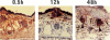
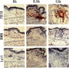
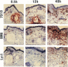
The kinetics of appearance of MIF+ cells was investigated in experimental contact dermatitis using a monoclonal antibody (7D10) against murine MIF which was reacted with cryostat sections of tissues and detected by the indirect immunoperoxidase test. Four groups of BALB/c mice were investigated: (1) sensitized with 2,4-dinitrofluorobenzene (DNFB); (2) unsensitized controls; (3) tolerized; (4) unsensitized. A challenge dose of DNFB was applied to the ear of animals of groups 1-3 and of croton oil to those of group 4. Three phases could be distinguished in group 1: (a) an initial vascular and exudative reaction; (b) an early cellular phase; and (c) a late cellular phase. At zero time rarely any T lymphocytes (Lyt 1+; Lyt 2+) were seen in all four groups. Within less than 30 min venous endothelial cells became strongly MIF+. This was followed by an influx of monocytes/macrophages reaching a maximum of 72 h in group 1 and a slight peak at 12 h in groups 2 and 3. At 16-24 h in all groups the endothelial reaction weakened while many 7D10+ macrophages appeared in group 1. By double-labelling it was shown that lymphocytes were 7D10-. The influx of lymphocytes, part of which carried the T cell receptor, began at 12 h, reaching a maximum at 72 h in group 1. In groups 2 and 3 only a weak lymphocytic infiltrate developed which declined at 24 h. Group 4 developed an inflammatory reaction after the initial phase with similar kinetics as in group 1. The data suggest that an immune inflammatory reaction is preceded by a nonspecific reaction of the vascular endothelium and the mononuclear phagocytic system and that MIF is playing a central role in these events.
Full text
PDF


Images in this article
Selected References
These references are in PubMed. This may not be the complete list of references from this article.
- Bloom B. R., Bennett B. Mechanism of a reaction in vitro associated with delayed-type hypersensitivity. Science. 1966 Jul 1;153(3731):80–82. doi: 10.1126/science.153.3731.80. [DOI] [PubMed] [Google Scholar]
- Burmeister G., Tarcsay L., Sorg C. Generation and characterization of a monoclonal antibody (1C5) to human migration inhibitory factor (MIF). Immunobiology. 1986 Jul;171(4-5):461–474. doi: 10.1016/S0171-2985(86)80077-8. [DOI] [PubMed] [Google Scholar]
- Farr A. G., Anderson S. K., Marrack P., Kappler J. Expression of antigen-specific, major histocompatibility complex-restricted receptors by cortical and medullary thymocytes in situ. Cell. 1985 Dec;43(2 Pt 1):543–550. doi: 10.1016/0092-8674(85)90183-7. [DOI] [PubMed] [Google Scholar]
- Kaufmann S. H., Hug E., De Libero G. Listeria monocytogenes-reactive T lymphocyte clones with cytolytic activity against infected target cells. J Exp Med. 1986 Jul 1;164(1):363–368. doi: 10.1084/jem.164.1.363. [DOI] [PMC free article] [PubMed] [Google Scholar]
- Knop J., Malorny U., Macher E. Selective induction of delayed hypersensitivity T-effector and T-suppressor lymphocytes in vitro by haptenized bone marrow-derived macrophages. Cell Immunol. 1984 Oct 15;88(2):411–420. doi: 10.1016/0008-8749(84)90174-6. [DOI] [PubMed] [Google Scholar]
- Knop J., Malorny U., Michels E., Sorg C. Selection of the delayed hypersensitivity T effector and T suppressor cell response by antigen-presenting macrophages. Immunobiology. 1984 Dec;168(3-5):246–259. doi: 10.1016/S0171-2985(84)80114-X. [DOI] [PubMed] [Google Scholar]
- Malorny U., Michels E., Sorg C. A monoclonal antibody against an antigen present on mouse macrophages and absent from monocytes. Cell Tissue Res. 1986;243(2):421–428. doi: 10.1007/BF00251059. [DOI] [PubMed] [Google Scholar]
- Michels E., Stenzinger W., Sorg C. Functional characteristics of murine macrophages responding to migration inhibitory factors. Eur J Immunol. 1984 Oct;14(10):902–905. doi: 10.1002/eji.1830141008. [DOI] [PubMed] [Google Scholar]
- Neumann C., Schlegel R., Steckel F., Sorg C. Detection of macrophage migration inhibitory factor by monoclonal antibody in Sézary syndrome. J Invest Dermatol. 1987 Jun;88(6):670–674. doi: 10.1111/1523-1747.ep12470326. [DOI] [PubMed] [Google Scholar]
- Platt J. L., Grant B. W., Eddy A. A., Michael A. F. Immune cell populations in cutaneous delayed-type hypersensitivity. J Exp Med. 1983 Oct 1;158(4):1227–1242. doi: 10.1084/jem.158.4.1227. [DOI] [PMC free article] [PubMed] [Google Scholar]
- Poulter L. W., Seymour G. J., Duke O., Janossy G., Panayi G. Immunohistological analysis of delayed-type hypersensitivity in man. Cell Immunol. 1982 Dec;74(2):358–369. doi: 10.1016/0008-8749(82)90036-3. [DOI] [PubMed] [Google Scholar]
- Reitamo S., Tolvanen E., Konttinen Y. T., Käyhkö K., Förström L., Salo O. P. Allergic and toxic contact dermatitis: inflammatory cell subtypes in epicutaneous test reactions. Br J Dermatol. 1981 Nov;105(5):521–527. doi: 10.1111/j.1365-2133.1981.tb00795.x. [DOI] [PubMed] [Google Scholar]
- Scheynius A., Fischer T., Forsum U., Klareskog L. Phenotypic characterization in situ of inflammatory cells in allergic and irritant contact dermatitis in man. Clin Exp Immunol. 1984 Jan;55(1):81–90. [PMC free article] [PubMed] [Google Scholar]
- Scheynius A., Klareskog L., Forsum U. In situ identification of T lymphocyte subsets and HLA-DR expressing cells in the human skin tuberculin reaction. Clin Exp Immunol. 1982 Aug;49(2):325–330. [PMC free article] [PubMed] [Google Scholar]