

Abstract
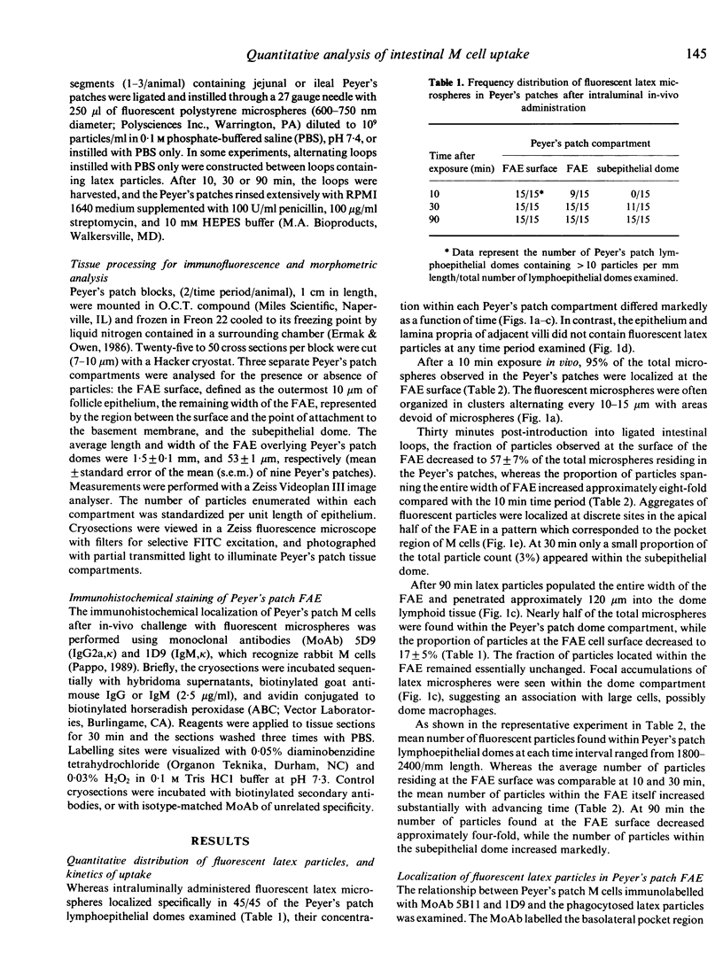
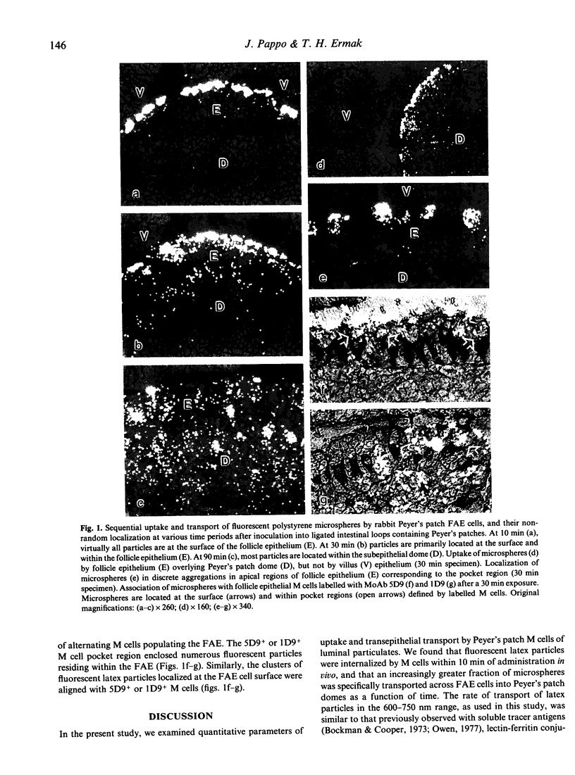
A quantitative, light microscopic morphometric model for uptake of particulates by Peyer's patch M cells was developed. Rabbit intestinal loops containing Peyer's patches were inoculated with fluorescent, non-degradable polystyrene microparticles (600-750 nm), and their localization in Peyer's patches was traced after varying time periods. The particles were localized sequentially at the FAE cell surface, spanning the entire width of FAE cells, and within the subepithelial dome as a function of time. The particles were associated with 5D9+ or 1D9+ M cells, but were not taken up or transported by villus epithelia. The kinetics suggested a synchronous wave of uptake and transepithelial transport. Quantitative analysis revealed a considerably greater uptake efficiency of polystyrene microspheres in comparison to other biological particles.
Full text
PDF
Selected References
These references are in PubMed. This may not be the complete list of references from this article.
- Abe K., Ito T. Qualitative and quantitative morphologic study of Peyer's patches of the mouse after neonatal thymectomy and hydrocortisone injection. Am J Anat. 1978 Feb;151(2):227–237. doi: 10.1002/aja.1001510206. [DOI] [PubMed] [Google Scholar]
- Bockman D. E., Cooper M. D. Pinocytosis by epithelium associated with lymphoid follicles in the bursa of Fabricius, appendix, and Peyer's patches. An electron microscopic study. Am J Anat. 1973 Apr;136(4):455–477. doi: 10.1002/aja.1001360406. [DOI] [PubMed] [Google Scholar]
- Ermak T. H., Owen R. L. Differential distribution of lymphocytes and accessory cells in mouse Peyer's patches. Anat Rec. 1986 Jun;215(2):144–152. doi: 10.1002/ar.1092150208. [DOI] [PubMed] [Google Scholar]
- Faulk W. P., McCormick J. N., Goodman J. R., Yoffey J. M., Fudenberg H. H. Peyer's patches: morphologic studies. Cell Immunol. 1970 Nov;1(5):500–520. doi: 10.1016/0008-8749(70)90038-9. [DOI] [PubMed] [Google Scholar]
- GRUSKAY F. L., COOKE R. E. The gastrointestinal absorption of unaltered protein in normal infants and in infants recovering from diarrhea. Pediatrics. 1955 Dec;16(6):763–769. [PubMed] [Google Scholar]
- Inman L. R., Cantey J. R. Specific adherence of Escherichia coli (strain RDEC-1) to membranous (M) cells of the Peyer's patch in Escherichia coli diarrhea in the rabbit. J Clin Invest. 1983 Jan;71(1):1–8. doi: 10.1172/JCI110737. [DOI] [PMC free article] [PubMed] [Google Scholar]
- Keljo D. J., Hamilton J. R. Quantitative determination of macromolecular transport rate across intestinal Peyer's patches. Am J Physiol. 1983 Jun;244(6):G637–G644. doi: 10.1152/ajpgi.1983.244.6.G637. [DOI] [PubMed] [Google Scholar]
- Kohbata S., Yokoyama H., Yabuuchi E. Cytopathogenic effect of Salmonella typhi GIFU 10007 on M cells of murine ileal Peyer's patches in ligated ileal loops: an ultrastructural study. Microbiol Immunol. 1986;30(12):1225–1237. doi: 10.1111/j.1348-0421.1986.tb03055.x. [DOI] [PubMed] [Google Scholar]
- Marcial M. A., Madara J. L. Cryptosporidium: cellular localization, structural analysis of absorptive cell-parasite membrane-membrane interactions in guinea pigs, and suggestion of protozoan transport by M cells. Gastroenterology. 1986 Mar;90(3):583–594. doi: 10.1016/0016-5085(86)91112-1. [DOI] [PubMed] [Google Scholar]
- Momotani E., Whipple D. L., Thiermann A. B., Cheville N. F. Role of M cells and macrophages in the entrance of Mycobacterium paratuberculosis into domes of ileal Peyer's patches in calves. Vet Pathol. 1988 Mar;25(2):131–137. doi: 10.1177/030098588802500205. [DOI] [PubMed] [Google Scholar]
- Neutra M. R., Phillips T. L., Mayer E. L., Fishkind D. J. Transport of membrane-bound macromolecules by M cells in follicle-associated epithelium of rabbit Peyer's patch. Cell Tissue Res. 1987 Mar;247(3):537–546. doi: 10.1007/BF00215747. [DOI] [PubMed] [Google Scholar]
- Owen R. L., Allen C. L., Stevens D. P. Phagocytosis of Giardia muris by macrophages in Peyer's patch epithelium in mice. Infect Immun. 1981 Aug;33(2):591–601. doi: 10.1128/iai.33.2.591-601.1981. [DOI] [PMC free article] [PubMed] [Google Scholar]
- Owen R. L., Apple R. T., Bhalla D. K. Morphometric and cytochemical analysis of lysosomes in rat Peyer's patch follicle epithelium: their reduction in volume fraction and acid phosphatase content in M cells compared to adjacent enterocytes. Anat Rec. 1986 Dec;216(4):521–527. doi: 10.1002/ar.1092160409. [DOI] [PubMed] [Google Scholar]
- Owen R. L., Jones A. L. Epithelial cell specialization within human Peyer's patches: an ultrastructural study of intestinal lymphoid follicles. Gastroenterology. 1974 Feb;66(2):189–203. [PubMed] [Google Scholar]
- Owen R. L., Pierce N. F., Apple R. T., Cray W. C., Jr M cell transport of Vibrio cholerae from the intestinal lumen into Peyer's patches: a mechanism for antigen sampling and for microbial transepithelial migration. J Infect Dis. 1986 Jun;153(6):1108–1118. doi: 10.1093/infdis/153.6.1108. [DOI] [PubMed] [Google Scholar]
- Owen R. L. Sequential uptake of horseradish peroxidase by lymphoid follicle epithelium of Peyer's patches in the normal unobstructed mouse intestine: an ultrastructural study. Gastroenterology. 1977 Mar;72(3):440–451. [PubMed] [Google Scholar]
- Pappo J., Steger H. J., Owen R. L. Differential adherence of epithelium overlying gut-associated lymphoid tissue. An ultrastructural study. Lab Invest. 1988 Jun;58(6):692–697. [PubMed] [Google Scholar]
- Sminia T., Janse E. M., Plesch B. E. Ontogeny of Peyer's patches of the rat. Anat Rec. 1983 Oct;207(2):309–316. doi: 10.1002/ar.1092070209. [DOI] [PubMed] [Google Scholar]
- Smith M. W., Jarvis L. G., King I. S. Cell proliferation in follicle-associated epithelium of mouse Peyer's patch. Am J Anat. 1980 Oct;159(2):157–166. doi: 10.1002/aja.1001590204. [DOI] [PubMed] [Google Scholar]
- Smith M. W., Peacock M. A. "M" cell distribution in follicle-associated epithelium of mouse Peyer's patch. Am J Anat. 1980 Oct;159(2):167–175. doi: 10.1002/aja.1001590205. [DOI] [PubMed] [Google Scholar]
- Warshaw A. L., Walker W. A., Cornell R., Isselbacher K. J. Small intestinal permeability to macromolecules. Transmission of horseradish peroxidase into mesenteric lymph and portal blood. Lab Invest. 1971 Dec;25(6):675–684. [PubMed] [Google Scholar]
- Wolf J. L., Rubin D. H., Finberg R., Kauffman R. S., Sharpe A. H., Trier J. S., Fields B. N. Intestinal M cells: a pathway for entry of reovirus into the host. Science. 1981 Apr 24;212(4493):471–472. doi: 10.1126/science.6259737. [DOI] [PubMed] [Google Scholar]