

Abstract
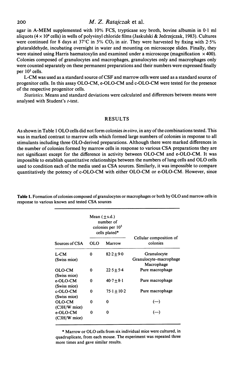
To test the hypothesis that the omental lymphoid organ (OLO) made by peritoneal milky spots is either a source of haemopoietic (macrophage) progenitors or growth factors we attempted to culture OLO cells in vitro in a variety of assay combinations. With the culture in vitro in semisolid agar it was found that OLO cells do not form granulocyte-macrophage or macrophage colonies in response to stimulants. However, when in the same assay marrow cells were used as the targets and OLO-related preparations as stimulants it was observed that marrow cells formed exclusively macrophage colonies. These marrow cells, in response to stimulants derived from other organs, produced granulocyte-macrophage, granulocyte and macrophage colonies. OLO-related preparations tested for macrophage-colony stimulating activity included partly purified medium conditioned by OLO cells derived from mice, either injected with endotoxin or not, and medium conditioned by OLO cells after 14 days, liquid culture in vitro. While these results were observed in Swiss mice, C3H/W mice, which are genetically endotoxin-unresponsive, failed to show this reaction. These data may suggest that the local production of macrophage-colony stimulating activity in the peritoneal cavity could be one physiological role for OLO. OLO is the first organ in adult mice identified to stimulate exclusively macrophage colony growth, and not granulocyte-macrophage or pure granulocyte colonies.
Full text
PDF
Selected References
These references are in PubMed. This may not be the complete list of references from this article.
- Apte R. N., Pluznik D. H. Genetic control of lipopolysaccharide induced generation of serum colony stimulating factor and proliferation of splenic granulocyte/macrophage precursor cells. J Cell Physiol. 1976 Oct;89(2):313–323. doi: 10.1002/jcp.1040890214. [DOI] [PubMed] [Google Scholar]
- Bradley T. R., Metcalf D. The growth of mouse bone marrow cells in vitro. Aust J Exp Biol Med Sci. 1966 Jun;44(3):287–299. doi: 10.1038/icb.1966.28. [DOI] [PubMed] [Google Scholar]
- Daems W. T., de Bakker J. M. Do resident macrophages proliferate? Immunobiology. 1982 Apr;161(3-4):204–211. doi: 10.1016/S0171-2985(82)80075-2. [DOI] [PubMed] [Google Scholar]
- Kaboth W., Ax E., Fischer H. Immunologische Studien am Omentum. II. Zur Immunmorphologie der "Plaque-bildenden" Milchflecken im Mäuseomentum. Z Naturforsch B. 1966 Aug;21(8):789–793. [PubMed] [Google Scholar]
- Kriegler A. B., Bradley T. R., Giap K. H., Hodgson G. S. Sources of murine macrophage colony-stimulating factor that also contain growth factors for primitive macrophage progenitor cells. Exp Hematol. 1984 Dec;12(11):844–849. [PubMed] [Google Scholar]
- Pelus L. M., Broxmeyer H. E., DeSousa M., Moore M. A. Heterogeneity among resident murine peritoneal macrophages: separation and functional characterization of monocytoid cells producing granulocyte-macrophage colony-stimulating factor (GM-CSF) and responding to regulation by lactoferrin. J Immunol. 1981 Mar;126(3):1016–1021. [PubMed] [Google Scholar]
- Sheridan J. W., Metcalf D. A low molecular weight factor in lung-conditioned medium stimulating granulocyte and monocyte colony formation in vitro. J Cell Physiol. 1973 Feb;81(1):11–23. doi: 10.1002/jcp.1040810103. [DOI] [PubMed] [Google Scholar]
- Sheridan J. W., Stanley E. R. Tissue sources of bone marrow colony stimulating factor. J Cell Physiol. 1971 Dec;78(3):451–460. doi: 10.1002/jcp.1040780314. [DOI] [PubMed] [Google Scholar]