Abstract
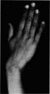
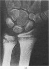
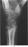
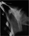
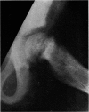
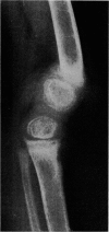
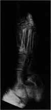
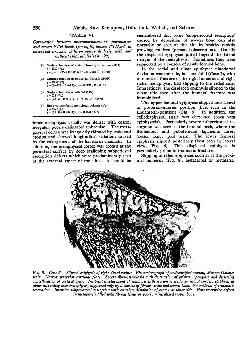
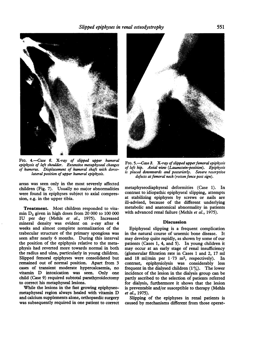
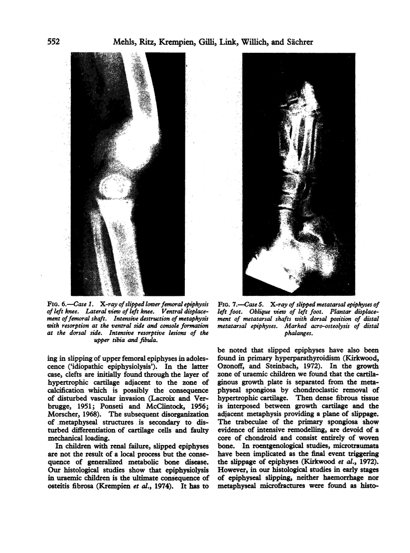
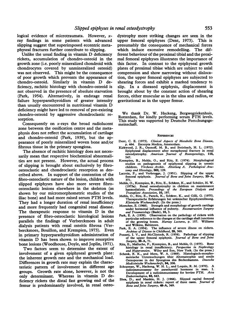
Clinical, biochemical, roentgenological, and histological features of slipped epiphyses (epiphysiolysis) in 11 out of 112 children with renal osteodystrophy have been analysed. Characteristic age-related patterns of involvement of different epiphyses are described. Quantitative measurements of iliac bone histology, serum parathyroid hormone levels, and clinical history show the presence of more advanced osteitis fibrosa in children with epiphysiolysis than in those without. A good correlation was found between serum parathormone levels and osteoclastic resorption, endosteal fibrosis as well as osteoid. Histological studies show that the radiolucent zone between the epiphyseal ossification centre and the metaphysis in x-rays is not caused by accumulation of cartilage and chondro-osteoid (as usually found in vitamin D deficiency rickets) but by the accumulation of woven bone and/or fibrous tissue. The response to vitamin D therapy in most cases was good. Parathyroidectomy was required in only one case.
Full text
PDF
Images in this article
Selected References
These references are in PubMed. This may not be the complete list of references from this article.
- Kirkwood J. R., Ozonoff M. B., Steinbach H. L. Epiphyseal displacement after metaphyseal fracture in renal osteodystrophy. Am J Roentgenol Radium Ther Nucl Med. 1972 Jul;115(3):547–554. doi: 10.2214/ajr.115.3.547. [DOI] [PubMed] [Google Scholar]
- Krempien B., Mehls O., Ritz E. Morphological studies on pathogenesis of epiphyseal slipping in uremic children. Virchows Arch A Pathol Anat Histol. 1974 Jan 30;362(2):129–143. doi: 10.1007/BF00432391. [DOI] [PubMed] [Google Scholar]
- LACROIX P., VERBRUGGE J. Slipping of the upper femoral epiphysis; a pathological study. J Bone Joint Surg Am. 1951 Apr;33-A(2):371–381. [PubMed] [Google Scholar]
- Mehls O., Krempien B., Ritz E., Schärer K., Schüler H. W. Renal osteodystrophy in children on maintenance haemodialysis. Proc Eur Dial Transplant Assoc. 1973;10(0):197–201. [PubMed] [Google Scholar]
- Morscher E. Strength and morphology of growth cartilage under hormonal influence of puberty. Animal experiments and clinical study on the etiology of local growth disorders during puberty. Reconstr Surg Traumatol. 1968;10:3–104. [PubMed] [Google Scholar]
- PARK E. A. The influence of severe illness on rickets. Arch Dis Child. 1954 Oct;29(147):369–380. doi: 10.1136/adc.29.147.369. [DOI] [PMC free article] [PubMed] [Google Scholar]
- PONSETI I. V., MCCLINTOCK R. The pathology of slipping of the upper femoral epiphysis. J Bone Joint Surg Am. 1956 Jan;38-A(1):71–83. [PubMed] [Google Scholar]
- Schenk R. K., Merz W. A. Histologisch-morphometrische Untersuchungen über Altersatrophie und senile Osteoporose in der Spongiosa des Beckenkammes. Dtsch Med Wochenschr. 1969 Jan 31;94(5):206–208. doi: 10.1055/s-0028-1108928. [DOI] [PubMed] [Google Scholar]
- Schopman W., Hackeng W. H., Lequin R. M. A radioimmunoassay for parathyroid hormone in man. I. Development of a radioimmunoassay for bovine PTH. Acta Endocrinol (Copenh) 1970 Apr;63(4):643–654. doi: 10.1530/acta.0.0630643. [DOI] [PubMed] [Google Scholar]
- Shea D., Mankin H. J. Slipped capital femoral epiphysis in renal rickets. Report of three cases. J Bone Joint Surg Am. 1966 Mar;48(2):349–355. [PubMed] [Google Scholar]
- Verberckmoes R., Bouillon R., Krempien B. Osteodystrophy of dialysed patients treated with vitamin D. Proc Eur Dial Transplant Assoc. 1973;10(0):217–226. [PubMed] [Google Scholar]
- Woodhouse N. J., Doyle F. H., Joplin G. F. Vitamin-D deficiency and primary hyperparathyroidism. Lancet. 1971 Aug 7;2(7719):283–286. doi: 10.1016/s0140-6736(71)91333-x. [DOI] [PubMed] [Google Scholar]


