Abstract
Angiogenesis, the process of development of a new microvasculature, is regulated by a balance of positive and negative factors. We show both in vivo and in vitro that the members of the human prolactin/growth hormone family, i.e., human prolactin, human growth hormone, human placental lactogen, and human growth hormone variant are angiogenic whereas their respective 16-kDa N-terminal fragments are antiangiogenic. The opposite actions are regulated in part via activation or inhibition of mitogen-activated protein kinase signaling pathway. In addition, the N-terminal fragments stimulate expression of type 1 plasminogen activator inhibitor whereas the intact molecules have no effect, an observation consistent with the fragments acting via separate receptors. The concept that a single molecule encodes both angiogenic and antiangiogenic peptides represents an efficient model for regulating the balance of positive and negative factors controlling angiogenesis. This hypothesis has potential physiological importance for the control of the vascular connection between the fetal and maternal circulations in the placenta, where human prolactin, human placental lactogen, and human growth hormone variant are expressed.
Prolactin (PRL), growth hormone (GH), and placental lactogen (PL) are homologous protein hormones believed to have arisen from a common ancestral gene (1). PRL participates in the regulation of reproduction, osmoregulation, and immunomodulation (2, 3) whereas GH is involved in regulating growth and morphogenesis (4). Human (h) GHs, unlike other mammalian GHs, bind to the PRL receptor and thus display PRL-like activity; however, hPRL does not bind to the hGH receptor (5). PRL and GH are produced mainly by the anterior pituitary in all vertebrates. PRL is expressed also in lymphocytes and in the decidua (6). The human placenta expresses two structural homologs of hGH, hPL and a variant of hGH (hGH-V) (7). hGH-V rather than pituitary hGH is believed to regulate maternal metabolism during the second half of pregnancy. hPL is somatotropic in fetal tissues and contributes to stimulating mammary cell proliferation (8). Rodent placentas express and secrete several proteins whose biological activities are more PRL-like than GH-like; these include proliferin (PLF) and a proliferin-related peptide (PRP) (9).
Members of the PRL/GH family and derived peptides have been reported to both stimulate and inhibit angiogenesis. PLF expressed during the first half of pregnancy in the mouse is angiogenic whereas PRP expressed later in gestation is antiangiogenic. These findings suggest that PLF and PRP might play a role in initiating and stopping placental neovascularization (9). Human GH was reported to be angiogenic in vitro (10) whereas both bovine and chicken GH were shown to be angiogenic in vivo (11). We have shown that the 16-kDa N-terminal fragments (16K) of rat PRL and hPRL are antiangiogenic both in vitro (12) and in vivo (13). Rat PRL is cleaved by cathepsin D (14) to yield a 16-kDa N-terminal fragment and a 7-kDa C-terminal fragment linked by a disulfide bridge (cleaved PRL). After reduction, the free N-terminal fragment is released (15). Cleaved PRL has been observed in mouse, rat, and human serum (16) whereas free 14- and 16-kDa PRL appear to be secreted in the hypothalamoneurohypophyseal system of the rat (17). Human GH is cleaved by plasmin, thrombin, and subtilisin, yielding similar fragments (18). Human 16K PRL and 16K hGH were observed in serum under nonreducing conditions (19), and the free 14-kDa hPRL fragment was observed in amniotic fluid and in maternal serum (20).
We now show that the members of the human PRL/GH family, i.e., hPRL, hPL, hGH, and hGH-V, stimulate vessel formation whereas their 16-kDa N-terminal fragments are antiangiogenic both in vivo and in vitro. We hypothesize that the intact and N-terminal fragments of the PRL/GH family members, through their opposite actions, are important physiological modulators of neovascularization.
MATERIALS AND METHODS
Production of Recombinant Proteins.
The cDNAs encoding hPRL, hPL, hGH, or hGH-V minus the corresponding signal peptide were inserted into the pT7L expression vector (21). An ATG was genetically engineered 5′ to the first codon of each cDNA. hPRL and hGH were cloned previously in our laboratory (22, 23). hGH-V was cloned by screening a placental cDNA library (CLONTECH). hPL was cloned by reverse transcription–PCR performed on RNA extracted from a primary culture of human syncytiotrophoblast cells. The intact proteins were produced and purified essentially as described (24).
The 16-kDa N-terminal fragments of the various hormones were produced either by cleavage (hPRL, hPL, and hGH-V) or by introducing a stop codon into the coding sequence (hGH). The DNA sequences from which the 16-kDa fragments were produced were constructed as follows. For the production of the 16-kDa fragment of hPRL (16K hPRL), the codon for Cys 58 (TGC) was mutated to a Ser codon (TCC), and the nucleotide sequence coding for amino acids 139–144 (Pro-Glu-Thr-Lys-Glu-Asn: CCT-GAA-ACC-AAA-GAA-AAT) in wild-type hPRL was replaced with the nucleotide sequence coding for the specific cleavage site of IgA protease (Pro-Arg-Pro-Pro-Thr-Pro: CCT-AGA-CCC-CCA-ACA-CCT). Cleavage occurs between Pro 142 and Thr 143. For 16K hPL, Cys 53 (TGC) was mutated to Ser (TCT), and Arg 133 (CGC) was mutated to Pro (CCC) to introduce a thrombin-specific cleavage site. A natural thrombin-specific cleavage site is present in 16K hGH-V at position Pro133-Arg134. Cleavage occurs after arginine. For 16K hGH, the codon for Cys 53 (TGT) was mutated to a Ser codon (TCT), and the codon for Arg 134 (CGG) was mutated to a stop codon (TAG). To generate 16K hPRL, 16K hPL, and 16K hGH-V, mutated hPRL, hPL and intact hGH-V were produced and purified as described (24) and then were cleaved enzymatically. For 16K hPRL, cleavage was performed with IgA protease (0.05%, 25°C, overnight; Boehringer Mannheim); for 16K hPL and 16K hGH-V, cleavage was performed with thrombin (0.3%, 25°C, overnight; Sigma). 16K hGH and all of the 16-kDa fragments obtained by cleavage were purified by ion-exchange chromatography (HiTrap Q, Amersham Pharmacia) except for 16K hPL, which was purified by hydrophobic chromatography (Phenyl Sepharose 6 Fast Flow, Amersham Pharmacia). The purity of each recombinant protein exceeded 95%. Endotoxin levels were measured by the Limulus amoebocyte lysate assay (Sigma).
Cell Culture.
Bovine brain capillary endothelial (BBCE) cells were isolated as described (25). The cells were grown and serially passed in low glucose DMEM supplemented with 10% fetal calf serum. Human recombinant basic fibroblast growth factor (bFGF) (Promega) was added (1 ng/ml) to the culture every other day. Experiments were initiated with confluent cells between passages 5 to 13.
In Vitro Endothelial Cell Proliferation Assay.
On day 1, confluent cell cultures were dispersed and plated at a density of 1 × 10⁴ cells per well (in 24-well plates) in 0.25 ml of low- glucose DMEM containing 10% fetal calf serum, 1 ng/ml bFGF, and increasing concentrations of the purified recombinant proteins as indicated. Wells containing cells plus medium plus serum without bFGF were included as basal-growth controls. On day 3, bFGF (1 ng/ml) and the purified recombinant proteins were added once again to the dishes. On day 4, the cells were incubated with 500,000 cpm of [3H]thymidine for 4 h, were washed in 5% trichloroacetic acid, were solubilized in NaOH, and were counted as described (13).
In Vivo Early-Stage Chicken Chorioallantoic Membrane (CAM) Assay.
On day 3 of development, fertilized chicken embryos were removed from their shells and were placed in plastic Petri dishes. On day 6, 5-mm disks of methylcellulose (0.5%, Sigma) containing 20 μg of recombinant purified protein and 2 μg of BSA were laid on the advancing edge of the chicken CAM as described (26). After a 48-h exposure, white India ink was injected into the chorioallantoic sac for photographic purposes.
In Vivo Late-Stage CAM Assay.
On day 10 of development, a window 1 cm wide was removed from the shell of the fertilized chicken embryos, and the membrane was allowed to drop as described (11, 27). Disks (5 mm) of methylcellulose (0.5%, Sigma) containing one of the purified recombinant proteins (10 μg of 16-kDa fragment or 20 μg of full length hormone) and 2 μg of BSA were placed on the chicken CAM. After closing the shell with tape, the egg was returned to the incubator. On day 14, white India ink was injected into the chorioallantoic sac for visualization of the vasculature.
Mitogen-Activated Protein Kinase (MAPK) Assays.
Cell lysates were prepared as described (28) from cells treated either for 5 min with 10 nM of the purified 16-kDa fragments and 250 pM bFGF or for 10 min with 10 nM of the intact hormones but without bFGF, or they were left untreated for control. The MAPK Westerns blots were performed as described (28). Cellular proteins were resolved by SDS/PAGE and were transferred to PVDF membrane (Boehringer Mannheim). Western blots were probed with an anti-phosphotyrosine mouse mAb (4G10, UBI, 1:2,000 dilution) and were stripped and reprobed with an anti-MAPK polyclonal antibody that recognizes both p42 and p44 MAPKs (erk1-CT, UBI, 1:10000 dilution). Antigen–antibody complexes were detected with horseradish peroxidase-coupled secondary antibodies and the enhanced chemiluminescence system (ECL, Amersham Pharmacia). The in-gel MAPK assay was performed as described (28). Approximately 20 μg of proteins from cell lysates were resolved by SDS/PAGE in gels containing 0.5 mg/ml myelin basic protein copolymerized in the running gel. After electrophoresis, the gel was denatured and renatured as described (28). Renatured myelin basic protein kinase activity was detected by incubating the gel for 60 min at room temperature in a reaction buffer containing 40 mM Hepes (pH 7.4), 2 mM DTT, 15 mM MgCl2, 300 μM sodium orthovanadate, 100 mM EGTA, 25 μM ATP, and 100 μCi of [γ-32P] ATP. The gels were dried and autoradiographed.
Assays of Type 1 Plasminogen Activator Inhibitor (PAI-1) Levels.
PAI-1 levels were measured by either Western blot analysis or ELISA. Confluent BBCE cell cultures were dispersed and plated at a density of 1 × 10⁵ cells per well (in 24-well plates) in 1 ml of DMEM containing 10% calf serum. Twenty-four hours after plating, the cells were treated for sixteen hours with the purified 16-kDa fragments or the full length hormones (10 nM) in serum-free DMEM. Untreated wells were left as controls.
The PAI-1 Western blots were performed as described (29). Twenty micrograms of cell lysates were resolved by SDS/PAGE (4–10%) and were transferred to a nitrocellulose membrane (Schleicher & Schüll). The blots were blocked for 1 h with 5% milk in Tris-buffered saline with 0.1% Tween-20 and were probed for 2 h with mouse anti-bovine PAI-1 mAb (GIBCO/BRL) at 1:2,000 dilution. The antigen–antibody complexes were detected with horseradish-peroxidase-conjugated secondary antibody and an enhanced chemiluminescence system (ECL; Amersham Pharmacia).
For the ELISA, 200 μl of conditioned medium were pipetted into 96-well Maxisorp immunoplates (Nunc). The plates were washed three times with 300 μl of PBS with 0.05% Tween-20 per well (washing step). Plates were saturated with PBS containing 0.25% BSA and 0.05% Tween-20 and were washed and incubated for 1 h at 37°C with 200 μl of mouse anti-bovine PAI-1 mAb (GIBCO/BRL) at 1:2,000 dilution (500 ng/ml). After washing, the plates were incubated for 1 h at 37°C with 200 μl of horseradish-peroxidase-conjugated rabbit anti-mouse secondary antibody (1:1,000 dilution) followed by addition of the peroxidase substrate o-phenylenediamine dihydrochloride (Sigma).
Immunodepletion Experiments.
These experiments were performed as described above for the PAI-1 level assays except that, before addition to the cultures, the 16-kDa fragments (20 and 50 nM) were preincubated for 30 min in DMEM containing 0.1 mg/ml BSA (DMEM/BSA) or in DMEM/BSA containing a specific neutralizing antiserum. The PAI-1 levels were measured by ELISA and were normalized relative to the cell protein content, measured by the Bradford (30) assay, to correct for the effects of the antisera on cell growth. Specific rabbit antisera for hPRL, hGH and hPL were obtained from A. F. Parlow (National Hormone and Pituitary Program). Antisera directed against hPRL, hGH, and hPL also recognized their respective 16-kDa fragment in Western blots (data not shown). The antiserum against hGH also recognized hGH-V and 16K hGH-V.
RESULTS
Production of Recombinant Proteins.
We produced and purified recombinant full-length (intact) hPRL, hPL, hGH, and hGH-V and the corresponding recombinant 16-kDa fragments (16K hPRL, 16K hPL, 16K hGH, and 16K hGH-V). Endotoxins expressed by Escherichia coli are known to inhibit endothelial cell proliferation. The endotoxin level for 1 ng of protein (EU) ranged from 0.000012 to 0.00125 EU. These levels are well below the lowest concentration capable of inhibiting bFGF stimulation of BBCE cells (13).
Capillary Endothelial Cell Proliferation.
We examined in vitro the effects of the full length hormones and 16-kDa fragments on the proliferation of BBCE cells induced by bFGF. As shown in Fig. 1A, all four 16-kDa peptides had a dose-dependent inhibitory effect on bFGF-induced BBCE cell proliferation. The concentration required for half-maximal inhibition (IC50) ranged from 1 to 2 nM for the various fragments. In contrast, both hGH and hGH-V overstimulated the bFGF-induced cell proliferation up to a maximum of 2-fold the level obtained with bFGF alone (EC50 = 3–4 nM; Fig. 1B). Intact hPL and hPRL had no significant effect.
Figure 1.
BBCE cell proliferation. Cells were treated with bFGF (1 ng/ml) and the recombinant proteins. The data are expressed as percentages of the stimulation obtained with bFGF alone, 0% being the basal growth level. (A) Inhibition of BBCE cell proliferation. ■, 16K hPRL; ♦, 16K hPL; ●, 16K hGH; ▴, 16K hGH-V. (B) Stimulation of BBCE cell proliferation in presence of bFGF. □, hPRL; ⋄, hPL; ○, hGH; ▵, hGH-V. Each point represents the mean of triplicate wells. The experiments were repeated at least three times, with similar results.
In Vivo Formation of Capillaries.
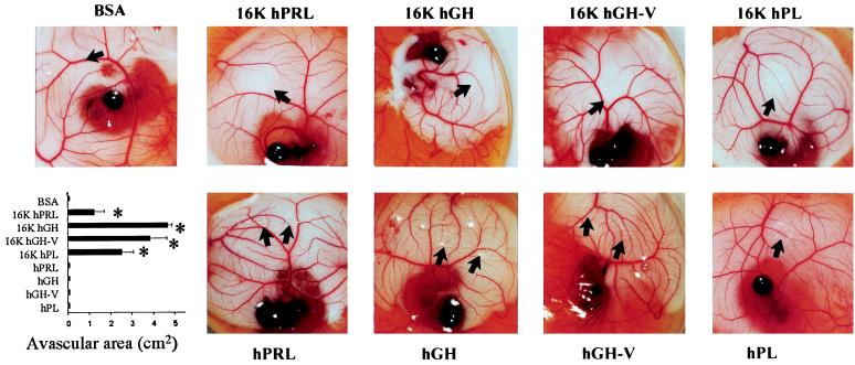
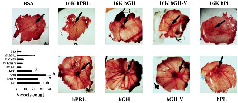
The in vivo activity of the intact molecules and fragments was examined in two different CAM assays. The CAM appears in the yolk sac at 48 h, grows rapidly over the next 6–8 days, and stops growing on day 10 (31). We performed an early-stage CAM bioassay (days 6–8) to assess each molecule’s effect on developing capillaries and a late-stage bioassay (days 10–14) to test their effect on nongrowing quiescent CAM. In the early-stage CAM assay, an avascular area was clearly present surrounding the disks containing the 16-kDa fragments (Fig. 2). The full length hormones had no effect in this bioassay. In the late-stage bioassay, the intact proteins stimulated new capillary and blood vessel formation, which could be observed emerging from the protein-containing disks whereas the 16-kDa fragments had no effect (Fig. 3).
Figure 2.
Early-stage CAM assay showing inhibition of angiogenesis in the CAM: Representative examples of CAM are taken from a typical experiment. The disks are visible by light reflection, and the black arrow shows the border of the disk or the border of the avascular area, if present. The lower left panel shows the quantification of the assay performed by measuring the area devoid of capillaries in the region surrounding the disk. Values are means ± SEM. ∗, P < 0.01 vs. BSA.
Figure 3.
Late-stage CAM assay showing stimulation of blood vessel formation in the CAM: Representative examples of CAM are taken from a typical experiment. The black arrow shows the border of the disk. The lower left panel shows the quantification of the assay performed by counting the number of blood vessels emerging from the disk. Vessels were scored in function of size from 1 (small) to 3 (large). Values are means ± SEM. ∗, P < 0.05 vs. BSA.
Activation of the MAPK Signaling Pathway.
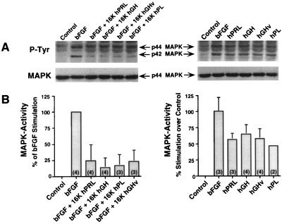
The signaling pathway by which 16K hPRL blocks the mitogenic activity of bFGF or vascular endothelial growth factor has been characterized partially (28). 16K hPRL inhibits the activation and tyrosine phosphorylation of MAPKs downstream of bFGF or vascular endothelial growth factor receptor phosphorylation. Western blot analyses and in vitro enzyme assays (Fig. 4) revealed that all of the 16-kDa fragments inhibited tyrosine phosphorylation and activation of MAPKs induced by bFGF. As shown in Fig. 4, treatment with all of the intact hormones induced the tyrosine phosphorylation and activation of MAPK in capillary endothelial cells.
Figure 4.
Fragments (16 kDa) inhibit bFGF-dependent MAPK tyrosine phosphorylation and activity whereas full length hormones stimulate these processes in the absence of bFGF. BBCE cells were treated for 5 min with the purified 16-kDa fragments (10 nM) and bFGF (250 pM) or for 10 min with the intact hormones (10 nM) but without bFGF or were left untreated for control. (A) P-Tyr, Western blots with the antityrosine antibody at the level of the MAPK p42 and p44; MAPK, the Western blot shown in the P-Tyr panel, stripped and reprobed with an anti-MAPK antibody. (B) In-gel MAPK activity. Numbers at the bottom of the histograms represent the number of experiments performed.
Regulation of PAI-1 Levels.
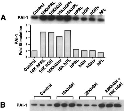
16K PRL was recently shown to stimulate the protein and mRNA levels of PAI-1 in capillary endothelial cells (29). This is consistent with the action of an antiangiogenic factor because PAI-1 limits degradation of the extracellular matrix by urokinase plasminogen activator, thus preventing angiogenesis (32). Activation of PAI-1 expression by 16K PRL is independent of the prior treatment of the cells with bFGF. All four 16-kDa fragments stimulated the cellular content of PAI-1 protein ≈4-fold in BBCE cells whereas the intact molecules had no effect on PAI-1 levels (Fig. 5A). A similar response was observed for PAI-1 secreted into the medium (data not shown). As shown in Fig. 5B, a 5-fold excess of hGH failed to inhibit the stimulation of PAI-1 expression induced by 16K hGH, providing direct evidence that the receptors mediating the actions of the intact molecules and N-terminal fragments are different.
Figure 5.
Stimulation of PAI-1 expression in BBCE cells by the 16-kDa fragments. Mouse antibovine PAI-1 Western blotting were performed on extracts of BBCE cells prestimulated by 20 nM of the recombinant proteins or were left untreated for control. (A) (Upper) Fragments (16 kDa) stimulate PAI-1 expression; full length hormones do not. (Lower) Quantification. (B) Competition between hGH (22K hGH) and 16K hGH. BBCE cells were treated with 20 nM 16K hGH (16K hGH lanes), with 20 nM hGH (22K hGH lanes), or with 20 nM both 16K hGH and hGH (16K hGH plus 22K hGH lanes) or were untreated (control lanes).
Immunodepletion Experiments.
The similarity of the four 16-kDa fragment biological responses raises the question of the presence of a possible contaminant that could be responsible for the effects. The presence of a contaminant at the same concentration in all of the 16-kDa fragment preparations is unlikely because they were prepared by using different production and purification protocols. Furthermore, 16K hPRL, 16K hGH-V, and 16K hPL were generated by cleavage of full length recombinant hPRL, hGH-V, and hPL, which showed opposite effects on angiogenesis. Nevertheless, we performed immunoneutralization experiments to demonstrate that antisera crossreacting with 16K hPRL, 16K hGH, 16K hGH-V, and 16K hPL were able to block the stimulation of PAI-1 levels induced by the fragments.
Treatment of BBCE cells with the four 16-kDa fragments increased PAI-1 accumulation in the conditioned media (Fig. 6A). This stimulation was abolished when the 16-kDa fragments were preincubated with their respective antisera before the addition to the cell cultures (Fig. 6A). The immunoneutralization of the stimulation of PAI-1 by 16K hPRL (Fig. 6B) and 16K hGH-V (Fig. 6C) is dose-dependent because increasing dilutions of the antisera progressively restore the full activity of the 16-kDa fragments. A similar dose-dependent inhibition was observed for the actions of 16K hGH and 16K hPL (data not shown).
Figure 6.
Immunodepletion experiments. PAI-1 levels were stimulated by the addition of 16-kDa fragments. The stimulation was inhibited after immunoneutralization of the 16-kDa fragments with their respective antisera. PAI-1 accumulation in the BBCE cell medium was measured by a mouse antibovine PAI-1 ELISA. (A) BBCE cells were treated with 50 nM of the indicated 16-kDa fragment in the presence or absence of its respective antiserum. (B) Stimulation of PAI-1 levels with 20 nM 16K hPRL was inhibited in a dose-dependent manner by the addition of increasing amounts of anti-hPRL antiserum. (C) Similarly, stimulation of PAI-1 levels by 20 nM 16K hGH-V was inhibited in a graded fashion by the anti-hGH antiserum. The data (in A) are expressed as optical density (O.D.) per microgram of cell protein, 0 being the level obtained with DMEM. The PAI-1 levels (in B and C) are expressed as the percentage of the stimulation obtained with the 16-kDa fragment alone, 0% being the level obtained with DMEM. Numbers at the bottom of the histogram represent the antiserum dilution in the well. Data represent the mean of duplicate measures (in A–C).
DISCUSSION
Our findings demonstrate that angiogenic activity is a general property of the members of the human PRL/GH family with the intact molecules being angiogenic and their N-terminal fragments being antiangiogenic. In vivo, intact and N-terminal fragments show opposite actions in two separate bioassays performed at different stages of CAM development. On growing capillaries, the 16-kDa fragments prevent angiogenesis, and the full length hormones have no effect, whereas on the quiescent vasculature, intact hormones stimulate blood vessel formation, and the 16-kDa fragments have no effect. In vitro, all four 16-kDa fragments inhibit BBCE cell proliferation whereas hGH and hGH-V are stimulatory. Of interest, intact hPRL and hPL are not stimulatory. The inability of observing an effect in BBCE cells could be explained by the recent preliminary observation that BBCE cells express and secrete a prolactin-like molecule and antibodies against bPRL inhibit BBCE cell growth (33). The presence of the endogenous hormone would mask the effects of the added hPRL and hPL. This explanation is consistent with the actions of hGH and hGH-V being mediated by GH receptors present on the BBCE cells whereas the actions of hPRL and hPL would be expected to be mediated via PRL receptors (5).
The opposite actions of the full length and cleaved hormones appear to be mediated by independent receptors rather than by competition for binding to the same receptor. A saturable, high-affinity binding site for 125I-labeled rat 16K PRL has been described on BBCE cell membranes. Intact GH and PRL do not compete for this receptor (34). On the other hand, 16K hPRL does not compete with hPRL for binding to the PRL receptor (data not shown). Data showing that the addition of excess hGH had no effect on the stimulation of PAI-1 expression by 16K hGH further supports the hypothesis that the N-terminal fragments act at separate receptors. To date, we have no data as to whether the 16-kDa fragments act at one receptor or multiple receptors. It appears that, if multiple receptors for the fragments exist, they signal via similar pathways because they all stimulate PAI-1 expression and inhibit the proliferative action of bFGF.
We have obtained preliminary data that the MAPK signaling pathway may mediate the related actions of the intact molecules and N-terminal fragments. 16K hPRL has been shown to inhibit activation of the MAPK signaling pathway by bFGF (28). The inhibition of MAPK activation by 16K hPRL appears to occur at the level of Ras activation (35). Although the Janus kinase/signal transducer and activator of transcription cascade is presumably the major signaling pathway for the PRL and GH receptors, activation of the MAPK pathway by GH and PRL has been reported in several biological systems also (36, 37). Stimulation of PAI-1 expression by the N-terminal fragments does not require the activation of bFGF receptors and would appear to be mediated by a more proximal signaling event not involving MAPK activation. This hypothesis is consistent with the finding that the intact molecules, which activate MAPK, have no effect on PAI-1 expression.
Angiogenesis is essential in several physiological processes, such as the formation of embryonic tissues or placental development, in which induction and cessation of angiogenesis appear to be highly regulated through the expression/repression of angiogenic/antiangiogenic factors (38). It has been suggested that, in the mouse placenta, initiation and cessation of vascularization correlates with the sequential expression of PLF and PRP (9). These related members of the PRL/GH family have been shown, respectively, to stimulate and inhibit angiogenesis.
Unlike the opposite actions of PLF and PRP, both the angiogenic and antiangiogenic actions of the PRL/GH family members reside within the same molecule. The idea that a fragment of a larger molecule is antiangiogenic agrees with studies using several other peptides (39). However, the peptides of the PRL/GH family are novel in that, in addition to the N-terminal fragments being antiangiogenic, the intact molecules are angiogenic. The ratio of intact to cleaved molecules would constitute an angiogenic switch regulated by the expression level of the protein and of the proteases responsible for their processing.
The angiogenic factors (intact hormones) can be cleaved by several enzymes (14, 18) to generate antiangiogenic factors (the 16-kDa fragments). The hormone and protease responsible for formation of a fragment should be located in the same compartment, and their levels should be correlated with angiogenic activity. Consistent with this hypothesis hPRL, hGH-V, hPL, cathepsin D—an enzyme that cleaves rat PRL (14) and hPRL (40)—and thrombin—an enzyme that cleaves hGH-V—are produced at the deciduoplacental interface (6–8). We hypothesize that regulation of the production and proteolytic cleavage of the PRL/GH family members could constitute a mechanism for modulating vascularization of the human placenta. Regulation of placental vascularization has important clinical implications because impairment of vascular development is observed in pregnancies complicated by preeclampsia and intrauterine fetal growth retardation (41).
Angiogenesis is essential for tumor growth and metastasis. It is generally assumed that proteases produced by tumors contribute to the release of angiogenic factors from stroma and are thus considered as positive modulators of angiogenesis (42). On the other hand, numerous cancers secrete cathepsin D (43), which, according to our model, could lead to the expression of the antiangiogenic factor, 16K hPRL. Simultaneous production by the tumor of an angiogenic factor and an antiangiogenic factor is not inconsistent with current thinking concerning tumor progression. It is now assumed that the tumor progression requires secretion of an angiogenic molecule for neovascularization of the primary tumor and, at the same time, an antiangiogenic factor involved in the inhibition of the growth of the micrometastases (44). Furthermore, the use of antiangiogenic factors to inhibit tumor progression is being tested widely. In addition to 16K PRL, this study identified three antiangiogenic factors that represent potential therapeutic agents for the treatment of cancer or other diseases whose etiology necessarily involves angiogenesis.
Acknowledgments
We acknowledge Prof. H. Vindevogel for his help with the CAM assays and Sylvie Devos for her excellent technical assistance in handling the eggs. We thank Dr. P. Jacquemin for providing RNA from human syncytiotrophoblast cells. We thank Dr. A. F. Parlow and the National Institute of Diabetes and Digestive and Kidney Disease’s National Hormone and Pituitary Program for providing specific antisera to hPRL, hGH, and hPL. We finally thank Dr. W. Fantl for critical reading of the manuscript. This work was supported by grants from the Human Frontier Science Program (to R.I.W. and J.A.M.), Deutsche Forschungsgesellschaft (to F.B.), Televie (to J.A.M.), and les Services Fédéraux des Affaires Scientifiques, Techniques et Culturelles de Belgique PAI P4/30 (to J.A.M.).
ABBREVIATIONS
- hPRL
human prolactin
- hGH
human growth hormone
- hPL
human placental lactogen
- hGH-V
human placental variant of growth hormone
- 16K
16-kDa N-terminal fragment
- MAPK
mitogen-activated protein kinase
- PAI-1
type 1 plasminogen activator inhibitor
- PLF
proliferin
- PRP
proliferin-related peptide
- BBCE cell
bovine brain capillary endothelial cell
- bFGF
basic fibroblast growth factor
- CAM
chicken chorioallantoic membrane
References
- 1.Nicoll C S, Mayer G L, Russel S M. Endocr Rev. 1986;7:169–203. doi: 10.1210/edrv-7-2-169. [DOI] [PubMed] [Google Scholar]
- 2.Clarke W C, Bern H A. In: Hormonal Proteins and Peptides. Li C H, editor. New York: Academic; 1980. pp. 105–107. [Google Scholar]
- 3.Murphy W J, Rui H, Longo D L. Life Sci. 1995;57:1–14. doi: 10.1016/0024-3205(95)00237-z. [DOI] [PubMed] [Google Scholar]
- 4.Lewis U J. Trends Endocrinol Metab. 1992;3:117–121. doi: 10.1016/1043-2760(92)90099-m. [DOI] [PubMed] [Google Scholar]
- 5.Goffin V, Shiverick K T, Kelly P A, Martial J A. Endocr Rev. 1996;17:385–410. doi: 10.1210/edrv-17-4-385. [DOI] [PubMed] [Google Scholar]
- 6.Ben-Jonathan N, Mershon J L, Allen D L, Steinmetz R W. Endocr Rev. 1996;17:638–669. doi: 10.1210/edrv-17-6-639. [DOI] [PubMed] [Google Scholar]
- 7.Soares M J, Faria T N, Roby K F, Deb S. Endocr Rev. 1991;12:402–423. doi: 10.1210/edrv-12-4-402. [DOI] [PubMed] [Google Scholar]
- 8.Walker W H, Fitzpatrick S L, Barrera-Saldana H A, Resendez-Perez D, Saunders G F. Endocr Rev. 1991;12:316–329. doi: 10.1210/edrv-12-4-316. [DOI] [PubMed] [Google Scholar]
- 9.Jackson D, Volpert O V, Bouck N, Linzer D I H. Science. 1994;266:1581–1584. doi: 10.1126/science.7527157. [DOI] [PubMed] [Google Scholar]
- 10.Rymaszewky Z, Cohen R H, Chomczynski P. Proc Natl Acad Sci USA. 1991;88:617–621. doi: 10.1073/pnas.88.2.617. [DOI] [PMC free article] [PubMed] [Google Scholar]
- 11.Gould J, Aramburo C, Capdevielle M, Scanes C G. Life Sci. 1995;56:587–594. doi: 10.1016/0024-3205(94)00491-a. [DOI] [PubMed] [Google Scholar]
- 12.Ferrara N, Clapp C, Weiner R I. Endocrinology. 1991;129:896–900. doi: 10.1210/endo-129-2-896. [DOI] [PubMed] [Google Scholar]
- 13.Clapp C, Martial J A, Guzman R C, Rentier-Delrue F, Weiner R I. Endocrinology. 1993;133:1292–1299. doi: 10.1210/endo.133.3.7689950. [DOI] [PubMed] [Google Scholar]
- 14.Baldocchi R A, Tan L, King D S, Nicoll C S. Endocrinology. 1993;133:935–938. doi: 10.1210/endo.133.2.8344226. [DOI] [PubMed] [Google Scholar]
- 15.Clapp C. Endocrinology. 1987;121:2055–2064. doi: 10.1210/endo-121-6-2055. [DOI] [PubMed] [Google Scholar]
- 16.Sinha Y N. Endocr Rev. 1995;16:354–369. doi: 10.1210/edrv-16-3-354. [DOI] [PubMed] [Google Scholar]
- 17.Torner L, Mejia S, Lopez-Gomez F J, Quintanar A, Martinez De La Escalera G, Clapp C. Endocrinology. 1995;136:5454–5460. doi: 10.1210/endo.136.12.7588295. [DOI] [PubMed] [Google Scholar]
- 18.Aston L, Li C H, Cheng C H K, Jibson M D. Biochemistry. 1981;20:7251–7258. doi: 10.1021/bi00528a031. [DOI] [PubMed] [Google Scholar]
- 19.Warner M D, Sinha Y N, Peabody C A. Horm Metab Res. 1993;25:425–429. doi: 10.1055/s-2007-1002137. [DOI] [PubMed] [Google Scholar]
- 20.Fukuoka H, Hamamoto R, Higurashi M. Horm Res. 1991;35:58–63. doi: 10.1159/000181931. [DOI] [PubMed] [Google Scholar]
- 21.L’hoir C, Renard A, Martial J A. Gene. 1990;89:47–52. doi: 10.1016/0378-1119(90)90204-5. [DOI] [PubMed] [Google Scholar]
- 22.Cooke N E, Coit D, Shine J, Baxter J, Martial J A. J Biol Chem. 1981;256:4007–4016. [PubMed] [Google Scholar]
- 23.Martial J A, Hallewell R A, Baxter J D, Goodman H M. Science. 1979;205:602–607. doi: 10.1126/science.377496. [DOI] [PubMed] [Google Scholar]
- 24.Paris N, Rentier-Delrue F, Defontaine A, Goffin V, Lebrun J J, Mercier L, Martial J A. Biotechnol Appl Biochem. 1990;12:436–449. [PubMed] [Google Scholar]
- 25.Gospodarowicz D, Massoglia S, Cheng J, Fujii D K. J Cell Biol. 1986;127:121–136. doi: 10.1002/jcp.1041270116. [DOI] [PubMed] [Google Scholar]
- 26.Taylor S, Folkman J. Nature (London) 1982;297:307–312. doi: 10.1038/297307a0. [DOI] [PubMed] [Google Scholar]
- 27.Risau W. Proc Natl Acad Sci USA. 1986;83:3855–3859. doi: 10.1073/pnas.83.11.3855. [DOI] [PMC free article] [PubMed] [Google Scholar]
- 28.D’Angelo G, Struman I, Martial J A, Weiner R I. Proc Natl Acad Sci USA. 1995;92:6374–6378. doi: 10.1073/pnas.92.14.6374. [DOI] [PMC free article] [PubMed] [Google Scholar]
- 29.Lee H, Struman I, Clapp C, Martial J A, Weiner R I. Endocrinology. 1998;139:3696–3703. doi: 10.1210/endo.139.9.6194. [DOI] [PubMed] [Google Scholar]
- 30.Bradford M M. Anal Biochem. 1976;72:248–254. doi: 10.1016/0003-2697(76)90527-3. [DOI] [PubMed] [Google Scholar]
- 31.Ausprunk D H, Kinghton D R, Folkman J. Dev Biol. 1974;38:237–248. doi: 10.1016/0012-1606(74)90004-9. [DOI] [PubMed] [Google Scholar]
- 32.Menashi S, Lu H, Soria C, Legrand Y. Baillieres Clin Haematol. 1993;6:559–576. doi: 10.1016/s0950-3536(05)80188-x. [DOI] [PubMed] [Google Scholar]
- 33.Clapp C, Lopez-Gomez F J, Nava G, Macotela J, Torner L, Duenas Z, Corbacho A, Arroyo A, Martinez De La Escalera G. The Endocrine Society Abstracts. Bethesda, MD: The Endocrine Society Press; 1996. p. 99. [Google Scholar]
- 34.Clapp C, Weiner R I. Endocrinology. 1992;130:1380–1386. doi: 10.1210/endo.130.3.1311239. [DOI] [PubMed] [Google Scholar]
- 35.D’Angelo G, Martini J-F, Weiner R I. The Endocrine Society Abstracts. Bethesda, MD: The Endocrine Society Press; 1997. p. 81. [Google Scholar]
- 36.Camarillo I G, Linebaugh B E, Rillema J A. Proc Soc Exp Biol Med. 1997;215:198–202. doi: 10.3181/00379727-215-44129. [DOI] [PubMed] [Google Scholar]
- 37.Winston L A, Hunter T. J Biol Chem. 1995;270:30837–30840. doi: 10.1074/jbc.270.52.30837. [DOI] [PubMed] [Google Scholar]
- 38.Bouck N, Stellmach V, Hsu S C. Adv Cancer Res. 1996;69:135–173. doi: 10.1016/s0065-230x(08)60862-3. [DOI] [PubMed] [Google Scholar]
- 39.Hanahan D, Folkman J. Cell. 1996;86:353–364. doi: 10.1016/s0092-8674(00)80108-7. [DOI] [PubMed] [Google Scholar]
- 40.Bentzien F, Vigne J-L, Weiner R I, Taylor R N. The Endocrine Society Abstracts. Bethesda, MD: The Endocrine Society Press; 1997. p. 143. [Google Scholar]
- 41.Friedman S A, Taylor R N, Roberts J M. In: Clinics in Perinatology. Sibai B M, editor. Philadelphia: Saunders; 1991. p. 661. [Google Scholar]
- 42.Liotta L A, Steeg P S, Stetler-Stevenson W G. Cell. 1991;64:327–336. doi: 10.1016/0092-8674(91)90642-c. [DOI] [PubMed] [Google Scholar]
- 43.Scharwtz M K. Clin Chim Acta. 1995;237:67–78. [Google Scholar]
- 44.O’Reilly M S, Holmgren L, Chen C, Folkman J. Nat Med. 1996;2:689–692. doi: 10.1038/nm0696-689. [DOI] [PubMed] [Google Scholar]


