Abstract
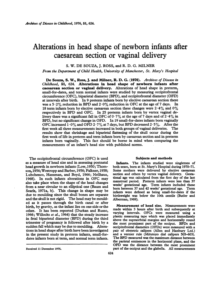
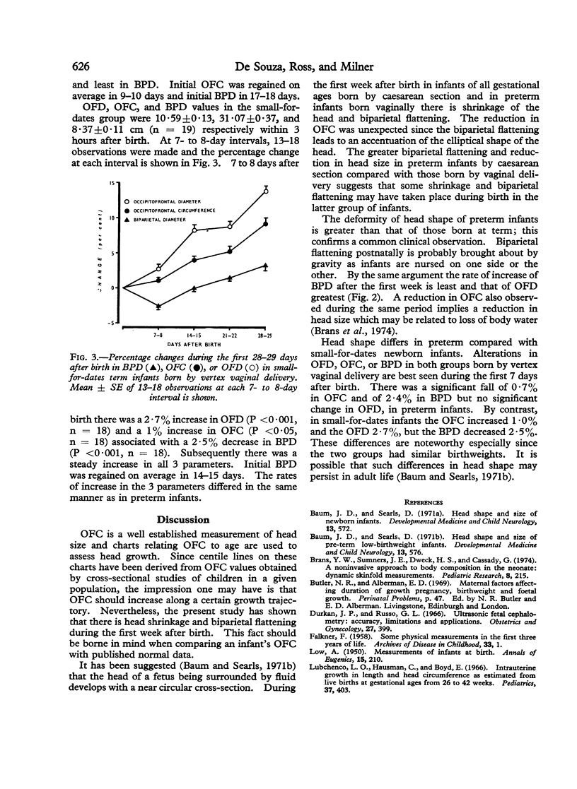
Alterations of head shape in preterm, small-for-dates, and term normal infants were studied by measuring occipitofrontal circumference (OFC), biparietal diameter (BPD), and occipitofrontal diameter (OFD) at intervals after birth. In 9 preterm infants born by elective caesarean section ther was a 5-2% reduction in BPD and 2-0% reduction in OFC at the age of 7 days. In 18 term infants born by elective caesarean section these changes were 2-4% and 0% respectively in BPD and OFC. In 25 preterm infants born by vertex vaginal delivery there was a significant fall in OFC of 0-7% at the age of 7 days and of 2-4% in BPD, but no significant change in OFD. In 19 small-for-dates infants born vaginally OFC increased 1-0% and OFD 2-7% at 7 days, but BPD decreased 2-5%. After the first week all three measurements increased in both groups of vaginal deliveries. The results show that shrinkage and biparietal flattening of the skull occur during the first week of life in preterm and term infants born by caesarean section and in preterm infants born vaginally. This fact should be borne in mind when comparing the measurements of an infant's head size with published norms.
Full text
PDF


Selected References
These references are in PubMed. This may not be the complete list of references from this article.
- Baum J. D., Searls D. Head shape and size of newborn infants. Dev Med Child Neurol. 1971 Oct;13(5):572–575. doi: 10.1111/j.1469-8749.1971.tb08319.x. [DOI] [PubMed] [Google Scholar]
- Baum J. D., Searls D. Head shape and size of pre-term low-birthweight infants. Dev Med Child Neurol. 1971 Oct;13(5):576–581. doi: 10.1111/j.1469-8749.1971.tb08320.x. [DOI] [PubMed] [Google Scholar]
- Brans Y. W., Sumners J. E., Dweck H. S., Cassady G. A noninvasive approach to body composition in the neonate: dynamic skinfold measurements. Pediatr Res. 1974 Apr;8(4):215–222. doi: 10.1203/00006450-197404000-00001. [DOI] [PubMed] [Google Scholar]
- Durkan J. P., Russo G. L. Ultrasonic fetal cephalometry: accuracy, limitations, and applications. Obstet Gynecol. 1966 Mar;27(3):399–403. [PubMed] [Google Scholar]
- FALKNER F. Some physical measurements in the first three years of life. Arch Dis Child. 1958 Feb;33(167):1–9. doi: 10.1136/adc.33.167.1. [DOI] [PMC free article] [PubMed] [Google Scholar]
- Lubchenco L. O., Hansman C., Boyd E. Intrauterine growth in length and head circumference as estimated from live births at gestational ages from 26 to 42 weeks. Pediatrics. 1966 Mar;37(3):403–408. [PubMed] [Google Scholar]
- Nellhaus G. Head circumference from birth to eighteen years. Practical composite international and interracial graphs. Pediatrics. 1968 Jan;41(1):106–114. [PubMed] [Google Scholar]
- THOMSON J. Infant growth. Arch Dis Child. 1956 Oct;31(159):382–389. doi: 10.1136/adc.31.159.382. [DOI] [PMC free article] [PubMed] [Google Scholar]


