

Abstract
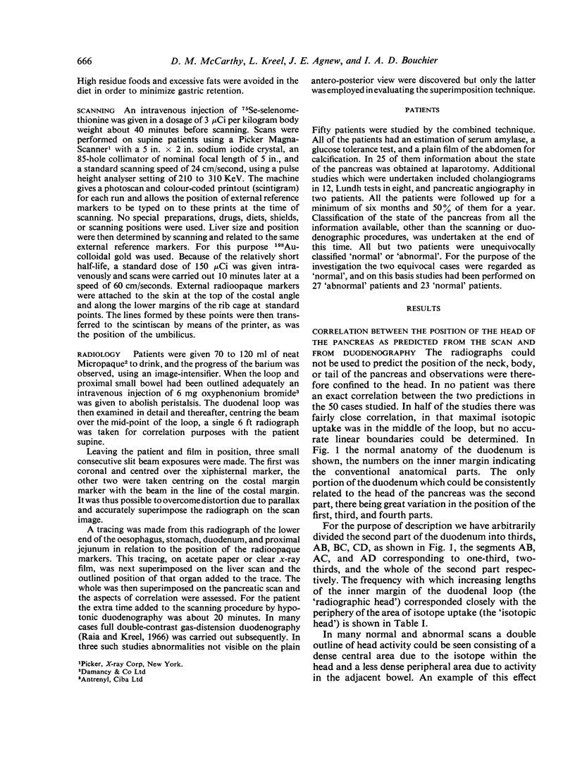
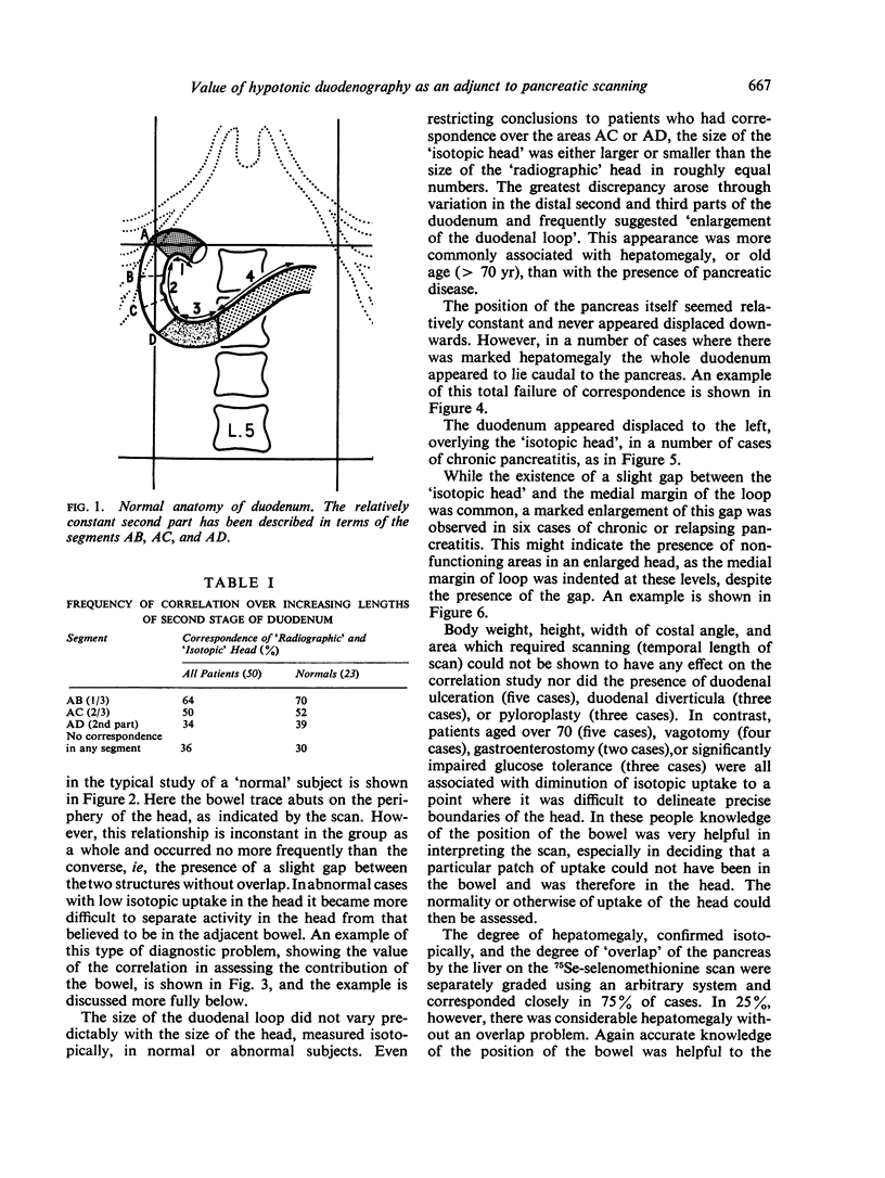
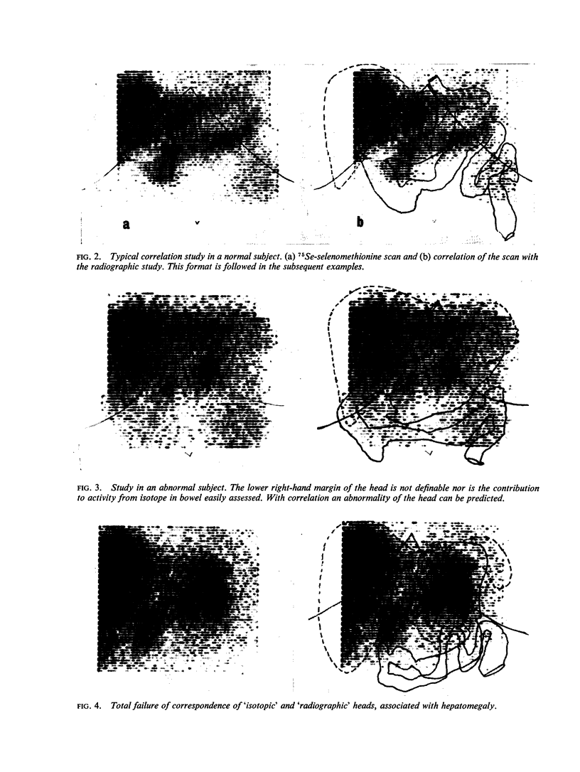
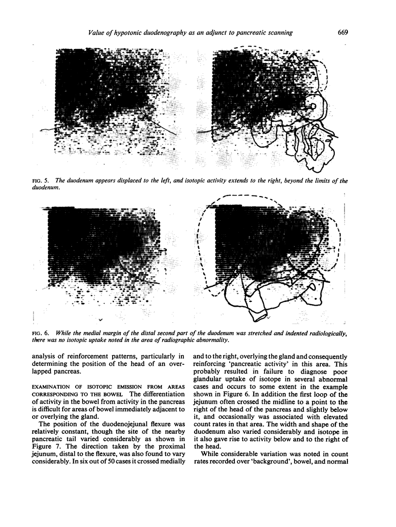
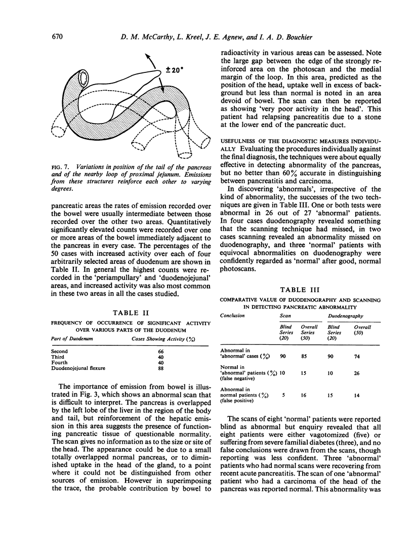
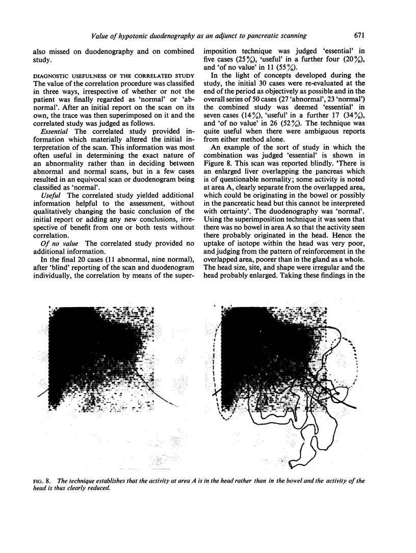
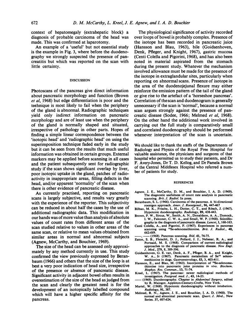
A technique has been developed which allows the position of the upper gastrointestinal tract, as determined radiographically, to be superimposed on the pancreatic photoscan, performed after an injection of 75Se-selenomethionine. Results of the technique applied to 27 `abnormal' and 23 `normal' subjects are described. The importance of the additional information gained by the combined technique is discussed in terms of normal variation and increased diagnostic yield, and the situations in which it was found most useful are described. The addition of correlated duodenography rendered scan reporting more objective in 50% of cases.
Full text
PDF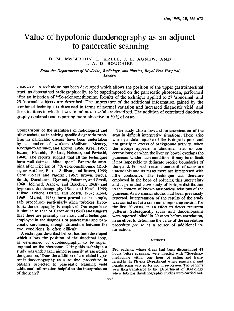
Images in this article
Selected References
These references are in PubMed. This may not be the complete list of references from this article.
- Beranbaum S. L. Carcinoma of the pancreas. A bi-directional roentgen approach. Am J Roentgenol Radium Ther Nucl Med. 1966 Feb;96(2):447–467. doi: 10.2214/ajr.96.2.447. [DOI] [PubMed] [Google Scholar]
- Bilbao M. K., Frische L. H., Dotter C. T., Rösch J. Hypotonic duodenography. Radiology. 1967 Sep;89(3):438–443. doi: 10.1148/89.3.438. [DOI] [PubMed] [Google Scholar]
- Colella A. C., Pigorini F. Experiences in pancreas scanning using 75-Se-selenomethionine. Br J Radiol. 1967 Sep;40(477):662–669. doi: 10.1259/0007-1285-40-477-662. [DOI] [PubMed] [Google Scholar]
- Eaton S. B., Fleischli D. J., Pollard J. J., Nebesar R. A., Potsaid M. S. Comparison of current radiologic approaches to the diagnosis of pancreatic disease. N Engl J Med. 1968 Aug 22;279(8):389–396. doi: 10.1056/NEJM196808222790801. [DOI] [PubMed] [Google Scholar]
- HANSSON E., BLAU M. INCORPORATION OF SE75-SELENOMETHIONINE INTO PANCREATIC JUICE PROTEINS IN VIVO. Biochem Biophys Res Commun. 1963 Sep 10;13:71–74. doi: 10.1016/0006-291x(63)90165-7. [DOI] [PubMed] [Google Scholar]
- Kreel L. The pancreas: newer radiological methods of investigation. Postgrad Med J. 1967 Jan;43(495):14–23. doi: 10.1136/pgmj.43.495.14. [DOI] [PMC free article] [PubMed] [Google Scholar]
- Lasky H. J. A new mammographic technic. Radiology. 1968 Aug;91(2):381–383. doi: 10.1148/91.2.381. [DOI] [PubMed] [Google Scholar]
- Melmed R. N., Agnew J. E., Bouchier I. A. The normal and abnormal pancreatic scan. Q J Med. 1968 Oct;37(148):607–624. [PubMed] [Google Scholar]
- Raia S., Kreel L. Gas-distension, double-contrast duodenography using the Scott-Harden gastroduodenal tube. Gut. 1966 Aug;7(4):420–424. doi: 10.1136/gut.7.4.420. [DOI] [PMC free article] [PubMed] [Google Scholar]
- Rodríguez-Antúnez A., Filson E. J., Sullivan B. H., Jr, Brown C. H. Photoscanning in diagnosis of carcinoma of the pancreas. Ann Intern Med. 1966 Oct;65(4):730–737. doi: 10.7326/0003-4819-65-4-730. [DOI] [PubMed] [Google Scholar]
- Sodee D. B. Pancreatic scanning. Radiology. 1966 Oct;87(4):641–645. doi: 10.1148/87.4.641. [DOI] [PubMed] [Google Scholar]